




Volume 195 - Issue 7
Interferon-α-related microscopic polyangiitis in a patient with chronic hepatitis C infection
Authors: Stephen Y Oh, Brett E Jones and Suran L Fernando
Med J Aust 2011; 195 (7): 385-386. || doi: 10.5694/mja11.10249
Published online: 3 October 2011
Published online: 3 October 2011
To the Editor: A 38-year-old man with genotype 1b chronic hepatitis C infection had been treated with 48 weeks of pegylated interferon (IFN)-α and ribavirin. Autoimmune serology performed just before treatment showed positive perinuclear antineutrophil cytoplasmic antibodies (ANCA) accompanied by an elevated antimyeloperoxidase antibody level (22 U/mL; reference range [RR], < 5 U/mL). Notably, the patient was ANCA-negative 12 months previously. The treatment course was uneventful.
One month after the completion of therapy, the patient presented with a subacute onset of fever, haemoptysis, breathlessness and generalised arthralgia.
Laboratory investigations demonstrated raised levels of C-reactive protein (128 mg/L; RR, < 5 mg/L) and creatinine (159 μmol/L; RR, 64–104 μmol/L), low albumin concentration (28 g/L; RR, 35–46 g/L), a low haemoglobin level (64 g/L; RR, 135–180 g/L), and microcytic hypochromic anaemia. Autoimmune serology showed persistence of a positive ANCA and an elevated antimyeloperoxidase antibody level (29 U/mL). A high-resolution computed tomography scan of the chest showed features of interstitial lung disease (Figure, A) and a renal biopsy demonstrated pauci-immune necrotising glomerulonephritis (Figure, B). These findings were consistent with a diagnosis of microscopic polyangiitis.
Despite treatment with intravenous cyclophosphamide and pulse methylprednisolone, the patient deteriorated and was admitted to the intensive care unit for ventilatory support, haemodialysis and plasmapheresis. This admission lasted 4 weeks and was complicated by line-related sepsis and persistent anaemia requiring multiple blood transfusions. The patient remains clinically well 14 months after discharge, with negative ANCA and negative hepatitis C virus RNA polymerase chain reaction consistent with a sustained viral response.
Autoimmune disease is a well recognised complication of IFN-α therapy in chronic hepatitis C infection. The clinical manifestations of IFN-α-related autoimmune disease can be either organ-specific (thyroiditis, psoriasis) or, less commonly, systemic (rheumatoid arthritis, lupus-like disease, sarcoidosis).1 IFN-α-based treatment in chronic hepatitis C infection unmasks silent autoimmune processes, or induces de novo autoimmune diseases or autoantibodies.2 A predisposition to autoimmunity, together with the presence of baseline auto-antibodies, has been demonstrated in most instances of IFN-α-mediated autoimmune diseases,1 as observed in our case.
Although rare, the diagnosis of microscopic polyangiitis needs to be considered in patients treated with IFN-α-based therapy for chronic hepatitis C infection presenting with skin rash, fevers, arthritis, an active urine sediment, renal failure or pulmonary haemorrhage. One could consider reducing the duration of therapy in patients who achieve negative RNA polymerase chain reaction at Week 4 of treatment. There is emerging evidence that 24 weeks of response-guided therapy in genotype 1b chronic hepatitis C infection is as effective as the standard-of-care treatment for 48 weeks in rapid responders.3 We also suggest that, in the presence of a positive ANCA at baseline screening, a chest x-ray and urinalysis be performed before initiating IFN-α-based treatment, and that patients be monitored clinically and with urinalysis during treatment. A chest x-ray should also be performed at the completion of treatment. The presence of auto-antibodies alone is not a contraindication to IFN-α therapy, but it does mandate careful monitoring and a high index of suspicion of an immune diathesis.
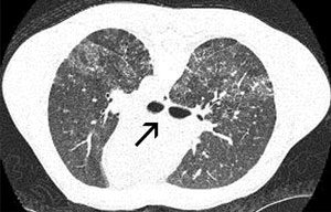 |
A: High-resolution computed tomography scan of the patients chest showing patchy foci of ground glass opacification and nodules bilaterally. |
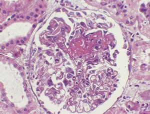 |
B: Renal biopsy showing focal crescentic necrotising glomerulonephritis (haematoxylin-eosin stain; original magnification x 400). |
Competing interests
No relevant disclosures.
References
- Dumoulin FL, Leifeld L, Sauerburch T, Spengler U. Autoimmunity induced by Interferon-αlpha therapy for chronic viral hepatitis. Biomed Pharmacother 1999; 53: 242-254.
- Dusheiko G. Side effects of alpha interferon in chronic hepatitis C. Hepatology 1997; 26 (3 Suppl 1): 112S-121S. CACEIEBI
- Yu ML, Dai CY, Huang JF, et al. Rapid virological response and treatment duration for chronic hepatitis C genotype 1 patients: a randomized trial. Hepatology 2008; 47: 1884-1893. CACFBEEE
Costs of Delivering the Hep B PAST Model of Care in Remote Australia: A Retrospective Pre–Post Study
Hoa Nguyen, Anh Le Tuan Nguyen, Paula Binks, Melita McKinnon, Emily Vintour-Cesar, Karen Wills, Nicola Stephens, Benjamin Cowie, Joshua S. Davis, Yuejen Zhao, Peter Nihill, Julie A. Campbell, Andrew J. Palmer, Jane Davies, Barbara de Graaff
Buruli Ulcer Acquired From Ringtail Possum Rescue
Daniel P. O'Brien, Naomi Clarke, Sara Bonnici, Mohammad A. Hussain
Q Fever Visceral Abscesses After Contact With Native Australian Marsupials
Samantha J. Laws, Enzo Binotto, James Stewart, Jennifer Ho, Trent Yarwood, Josh Hanson, Simon Smith
Hospital-Admitted Injection-Related Infections Among Incarcerated People Who Inject Drugs in Australia: A Retrospective Cohort Study
Andrew Palmer, Matthew Carter, Jeremy Yeo, Cecilia Shim, Jason Connor, Jeremy Hayllar, Gerald Holtmann, Naomi Moy, Elliott G. Playford, Naomi Runnegar, Paul J. Clark
The Live Attenuated Influenza Vaccine in Australia: An Additional Tool for Influenza Prevention
Cyra Patel, Alexis Pillsbury, Tran Nguyen, Xia Wang, Helen E. Quinn, Clayton K. Chiu, Allen C. Cheng, Katie L. Flanagan, Zhicheng Wang
Reflex Testing for Hepatitis D Infection: A Unique Opportunity to Reduce Hepatitis D-Related Chronic Liver Disease Deaths in Australia
Jessica Howell, Lauren Andersson, Miriam T. Levy, James O'Beirne, Leon Adams, Katharine Irvine, Avik Majumdar, Golo Ahlenstiel, Kathy Jackson, Krispin Hajkowicz, Joseph Doyle, Jane Davies, Sarah Cherian, Wayne Dimech, Alexander J. Thompson