




Medical education
Lessons from practice
Volume 220 - Issue 6
PEG‐GCSF‐induced aortitis in a patient with breast cancer: distinguishing between infective and immune‐mediated aortitis
Authors: Luigi Zolio, Prudence A Francis and Nava Ferdowsi
Med J Aust 2024; 220 (6): 307-309. || doi: 10.5694/mja2.52237
Published online: 1 April 2024
Published online: 1 April 2024
A 64-year-old woman presented with non-neutropenic fever and back pain one day after the first cycle of neoadjuvant chemotherapy
Clinical record
A 64‐year‐old woman presented with non‐neutropenic fever and back pain one day after the first cycle of neoadjuvant chemotherapy (doxorubicin–cyclophosphamide) for locally advanced breast cancer. She had also received pegylated granulocyte colony stimulating factor (PEG‐GCSF) one day after chemotherapy to enable dose‐dense chemotherapy scheduling. She was originally treated with intravenous antibiotics and discharged home on oral amoxicillin–clavulanate, with improvement in symptoms.
Her symptoms recurred on day 8 prompting admission. Initial investigations demonstrated raised white cell count with neutrophilia, extremely elevated C‐reactive protein (682mg/L; reference interval, <5mg/L), and negative blood and urine cultures (Box 1). Chest x‐ray was unremarkable. The patient received five days of intravenous broad‐spectrum antibiotics (piperacillin–tazobactam) with no resolution of fevers but no clinical deterioration.
A computed tomography (CT) scan of the abdomen and pelvis demonstrated smooth circumferential aortic mural thickening, stranding surrounding the origin of coeliac axis, superior mesenteric artery and bilateral renal arteries, suggestive of aortitis. There was no stenosis or aneurysmal dilatation, and intra‐abdominal organ appearance was normal.
Infectious disease consultation postulated a non‐infective cause, with no organism found and imaging features not supportive of infection. Rheumatology review identified no symptoms or signs of autoimmune disease associated with aortitis, including giant cell arteritis. A diagnosis of PEG‐GCSF‐induced aortitis was considered.
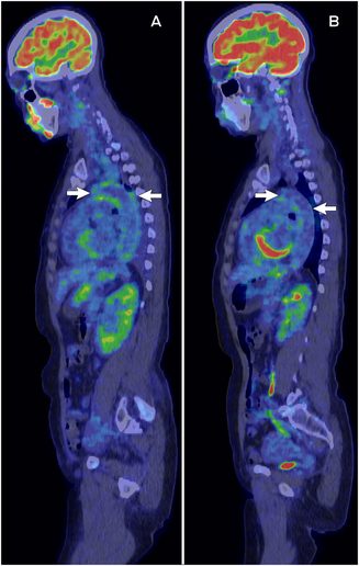
The patient commenced high dose oral prednisolone 50mg per day (1mg/kg), with rapid improvement in fever and inflammatory markers, enabling discharge from hospital on the following day. Positron emission tomography (PET)/CT completed within one week demonstrated moderate metabolic activity from the aortic arch, descending thoracic and abdominal aorta (Box 2, A).
On outpatient follow‐up, the patient remained asymptomatic of aortitis and proceeded with further cycles of non‐dose‐dense doxorubicin–cyclophosphamide chemotherapy without PEG‐GCSF, with no subsequent flare of symptoms. A re‐staging PET/CT completed after cessation of steroids (but ongoing chemotherapy) showed tumour response and resolution of aortitis (Box 2, B).
Discussion
This case highlights aortitis as a differential diagnosis for pyrexia of unknown origin, and the importance of distinguishing infective and non‐infective causes of aortitis to determine appropriate treatment. Aortitis may present with fevers, constitutional symptoms, neck, chest and/or abdominal pain, upper or lower limb claudication. It may be secondary to infective or immune‐mediated causes (Box 3).1 Although clinical features may be non‐specific, distinction is made through laboratory, imaging and, rarely, histological results. Imaging findings specific to infection include mycotic aneurysms, gas formation and focal crescenteric involvement, while immune‐mediated causes often appear with diffuse mural thickening and contiguous involvement of longer sections of the aorta.2
The patient's undifferentiated clinical presentation prompted empiric treatment and investigation for infection. However, once aortitis was recognised, negative microbial cultures and serology, imaging features, and non‐improvement with antibiotics suggested a non‐infective cause.
The case was also unusual for typical causes of immune‐mediated aortitis given the abrupt onset and preceding chemotherapy. A paraneoplastic cause was considered unlikely because symptoms began after initiation of chemotherapy and resolved rapidly after initiation of steroids within the first chemotherapy cycle. The cause of aortitis was attributed to GCSF due to abrupt onset, unusually high inflammatory markers, exclusion of other causes, and improvement with corticosteroids, with no recurrence of symptoms after taper and cessation.
GCSF‐induced aortitis is a rare complication of GCSF in patients receiving treatment for solid organ malignancies (breast, ovarian, uterine) and lymphoma. GCSF is being used more frequently with curative breast cancer chemotherapy to enable dose‐dense scheduling, which is associated with better cancer outcomes.3 Most reported cases have occurred in women aged 60years or older. Taxane agents are the most associated chemotherapeutic class. Most cases occur within ten days of GCSF administration and present with fever unresponsive to antibiotics, accompanied by neck, chest or abdominal pain. The thoracic aorta is the most commonly involved aortic segment. GCSF may lead to fever and high inflammatory markers through stimulation of interleukin (IL)‐1, IL‐6 and prostaglandins. Treatment includes discontinuation of GCSF and high dose oral corticosteroids (1mg/kg), tapered over several weeks, but some patients may improve solely with clearance of GCSF. Prognosis with treatment is usually benign,4 although complications of aortitis may include aneurysmal dilatation and death.1
Treatment for infective aortitis consists of targeted antimicrobial treatment. Immune‐mediated aortitis is treated with high dose intravenous or oral corticosteroids (1mg/kg) followed by tapering, with introduction of steroid‐sparing agents in persistent or treatment‐refractory cases.5 The choice of immunosuppressants is dependent on the underlying diagnosis and other end‐organ manifestations. Disease is monitored using clinical parameters, relevant laboratory markers (erythrocyte sedimentation rate, C‐reactive protein, angiotensin‐converting enzyme levels, antineutrophil cytoplasmic antibody titres) and imaging (PET/CT, CT and/or magnetic resonance imaging with contrast angiography).1 Exclusion of an infective cause is critically important before immunosuppression.
Lessons from practice
- Aortitis is a differential diagnosis for pyrexia of unknown origin, but its clinical features (fever, systemic symptoms, chest and abdominal pain) are non‐specific for its underlying aetiology.
- Causes of aortitis may be classified as infective and immune‐mediated. Granulocyte colony stimulating factor (GCSF) is a rare cause of immune‐mediated aortitis in patients with malignancy receiving GCSF as part of chemotherapy treatment.
- Exclusion of infective causes is critical before immunosuppression, through a combination of laboratory and imaging investigations.
Box 1 – Clinical record: relevant laboratory investigation findings
|
Investigation |
Measurements |
Reference interval |
|||||||||||||
|
|
|||||||||||||||
|
Full blood examination |
|
|
|||||||||||||
|
Hb (g/L) |
114 |
115–165 |
|||||||||||||
|
WCC (×109) |
11.4 |
4–11 |
|||||||||||||
|
Neutrophils (×109) |
10.1 |
2–7.5 |
|||||||||||||
|
Lymphocytes (×109) |
0.4 |
1–4 |
|||||||||||||
|
Platelets (×109) |
236 |
150–400 |
|||||||||||||
|
Creatinine, electrolytes and urea |
|
|
|||||||||||||
|
Creatinine (μmol/L) |
61 |
45–90 |
|||||||||||||
|
eGFR (mL/min/1.73m2) |
>90 |
>90 |
|||||||||||||
|
Urea (mmol/L) |
4.1 |
2.9–8.2 |
|||||||||||||
|
Liver function tests |
|
|
|||||||||||||
|
Albumin (g/L) |
24 |
35–50 |
|||||||||||||
|
ALP (U/L) |
186 |
30–110 |
|||||||||||||
|
GGT (U/L) |
124 |
0–35 |
|||||||||||||
|
ALT (U/L) |
38 |
<36 |
|||||||||||||
|
Bilirubin (μmol/L) |
12 |
1–20 |
|||||||||||||
|
C‐reactive protein (mg/L) |
682 |
<5 |
|||||||||||||
|
Erythrocyte sedimentation rate (mm/h) |
137 |
0–20 |
|||||||||||||
|
Troponin I (ng/L) |
6 |
<16 |
|||||||||||||
|
Autoimmune serology |
|
|
|||||||||||||
|
ANA |
Negative |
na |
|||||||||||||
|
ENA |
Negative |
na |
|||||||||||||
|
ANCA |
Negative |
na |
|||||||||||||
|
RF |
28 |
<14 |
|||||||||||||
|
IgG4 (g/L) |
0.05 |
0.04–0.86 |
|||||||||||||
|
Blood borne virus serology |
|
|
|||||||||||||
|
HBsAb, HBcAb, HBsAg |
Negative |
na |
|||||||||||||
|
HCV antibody |
Negative |
na |
|||||||||||||
|
HIV antibody |
Negative |
na |
|||||||||||||
|
Syphilis serology |
|
|
|||||||||||||
|
TPPA |
Reactive |
na |
|||||||||||||
|
RPR |
Non‐reactive |
na |
|||||||||||||
|
Urinary Streptococcus pneumoniae antigen |
Negative |
na |
|||||||||||||
|
Blood cultures |
Negative |
na |
|||||||||||||
|
Urine microscopy and culture |
Negative |
na |
|||||||||||||
|
Stool MCS/PCR |
No leukocytes/erythrocytes |
|
|||||||||||||
|
Faecal pathogen PCR |
Negative |
na |
|||||||||||||
|
Clostridium difficile toxin |
Negative |
na |
|||||||||||||
|
|
|||||||||||||||
|
ANA = antinuclear antibody; ANCA = antineutrophil cytoplasmic antibody; ALP = alkaline phosphatase; ALT = alanine transaminase; eGFR = estimated glomerular filtration rate; ENA = extractable nuclear antigen; GGT = γ‐glutamyl transferase; Hb = haemoglobin; HBcAb = hepatitis B core antibody; HBsAb = hepatitis B surface antibody; HBsAg = hepatitis B surface antigen; HCV = hepatitis C virus; HIV = human immunodeficiency virus; MCS = microscopy, culture and sensitivity; na = not applicable; PCR = polymerase chain reaction; RF = rheumatoid factor; RPR = rapid plasma reagin; TPPA = |
|||||||||||||||
Box 2 – Positron emission tomography/computed tomography (PET/CT) demonstrating aortic inflammation at the start of steroid treatment (A) and subsequent resolution after cessation of treatment (B)
Box 3 – Distinguishing between infective and immune‐mediated causes of aortitis
|
Aortitis characteristics |
Infective |
Immune‐mediated |
|||||||||||||
|
|
|||||||||||||||
|
Causes1 |
|
|
|||||||||||||
|
Laboratory investigations1 |
|
|
|||||||||||||
|
Imaging findings (CT/MRI/PET)2 |
|
|
|||||||||||||
|
|
|||||||||||||||
|
ACE = angiotensin‐converting enzyme; ANCA = antineutrophil cytoplasmic antibody; ASO = antistreptolysin O; CCP = cyclic citrullinated peptide; CT = computed tomography; CRP = C‐reactive protein; ESR = erythrocyte sedimentation rate; FDG = fluorodeoxyglucose; GCSF = granulocyte colony stimulating factor; HIV = human immunodeficiency virus; HLA‐B27 = human leukocyte antigen B27; MRI = magnetic resonance imaging; PET = positron emission tomography. |
|||||||||||||||
Competing interests
Prudence Francis received an honorarium for giving a lecture at a meeting organised by Eli Lilly.
References
- Pugh D, Grayson P, Basu N, Dhaun N. Aortitis: recent advances, current concepts and future possibilities. Heart 2021; 107: 1620–1629.
- Murphy DJ, Keraliya AR, Agrawal MD, et al. Cross‐sectional imaging of aortic infections. Insights Imaging 2016; 7: 801–818.
- Early Breast Cancer Trialists’ Collaborative Group (EBCTCG). Increasing the dose intensity of chemotherapy by more frequent administration or sequential scheduling: a patient‐level meta‐analysis of 37298 women with early breast cancer in 26 randomised trials. Lancet 2019; 393: 1440–1452.
- Hoshina H, Takei H. Granulocyte‐colony stimulating factor‐associated aortitis in cancer: a systematic literature review. Cancer Treat Res Commun 2021; 29: 100454.
- Maz M, Chung SA, Abril A, et al. 2021 American College of Rheumatology/Vasculitis Foundation guideline for the management of giant cell arteritis and Takayasu arteritis. Arthritis Rheumatol 2021; 73: 1349–1365.
Provenance: Not commissioned; externally peer reviewed.
The epidemiology of haemodialysis catheter infections in Australia, 2016–20: a prospective cohort study
Benjamin Lazarus, Kevan R Polkinghorne, Martin P Gallagher, Jayson Catiwa, Nicholas A Gray, Sarah Coggan, Kathryn R Higgins, Girish Talaulikar, Stephen P McDonald AM, Sradha Kotwal
30/60/90 National stroke targets and stroke unit access for all Australians: it's about time
For the 30/60/90 National Stroke Targets Taskforce
A summary of the 2023 Society of Obstetric Medicine of Australia and New Zealand (SOMANZ) hypertension in pregnancy guideline
Renuka Shanmugalingam, Helen L Barrett, Amanda Beech, Lucy Bowyer, Tim Crozier, Amanda Davidson, Marloes Dekker Nitert, Kerrie Doyle, Luke Grzeskowiak, Nicole Hall, Hicham Ibrahim Cheikh Hassan, Annemarie Hennessy, Amanda Henry, David Langsford, Vincent WS Lee, Zachary Munn, Michael J Peek, Joanne M Said, Helen Tanner, Rachel Taylor, Meredith Ward, Jason Waugh, Linda LY Yen, Ellie Medcalf, Katy JL Bell, Deonna Ackermann, Robin Turner, Angela Makris
Cough in Children and Adults: Diagnosis, Assessment and Management (CICADA). Summary of an updated position statement on chronic cough in Australia
David A Prentice
Catheter‐related superior vena cava syndrome: an increasing problem
John J Harvey, John I Vrazas, Elhamy Bekhit, Chris Barnes, Philip J Robinson
Australian evidence‐based guidelines for the prevention and management of diabetes‐related foot disease: a guideline summary
Peter A Lazzarini, Anita Raspovic, Jenny Prentice, Robert J Commons, Robert A Fitridge, James Charles, Jane Cheney, Nytasha Purcell, Stephen M Twigg