




Medical education
Lessons from practice
Volume 217 - Issue 10
Hypercalcaemia in an immunocompromised patient: consider Pneumocystis jirovecii pneumonia
Authors: Karen Bromley, Jessica Phillips and Ashley Irish
Med J Aust 2022; 217 (10): 517-518. || doi: 10.5694/mja2.51761
Published online: 21 November 2022
Published online: 21 November 2022
A 71-year-old man with a history of 6 weeks of generalised decline presented for outpatient clinic assessment
Clinical record
A 71‐year‐old man with a history of 6weeks of generalised decline presented for outpatient clinic assessment. Initial symptoms included fatigue, weakness, and anorexia causing 6kg weight loss. His medical history was significant for a live‐related renal transplant 20years earlier for membranous glomerulonephritis, insulin‐dependent type 2 diabetes mellitus, a right native nephrectomy 12‐months prior for clear cell renal cell carcinoma (Grade 3, pT1a), and multiple basal and squamous cell carcinomas of the skin. Maintenance immunosuppression was with mycophenolate mofetil 1g twice a day and cyclosporine 50mg twice a day without prednisolone. He had received one infusion of rituximab 800mg 4 months earlier for biopsy‐proven recurrent membranous glomerulonephritis. Further history and examination did not reveal any localising symptoms or signs.
Biochemistry showed new hypercalcaemia (corrected calcium, 3.04mmol/L; reference interval [RI], 2.1–2.6mmol/L), lower normal range parathyroid hormone (3.2pmol/L; RI, 1.6–9pmol/L), normal 25‐hydroxyvitamin D level (71nmol/L; RI, >50nmol/L; 1,25‐dihydroxyvitamin D level unavailable), and stable renal function (creatinine, 143μmol/L; RI, 60–110μmol/L). C‐reactive protein was raised (102mg/L; RI, <5mg/L). Total white‐cell count was normal (8.06×10⁹/L; RI, 4–11×109/L), with absolute neutrophils 5.31×10⁹/L (RI, 2.0–7.5×10⁹/L) and total lymphocytes 1.81×10⁹/L (RI, 1.21–4×10⁹L).
The patient's symptoms were judged to be from hypercalcaemia, and treatment with intravenous hydration and pamidronate was started with initial benefit. However, symptomatic hypercalcaemia recurred with each attempt to wean therapy. Given his biochemical findings and history, malignancy was considered a possible causative aetiology, but whole‐body computer tomography imaging was not diagnostic. A chest computed tomography scan showed bilateral ill‐defined ground glass infiltrates (Box), with an induced sputum polymerase chain reaction positive for Pneumocystis jirovecii (DNA, 952000copies/mL).
P. jirovecii pneumonia (PJP) was diagnosed, despite no respiratory signs or symptoms except for intermittent oxygen desaturation to 90–95% on room air at rest. Antibiotic treatment for PJP was commenced, initially with trimethoprim–sulfamethoxazole 160mg/800mg three times a day, but subsequently changed to oral clindamycin and atovaquone due to trimethoprim–sulfamethoxazole‐associated acute kidney injury. In addition, a short term weaning course of prednisolone 25mg daily was commenced. There was rapid normalisation of calcium and resolution of his symptoms over a 4‐week period.
Discussion
PJP is an opportunistic infection in immunocompromised hosts caused by the P. jirovecii fungus. Prophylaxis with trimethoprim–sulfamethoxazole is typically given for 6–12 months following a renal transplant when immunosuppression is highest, then is ceased and only recommenced if increased immunosuppression is restarted.1
However, there have been reported cases of PJP beyond this initial post‐transplant period.1,2 A retrospective study of 70 kidney transplant patients with PJP found infection could occur at any point from cessation of prophylaxis to 20years after the transplant, peaking at 12–24 months after the transplant and again at 6–18 months following an episode of acute transplant rejection.2 Compared with patients without infection, those who developed PJP were older, had lower total lymphocyte counts, greater rates of mammalian target of rapamycin inhibitor use, and more episodes of acute rejection.2
Late onset PJP is not restricted to renal transplant recipients. A systematic review and meta‐analysis of 880 solid organ transplant recipients diagnosed with PJP identified risk factors for PJP to include lymphopenia, cytomegalovirus‐related illness, BK virus, human leukocyte antigen mismatch greater than three, allograft rejection, and rituximab or polyclonal antibody use.1
Treatment with rituximab, a monoclonal antibody to CD20, has also been associated with PJP, likely due to the role of B cells in forming an immune response against P. jirovecii infection. A recent case–control study of 3524 patients receiving rituximab found the one‐year incidence of PJP following rituximab administration in the control group to be 4.11 per 100 person years, reduced to 1.47 with trimethoprim–sulfamethoxazole prophylaxis.3
Although it typically presents with lower respiratory tract symptoms and fever, the primary presentation of PJP can be non‐specific. In a retrospective study of 49 transplant patients with established PJP, 12.2% presented with only generalised decline, without fevers or respiratory symptoms. Furthermore, 37% of patients were hypercalcaemic, with these patients more likely to present with isolated general decline.4
PJP is known to form granulomatous tissue, with granulomatous tissue shown to cause hypercalcaemia through extrarenal production of 1,25‐dihydroxyvitamin D in diseases such as sarcoidosis and tuberculosis.4,5 From case examples and comparisons with these other granulomatous diseases, it has been deduced that hypercalcaemia in PJP is also due to ectopic 1,25‐dihydroxyvitamin D production.4,5 Hypercalcaemia associated with granulomatous disease generally responds well to treatment with corticosteroids.5
In conclusion, PJP is a potentially life‐threatening infection occurring in immunosuppressed individuals. Despite being typically described as a respiratory illness occurring in the immediate post‐transplant period, PJP can present with non‐specific symptoms and distantly after the transplant, particularly following a period of increased immunosuppression. PJP should be considered as a potential diagnosis in all patients with a history of solid organ transplant and non‐specific symptoms, especially those with hypercalcaemia or relevant risk factors.
Lessons from practice
- Pneumocystis jirovecii pneumonia (PJP) is a fungal infection occurring in immunocompromised patients which can present without fever or typical respiratory symptoms, requiring a higher index of suspicion for diagnosis.
- Hypercalcaemia can be the presenting feature of PJP, and is deduced to be due to ectopic production of 1,25‐dihydroxyvitamin D by granulomatous tissue.
- PJP can occur many years after solid organ transplant, with risk factors for later development including increased age, lower absolute lymphocyte count and increased immunosuppression, such as with rituximab.
Box – Radiological appearance of Pneumocystis jirovecii pneumonia
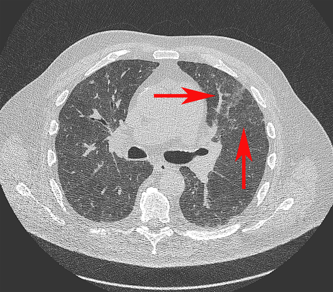
Chest computed tomography scan showing ill‐defined ground glass infiltrates and associated interlobular septal thickening (arrows). When combined with an appropriate clinical history and induced sputum positive for P. jirovecii, this was diagnostic for P. jirovecii pneumonia.
Competing interests
No relevant disclosures.
References
- Permpalung N, Kittipibul V, Mekraksakit P, et al. A comprehensive evaluation of risk factors for Pneumocystis jirovecii pneumonia in adult solid organ transplant recipients: a systematic review and meta‐analysis. Transplantation 2021; 105: 2291‐2306.
- Kamanski H, Belliere J, Burguet L, et al. Identification of predictive markers and outcomes of late‐onset Pneumocystis jirovecii pneumonia in kidney transplant recipients. Clin Infect Dis 2020; 73: 1456‐1463.
- Park JW, Curtis JR, Jun KI et al. Primary prophylaxis for Pneumocystis jirovecii pneumonia in patients receiving rituximab. Chest 2022; 161: 1201‐1210.
- Hamroun A, Lenain R, Nguyen LB et al. Hypercalcemia is common during Pneumocystis pneumonia in kidney transplant recipients. Nature 2019; 9: 12508.
- Tebben PJ, Ravinder JS, Kumar R. Vitamin D‐mediated hypercalcemia: mechanisms, diagnosis, and treatment. Endocr Rev 2016; 37; 521‐547.
Provenance: Not commissioned; externally peer reviewed.
Rhabdomyolysis After COVID-19
Navya Kataria, Abel Devadass, Lana Sundac, Laura De Souza, Nabeel Sheikh, Samantha Ng, Scott Campbell, Yishen Wang, Ross S. Francis
Technological advances in the search for a CURE for asthma
Sara Quon, Timothy M Johanson, Ridhima Wadhwa, Alen Faiz, Anthony Flynn, Gary P Anderson, Amanda J Cox, Nicholas P West, Michael P Menden, Rhys S Allan
The CURE Asthma roadmap
Gary P Anderson, Anthony Flynn, Phil G Bardin, John D Blakey, Shyamali C Dharmage, Paul Foster, Peter G Gibson, Adam Jaffe, Alan James, Christine R Jenkins, Sundram Sivamalai, Peter D Sly, Guy B Marks, Vanessa M McDonald, Judy Wetttenhall
The first live term birth following uterus transplantation in Australia
Rebecca Deans, Brigitte Gerstl, Antonia W Shand, Sarah Lyons, Aaron Budden, Helen L Barrett, Grant Luxton, Mangalee Fernando, Kenneth Yong, Karen Keung, Kaushalya Arulpragasam, Henry Pleass, King Man Wan, Eva Kehag, Jana‐Emily Pittman, Mianna Lotz, Maria Fenn, Erin Nesbitt‐Hawes, Lily Byun, Katrina Tang, Mats Brannstrom, Jason Abbott
The public health impacts of mining in Australia
Javier Cortes‐Ramirez, Ruby N Michael, Leisa‐Maree Toms, Melissa Haswell
Lingual Raynaud phenomenon
Michael Taggart, Mina John