




Medical education
Snapshot
Volume 215 - Issue 3
Isolated intrathoracic kidney
Authors: Jessica Phillips and Jonathan Foo
Med J Aust 2021; 215 (3): 108-108.e1. || doi: 10.5694/mja2.51164
Published online: 2 August 2021
Published online: 2 August 2021
A 75-year-old woman presented to her general practitioner with several weeks of dyspnoea
A 75‐year‐old woman presented to her general practitioner with several weeks of dyspnoea. Examination revealed decreased air entry in the right lung base.
After some initial investigations, she underwent computed tomography of the chest, which showed a right‐sided diaphragmatic hernia containing the ipsilateral kidney (Figure, A [coronal view] and B [sagittal view]). Renal function was normal and renal perfusion scanning (99mTc MAG3) estimated a functional contribution of 62% from the right kidney.
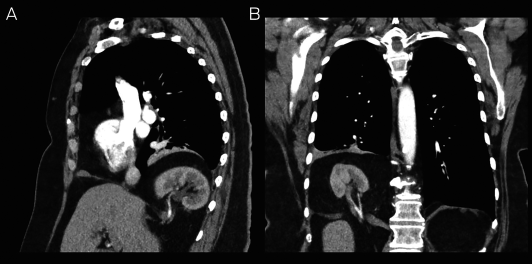
The presence of an intrathoracic kidney is a rare complication of diaphragmatic hernia.1 The follow‐up and management plan needs to be individualised and should consider the patient’s symptoms, the possibility of incarceration of the hernia contents, and the risks of surgical intervention. In this case, it was an incidental imaging finding unrelated to the patient’s presenting symptom. Her dyspnoea resolved without treatment and imaging appearances were stable one year after her initial presentation.
Competing interests
No relevant disclosures.
References
- Shah AD, Ajay S, Adalia M, Rathi A. Bochdalek hernia with intrathoracic kidney. Lung India 2012; 29: 373–375.
Provenance: Not commissioned; externally peer reviewed.
Rapid Growth in Diagnostic CT Knee Imaging in Australia: Implications for Imaging Stewardship
Hanwen Chen, Rachelle Buchbinder, Sean Docking, Ian A Harris
Disease‐specific survival for people with chronic myeloid leukaemia, South Australia, 1980–2020: a retrospective cohort study
Kerri Beckmann, Brendon J Kearney, Naranie Shanmuganathan, David Yeung, Tim Hughes
Technological advances in the search for a CURE for asthma
Sara Quon, Timothy M Johanson, Ridhima Wadhwa, Alen Faiz, Anthony Flynn, Gary P Anderson, Amanda J Cox, Nicholas P West, Michael P Menden, Rhys S Allan
Who gets asthma, and why?
Denby J Evans, Peter D Sly, Paul Foster, Chantal Donovan
The first Australian evidence‐based guidelines on male infertility
Darren J Katz, Liza O’Donnell, Robert I McLachlan, Tim J Moss, Clare V Boothroyd, Veena Jayadev, Sarah R Catford
Sex differences in management and outcomes of people with ST‐elevation myocardial infarction, New South Wales, 2011–2020: a retrospective cohort study
Samia Kazi, Simone Marschner, Haeri Min, Desi Quintans, James JH Chong, Ehsan Khan, David B Brieger, Clara K Chow