




Medical education
Lessons from practice
Volume 211 - Issue 11
Diagnosis of West Nile virus encephalitis in a returned traveller
Authors: Naomi CA Whyler, Jasmine C Teng, David J Brewster, Ruth Chin, Ian Cox, Julian Druce, Henry M Prince, David A Sheffield, Eugene Teh and Vineet Sarode
Med J Aust 2019; 211 (11): 501-502.e1. || doi: 10.5694/mja2.50416
Published online: 9 December 2019
Published online: 9 December 2019
A 63- year- old woman developed chills, vomiting and diarrhoea the day after returning from 2 months’ travel in South East Europe
Clinical record
A 63‐year‐old woman developed chills, vomiting and diarrhoea the day after returning from 2 months' travel in South East Europe. Travel included a Danube River cruise trip and time in Serbia, Croatia, Germany, Austria, Slovakia and Hungary. She visited rural areas and sustained mosquito bites, but reported no unpasteurised food consumption, animal contact or bites. Pre‐travel consultation was not undertaken. Past medical history included follicular lymphoma in remission, with rituximab‐induced hypogammaglobulinaemia.
She was hospitalised on day 3 of her illness. On examination, she was febrile (39.1°C), dehydrated but cognitively intact, without meningism, rash or arthritis. Blood pressure of 80/60 mmHg responded to intravenous hydration. Investigations including full blood examination, biochemistry, liver function, C‐reactive protein and stool cultures were unremarkable. Initial presumptive diagnosis was infectious gastroenteritis, and empirical ciprofloxacin was administered.
On day 5, she developed confusion, nominal aphasia and slow affect. Brain computed tomography and magnetic resonance imaging (MRI) were unremarkable. Cerebrospinal fluid (CSF) analysis revealed mild lymphocytic pleocytosis (24 × 106/L; reference interval [RI], 0–5 × 10⁶/L), raised erythrocytes (224 × 106/L; RI, < 1 × 10⁶/L) and a mildly raised protein level (0.54 g/L; RI, 0.15–0.45 g/L), consistent with infection rather than traumatic tap. Aciclovir, ampicillin and piperacillin–tazobactam were administered to cover gram‐positive and gram‐negative bacterial and viral meningoencephalitis, including listeria and nosocomial pathogens given deterioration 48 hours after admission. Intravenous immunoglobulin, the patient's longstanding treatment for hypogammaglobulinaemia, was administered.
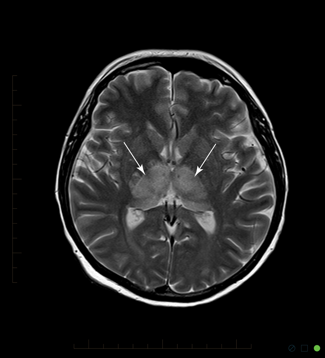
Intubation for mechanical ventilation was performed on day 10, due to worsening symptoms of fluctuating conscious state and uncoordinated motor responses. MRI identified new symmetrical bilateral thalamic, mesial temporal and brainstem signal abnormality (Box 1). Electroencephalography showed subclinical seizure activity.
Initial laboratory investigations were negative: CSF bacterial cultures, viral polymerase chain reaction (PCR) for enterovirus and herpes viruses (herpes simplex virus types 1 and 2, varicella virus and cytomegalovirus), cryptococcal antigen, fungal cultures, and treponemal and human immunodeficiency virus serology.
Imported flavivirus meningoencephalitis including West Nile virus and tick‐borne encephalitis virus were considered. Negative flavivirus serology was difficult to interpret due to hypogammaglobulinaemia. Flavivirus PCR testing was performed on CSF (day 14), whole blood (days 9 and 11) and urine (days 12 and 24). All results were negative except day 12 urine flavivirus PCR; DNA sequencing of this sample identified West Nile virus imported type with the closest match being West Nile virus Bulgaria 2015, confirmed by positive urine viral culture.
Treatment was supportive, with phenytoin, levetiracetam, intravenous immunoglobulin (total 1 g/kg), ventilatory support and tracheostomy.
Despite oedema improving on day 14 MRI, neurological status plateaued: the patient continued to exhibit persistent central apnoeas, decerebrate posturing and reduced consciousness level. Day 28 MRI demonstrated improvement of thalamic and midbrain oedema, but showed new bilateral, punctate foci of marked T2 hyperintensity in midbrain corticospinal tracts, consistent with a diagnosis of severe acquired brain injury secondary to encephalitis. Given limited prognosis, the patient was palliated and died on day 32.
Discussion
This is one of two reported cases of imported West Nile virus infection into Australia causing neuroinvasive disease. In 2009, Rogers and colleagues described an Australian laboratory‐confirmed imported West Nile virus infection in a visiting Israeli tourist.1
West Nile virus is a mosquito‐borne neurotropic flavivirus endemic to North America, Africa, Asia, Europe, and Northern Australia (Kunjin subtype).2,3 Its transmission cycle includes bird species as amplifier hosts, mosquito vectors, and mammals including humans as dead‐end hosts.2 Most human infections are asymptomatic, but 20% develop influenza‐like symptoms including malaise and fever, and < 1% develop West Nile virus neuroinvasive disease with encephalitis, meningitis or acute flaccid paralysis.2 West Nile virus Kunjin subtype causes milder disease, lower rates of West Nile virus neuroinvasive disease, and no reported deaths.2,3 Exposure is hypothesised to protect against other West Nile virus subtypes.3 Risk factors for severe disease include advanced age, chronic conditions and immunosuppression, with mortality rates of 3–19%.2
Travel history is essential for diagnosis, and real time monitoring resources describing local West Nile virus outbreaks in endemic areas are useful. The European Centre for Disease Prevention and Control reported a 7.2‐fold increase in West Nile virus cases from 2017 to 20184 and aided diagnosis in our case.
Rogers and colleagues relied on paired flavivirus serology for diagnosis,1 whereas our case relied on urine flavivirus PCR for diagnosis. Flavivirus diagnosis can be difficult due to low viraemia levels, potential for serological cross‐reactivity,5 or false negative results in the presence of hypogammaglobulinaemia. Emerging evidence suggests flavivirus PCR may be detectable for a number of weeks in urine and whole blood,5 particularly in neuroinvasive disease,6 compared with serum, which may remain positive for up to a week after symptom onset.5
Radiological changes can help narrow down the differential diagnoses of encephalitis. In our case, thalamic MRI changes supported the diagnosis of West Nile virus. Bilateral thalamic and basal ganglia involvement is reported with specific encephalitis aetiologies, particularly respiratory viruses, Mycobacterium species, and arboviruses including West Nile virus.7
West Nile virus management is predominantly supportive, although intravenous immunoglobulin with high anti‐West Nile virus antibody titres may provide benefit.8 Box 2 summarises current avenues of pharmacotherapy.
Our case highlights the importance of travel history, knowledge of endemic disease outbreaks, appropriate sampling to increase diagnostic yield, and radiology to assist diagnosis of undifferentiated infection in returned travellers.
Lessons from practice
- The evolving spectrum of disease in an era of increasing international travel and climate change presents new challenges to local clinicians. Use of health monitoring internet resources can help identify outbreaks at travel destinations to aid diagnosis and management of undifferentiated infection in returned travellers.
- Use of flavivirus serology for diagnosis is challenging in the presence of hypogammaglobulinaemia, and may produce false negatives. Close liaison with a laboratory for appropriate tests is essential.
- Consider flavivirus polymerase chain reaction testing of urine, which remains positive for longer than serum/plasma. Urine and whole blood polymerase chain reaction specimens are increasingly recognised as the best basis for molecular flavivirus diagnosis.
- MRI imaging demonstrating typical features of bilateral thalamic and basal ganglia changes supports a diagnosis of West Nile virus encephalitis and can assist with diagnosis, management and prognostication.
Box 1 – T2‐weighted magnetic resonance imaging (day 10), showing bilateral thalamic hyperintensity (arrows)
Box 2 – Current pharmacotherapy for West Nile virus
|
Pharmacotherapy |
Current research avenues |
||||||||||||||
|
|
|||||||||||||||
|
Vaccines |
Several equine vaccines are available; with some human vaccines in clinical trial9 |
||||||||||||||
|
Ribavirin |
In vitro activity has been demonstrated, but ribavirin has been associated with higher mortality when used in human West Nile virus outbreaks10 |
||||||||||||||
|
Mycophenolic acid |
In vitro activity has been demonstrated, but immunosuppressive effects have been seen with human use10 |
||||||||||||||
|
Intravenous immunoglobulin |
Case reports suggest improved outcomes particularly for West Nile neuroinvasive disease requiring respiratory support;8 benefit may be due to the presence of high titres of West Nile virus antibodies in the sourced intravenous immunoglobulin8 |
||||||||||||||
|
Targeted antiviral therapy |
Candidates for viral entry/fusion inhibitors, protease inhibitors and other viral replication inhibitors are in early stages of research9 |
||||||||||||||
|
Monoclonal antibodies |
Humanised anti‐West Nile virus monoclonal antibodies appear promising for post‐exposure therapy, and are currently undergoing clinical trial evaluation9 |
||||||||||||||
|
|
|||||||||||||||
|
|
|||||||||||||||
Competing interests
No relevant disclosures.
References
- Rogers BA, Hueston L, Ratnam I. Imported West Nile virus encephalitis in an Israeli tourist. Med J Aust 2009; 191: 232–234. https://www.mja.com.au/journal/2009/191/4/imported-west-nile-virus-encephalitis-israeli-tourist
- Chancey C, Grinev A, Volkova E, Rios M. The global ecology and epidemiology of West Nile virus. BioMed Res Int 2015; 376230: 1–20.
- Mackenzie JS, Hall RA, Smith DW. West Nile virus: is there a message for Australia? Med J Aust 2003; 178: 5–6. https://www.mja.com.au/journal/2003/178/1/west-nile-virus-there-message-australia
- European Centre for Disease Prevention and Control. Epidemiological update: West Nile virus transmission season in Europe, 2018. https://ecdc.europa.eu/en/news-events/epidemiological-update-west-nile-virus-transmission-season-europe-2018 (viewed July 2019).
- Lustig Y, Sofer D, Bucris ED, Mendelson E. Surveillance and diagnosis of West Nile virus in the face of flavivirus cross‐reactivity. Front Microbiol 2018; 9: 2421.
- Barzon L, Pacenti M, Franchin E, et al. Excretion of West Nile virus in urine during acute infection. J Infect Dis 2013; 208: 1086–1092.
- Beattie GC, Glaser CA, Sheriff H, et al. Encephalitis with thalamic and basal ganglia abnormalities: etiologies, neuroimaging, and potential role of respiratory viruses. Clin Infect Dis 2013; 56: 825–832.
- Shimoni Z, Bin H, Bulvik S, et al. The clinical response of West Nile virus neuroinvasive disease to intravenous immunoglobulin therapy. Clin Pract 2012; 2: e18.
- Acharya D, Bai F. An overview of current approaches toward the treatment and prevention of West Nile virus infection. Methods Mol Biol 2016; 1435: 249–291.
- David S, Abraham AM. Epidemiological and clinical aspects on West Nile virus, a globally emerging pathogen. Infect Dis 2016; 48: 571–585.
Provenance: Not commissioned; externally peer reviewed.
Costs of Delivering the Hep B PAST Model of Care in Remote Australia: A Retrospective Pre–Post Study
Hoa Nguyen, Anh Le Tuan Nguyen, Paula Binks, Melita McKinnon, Emily Vintour-Cesar, Karen Wills, Nicola Stephens, Benjamin Cowie, Joshua S. Davis, Yuejen Zhao, Peter Nihill, Julie A. Campbell, Andrew J. Palmer, Jane Davies, Barbara de Graaff
Buruli Ulcer Acquired From Ringtail Possum Rescue
Daniel P. O'Brien, Naomi Clarke, Sara Bonnici, Mohammad A. Hussain
Q Fever Visceral Abscesses After Contact With Native Australian Marsupials
Samantha J. Laws, Enzo Binotto, James Stewart, Jennifer Ho, Trent Yarwood, Josh Hanson, Simon Smith
Hospital-Admitted Injection-Related Infections Among Incarcerated People Who Inject Drugs in Australia: A Retrospective Cohort Study
Andrew Palmer, Matthew Carter, Jeremy Yeo, Cecilia Shim, Jason Connor, Jeremy Hayllar, Gerald Holtmann, Naomi Moy, Elliott G. Playford, Naomi Runnegar, Paul J. Clark
The Live Attenuated Influenza Vaccine in Australia: An Additional Tool for Influenza Prevention
Cyra Patel, Alexis Pillsbury, Tran Nguyen, Xia Wang, Helen E. Quinn, Clayton K. Chiu, Allen C. Cheng, Katie L. Flanagan, Zhicheng Wang
Reflex Testing for Hepatitis D Infection: A Unique Opportunity to Reduce Hepatitis D-Related Chronic Liver Disease Deaths in Australia
Jessica Howell, Lauren Andersson, Miriam T. Levy, James O'Beirne, Leon Adams, Katharine Irvine, Avik Majumdar, Golo Ahlenstiel, Kathy Jackson, Krispin Hajkowicz, Joseph Doyle, Jane Davies, Sarah Cherian, Wayne Dimech, Alexander J. Thompson