




Medical education
Lessons from practice
Volume 210 - Issue 9
A rare red eye: cavernous sinus dural arteriovenous fistula masquerading as conjunctivitis and sinusitis
Authors: Stuart Campbell, Emma Hack and Babu Philip
Med J Aust 2019; 210 (9): 398-399.e1. || doi: 10.5694/mja2.50157
Published online: 20 May 2019
Published online: 20 May 2019
A 77-year-old woman with a background of hypertension, diet-controlled type 2 diabetes and bilateral cataract surgery was referred by her GP to the emergency department
Clinical record
A 77‐year‐old woman with a background of hypertension, diet‐controlled type 2 diabetes and bilateral cataract surgery was referred by her general practitioner to the emergency department of the Royal Darwin Hospital with a progressive history of red eyes, lacrimation, rhinorrhoea and, more recently, diplopia. The patient first sought medical attention 6 weeks prior at symptom onset. Her GP had initially diagnosed and managed the presentation as sinusitis and, after failed resolution, as viral conjunctivitis. There was no history of trauma. Following onset of new diplopia, the patient was referred to hospital.
Her regular medications were atorvastatin 40 mg daily, felodipine 10 mg daily and atenolol 100 mg daily. Examination revealed blood pressure 183/84 mmHg and bilateral injected conjunctivae with a corkscrew appearance of the scleral vessels, left‐sided proptosis, diplopia and abducens nerve palsy (Box, A). There was no palpable thrill or audible bruit over either eye, and the remainder of the examination, including peripheral neurological assessment, was unremarkable.
The initial investigation focused on excluding cavernous sinus thrombosis, orbital collection, glaucoma and Graves disease. Routine blood tests were normal, as were inflammatory markers, antithyroid and autoimmune antibodies. Brain magnetic resonance imaging scan excluded stroke, and the globe pressures were normal. Initial computed tomography (CT) angiography found an enlarged cavernous sinus but no other pathology. Subsequent time‐resolved CT angiography demonstrated early arterial phase contrast flow into the cavernous sinus, raising suspicion for a cavernous sinus dural arteriovenous fistula (CS‐DAVF).
The patient was transferred interstate to neurointerventional radiology for further assessment and treatment. Digital subtraction angiography demonstrated both internal and external carotid contributions into the cavernous sinus (Box, B, D and E). The connection was eliminated via transorbital cavernous sinus embolisation.1 Six months later, the patient's red eye had resolved, although the abducens nerve palsy and diplopia persisted and required surgical intervention with transposition of superior and inferior rectus muscles to the lateral rectus (Jensen procedure).
Discussion
A communication between the carotid arterial system and the cavernous sinus is a rare and sight threatening cause of red eye. This pathology, termed CS‐DAVF, historically categorised as an indirect carotid‐cavernous fistula (CCF), is a challenging diagnosis.
CS‐DAVF are formed by the communication of the dural branches of the internal and external carotid arteries with the cavernous sinus. This communication is hypothesised to be due to the thrombolytic response to a cavernous sinus thrombosis.2,3 In contrast, direct CCF are formed by trauma or aneurysmal rupture to the intercavernous portion of the internal carotid artery itself, resulting in a high flow, direct CCF. These entities have classically been combined in classification schema, but there is debate advocating for their separation due to the substantially differing pathophysiology behind each disorder.4
CS‐DAVF is a rare clinical entity predominately seen in older women or patients with hypertension, while trauma or connective tissue disease are risk factors for direct CCF.5 The natural history of the disease is variable and relates to the interplay between fistula location and patient anatomy.2 Typical signs include progressive chemosis, exophthalmos and corkscrewing of the scleral vessels. Diplopia may result from either oculomotor cranial nerve involvement or venous congestion. As the flow through the fistula progresses, these signs become more evident and there is potential for facial pain and glaucoma. Ophthalmic signs include central retinal vein occlusion, retinal detachment and optic disc swelling.2 Signs may be ipsilateral, contralateral or bilateral due to intercavernous communication and individual drainage patterns.3
Common symptoms include red eyes and possibly congestion; a headache may be also present. Uncommon presentations include epistaxis or otorrhagia.3 Posteriorly draining fistulae predispose the patient to intracranial haemorrhage and may lack red eyes or other external findings. Post‐traumatic direct CCF (eg, due to motor vehicle accident) may be overlooked due to distracting injuries.
The gold standard diagnostic test is digital subtraction angiography of the external and internal carotids bilaterally and subsequent demonstration of contrast flow into the cavernous sinus, but access to this procedure is limited to quaternary centres. Plain or contrast CT may reveal superior optic vein enlargement, and additional time‐gated modality may better demonstrate early filling of this superior optic vein, supporting clinical suspicion of CS‐DAVF.
The wide variety of signs and presentations mean that this condition may be misdiagnosed and treatment delayed. This is particularly true of CS‐DAVF, which may progress insidiously until potentially irreversible complications such as diplopia and visual loss are present. Therefore, patients with CS‐DAVF manifesting as a red eye require a high index of suspicion to ensure timely diagnosis, which may be complicated by the lack of specialised diagnostic and treatment resources in rural and remote Australia.
Lessons from practice
- Cavernous sinus dural arteriovenous fistula (CS‐DAVF) is a rare cause of red eyes that can result in irreversible visual impairment, cranial nerve palsies and, less commonly, intracranial haemorrhage if not treated promptly.
- The presence of worsening or refractory red eyes with worrisome features including diplopia, cranial nerve palsies and/or headache should alert the clinician to consider CS‐DAVF.
- Although diagnosis requires digital subtraction angiography, certain findings on computed tomography modalities including early filling or enlargement of the superior ophthalmic vein are suggestive of CS‐DAVF.
- Specialist neurointerventional services are required to diagnose and manage CS‐DAVF — these services may be unavailable in rural and remote Australia, resulting in delays to optimal treatment.
Box – Digital subtraction angiograms showing cavernous sinus dural arteriovenous fistula
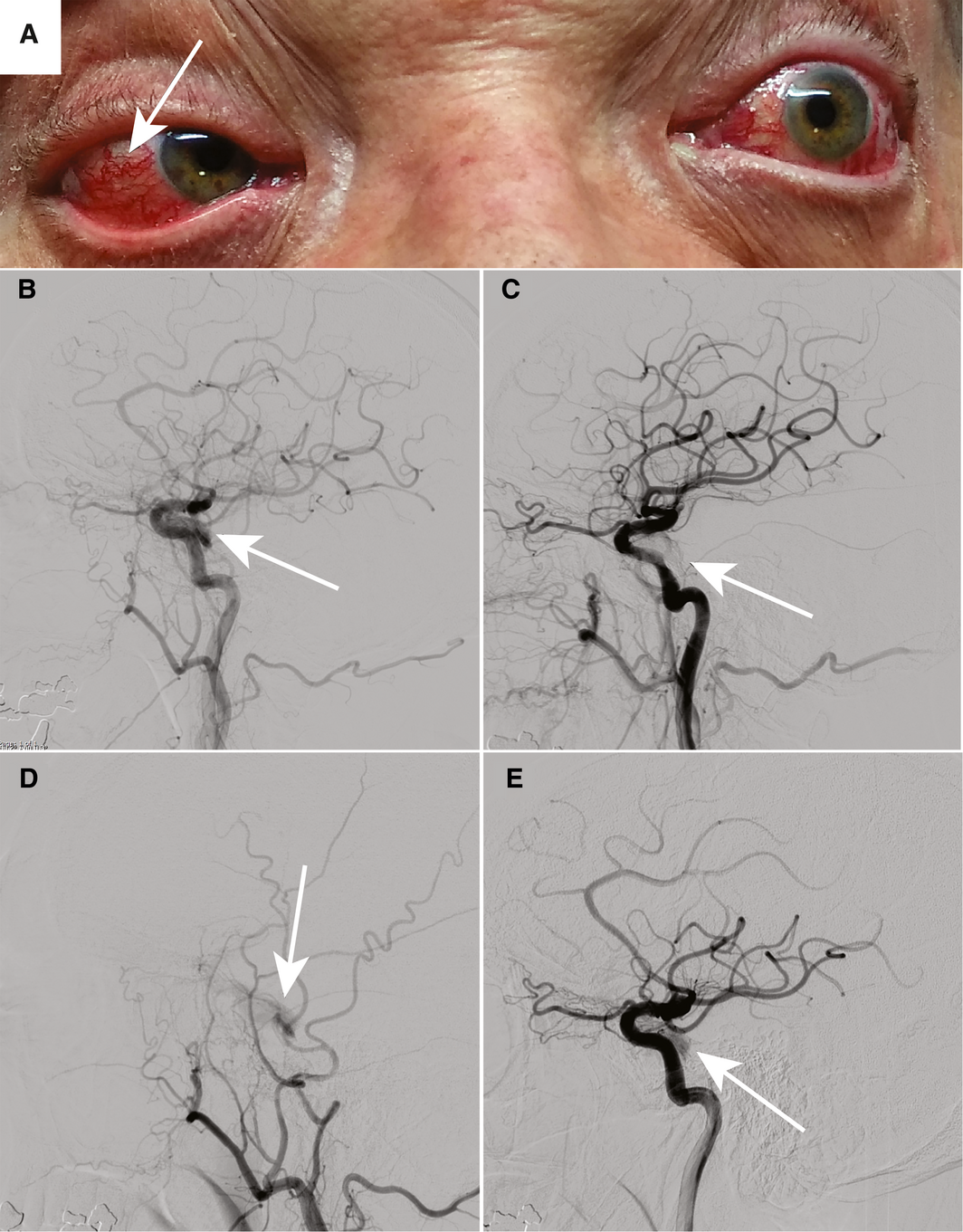
The patient's eyebrows are held up and she is asked look to the left (A) — note the injected conjunctivae with tortuous vessels consistent with arterialisation of the veins (arrow). Also shown is a near complete abducens nerve palsy of the left eye with a failure to abduct. There is the impression of left‐sided proptosis. Lateral digital subtraction angiograms of the left common carotid pre‐embolisation (B) and post‐embolisation (C). The arrow in B points out contrast flowing through the cavernous sinus, while in C, post‐embolisation, the cavernous sinus is not opacified, indicating successful obliteration of the fistulous communication. D and E — selective angiograms of the external carotid artery and internal carotid artery, respectively — illustrate contrast progression into the cavernous sinus via dural branches (arrows). ◆
Competing interests
No relevant disclosures.
References
- Wenderoth J. Novel approaches to access and treatment of cavernous sinus dural arteriovenous fistula (CS‐DAVF): case series and review of the literature. J Neurointerv Surg 2017; 9: 290–296.
- Miller NR. Dural carotid‐cavernous fistulas: epidemiology, clinical presentation, and management. Neurosurg Clin N Am 2012; 23: 179–192.
- Korkmazer B, Kocak B, Tureci E, et al. Endovascular treatment of carotid cavernous sinus fistula: a systematic review. World J Radiol 2013; 5: 143–155.
- Wenderoth J. Proposal for an improved classification system for cavernous sinus dural arteriovenous fistula (CS‐DAVF). J Neurointerv Surg 2017; 9: 220.
- Rodrigues T, Willinsky R, Agid R, et al. Management of dural carotid cavernous fistulas: a single‐centre experience. Eur Radiol 2014; 24: 3051–3058.
Provenance: Not commissioned; externally peer reviewed.
The epidemiology of haemodialysis catheter infections in Australia, 2016–20: a prospective cohort study
Benjamin Lazarus, Kevan R Polkinghorne, Martin P Gallagher, Jayson Catiwa, Nicholas A Gray, Sarah Coggan, Kathryn R Higgins, Girish Talaulikar, Stephen P McDonald AM, Sradha Kotwal
30/60/90 National stroke targets and stroke unit access for all Australians: it's about time
For the 30/60/90 National Stroke Targets Taskforce
A summary of the 2023 Society of Obstetric Medicine of Australia and New Zealand (SOMANZ) hypertension in pregnancy guideline
Renuka Shanmugalingam, Helen L Barrett, Amanda Beech, Lucy Bowyer, Tim Crozier, Amanda Davidson, Marloes Dekker Nitert, Kerrie Doyle, Luke Grzeskowiak, Nicole Hall, Hicham Ibrahim Cheikh Hassan, Annemarie Hennessy, Amanda Henry, David Langsford, Vincent WS Lee, Zachary Munn, Michael J Peek, Joanne M Said, Helen Tanner, Rachel Taylor, Meredith Ward, Jason Waugh, Linda LY Yen, Ellie Medcalf, Katy JL Bell, Deonna Ackermann, Robin Turner, Angela Makris
Cough in Children and Adults: Diagnosis, Assessment and Management (CICADA). Summary of an updated position statement on chronic cough in Australia
David A Prentice
PEG‐GCSF‐induced aortitis in a patient with breast cancer: distinguishing between infective and immune‐mediated aortitis
Luigi Zolio, Prudence A Francis, Nava Ferdowsi
Catheter‐related superior vena cava syndrome: an increasing problem
John J Harvey, John I Vrazas, Elhamy Bekhit, Chris Barnes, Philip J Robinson