




Medical education
Lessons from practice
Volume 208 - Issue 7
A severe case of Mycobacterium ulcerans (Buruli ulcer) osteomyelitis requiring a below-knee amputation
Authors: Michael J Loftus, Nicola Kettleton-Butler, Denton Wade, R Michael Whitby and Paul DR Johnson
Med J Aust 2018; 208 (7): 290-291. || doi: 10.5694/mja17.01158
Published online: 16 April 2018
Published online: 16 April 2018
About 80-90% of patients with Buruli ulcer acquired in Far North Queensland present with an ulcer
Clinical record
In July 2012, a 68-year-old man presented to his general practitioner in Brisbane with a swollen and painful right ankle. He was otherwise well apart from recent excision 10 months previously of a left forearm Mycobacterium ulcerans nodule; initially diagnosed on histopathological examination and confirmed on retrospective polymerase chain reaction (PCR) testing of paraffin sections. This infection was likely acquired during residence in Far North Queensland between February 2010 and November 2011.
Initial ankle x-rays and ultrasound revealed only synovitis; blood tests showed elevated C-reactive protein (52 mg/L; reference interval, 0–5 mg/L) and normal uric acid levels. The patient failed to respond to non-steroidal anti-inflammatory drugs, and experienced only temporary pain relief with a short course of oral prednisolone and then an intra-articular steroid injection in August 2012. Joint aspiration for persisting pain in October 2012 showed a heavily bloodstained fluid with numerous leucocytes (formal cell count not performed), and absence of bacteria or crystals. Magnetic resonance imaging confirmed synovitis of the ankle joint with diffuse bone marrow oedema (Box). Extensive testing for autoimmune and vector-borne diseases revealed only past exposure to Ross River virus and positive human leucocyte antigen B27 (HLA-B27). A synovial biopsy performed in November 2012 showed scant leucocytes and was culture negative; however, there was histological evidence of granulomatous inflammation. Given the patient’s recent history, dual therapy for M. ulcerans was commenced (clarithromycin 500 mg twice a day and rifampicin 600 mg daily).
In December 2012, he developed skin ulceration for the first time at the biopsy site and underwent extensive debridement including of involved bone, requiring bone and skin autografts. Operative specimens showed granulomas in bone, and M. ulcerans infection was confirmed contemporaneously by PCR.
Despite prolonged antibiotic therapy and normalisation of inflammatory markers by March 2013, the patient experienced persistent pain, and x-rays showed failure of the bone graft to incorporate into the talus. After extensive multidisciplinary discussions involving the patient, a below knee amputation was performed in June 2013.
Three weeks after the below knee amputation, and after 8 months of uninterrupted medical therapy, antibiotics were ceased. Within a month, the patient developed a new nodule over his left elbow. This was excised in August, no acid-fast bacilli were seen, but granulomatous inflammation was present; retrospective PCR testing on paraffin sections was positive for M. ulcerans. He recommenced dual antibiotic therapy for a further 8 months, ceasing in March 2014 with no subsequent disease recurrence.
The patient has adapted well to his prosthesis, regained full mobility and is pain-free 4 years after his amputation.
Buruli ulcer — also known in Australia as Bairnsdale ulcer or Daintree ulcer — is caused by the toxin-producing environmental pathogen M. ulcerans.
About 80–90% of patients with Buruli ulcer acquired in Far North Queensland present with an ulcer.1 When there is no ulcer present, the diagnosis of Buruli ulcer can be challenging. Atypical presentations may include nodules, plaque, oedematous lesions or osteomyelitis.
The case we describe is a highly unusual non-ulcerative recurrence in an anatomically separate site 10 months after surgical resection of a left forearm ulcer. While small Buruli ulcer lesions are frequently successfully managed with surgery alone, there is an increased risk of disease recurrence without antibiotics,2,3 usually near the site of initial excision.
Osteomyelitis is an uncommon manifestation of Buruli ulcer, only occurring in 6% of patients in a large cohort from Benin4 and has been rare in Australia. In this case, differential diagnoses of musculoskeletal injury, cellulitis, gout and seronegative arthritis were considered and investigated before the diagnosis was made. We propose that PCR for M. ulcerans on synovial fluid in October 2012 may have confirmed the diagnosis earlier due to the known sensitivity of this method; however, by then, extensive bone destruction was already present. Amputation for Buruli ulcer is rarely required but, in this case, it successfully resolved the patient’s pain and immobility which had not improved despite antibiotics and surgical debridement.
Antibiotic treatment based on rifampicin and a companion drug (usually clarithromycin) for 8 weeks effectively kills M. ulcerans cells, but surgery may still be required to remove necrotic tissue and repair skin defects.5 Paradoxical reactions — clinical worsening after commencement of antibiotics — are also a challenge for clinicians and patients.6-8 We believe that the two left forearm lesions likely arose from a single common inoculation event and that the second forearm recurrence probably represented a paradoxical reaction to residual dead bacterial cells, and, in retrospect, it probably would have resolved without further antibiotics. The right ankle is likely to have become infected from seeding of live bacteria via the blood from the initial infection or from a separate episode of unnoticed percutaneous inoculation.
Lessons from practice
-
Buruli ulcer may present in an atypical fashion, with manifestations including nodules, plaque, oedematous lesions, septic arthritis or osteomyelitis.
-
Buruli ulcer should be considered in anyone with compatible symptoms who has resided in or visited an endemic region within the previous year. The two most active endemic regions in Australia are in Victoria (Mornington and Bellarine peninsulas), and the Daintree region in Far North Queensland.
-
Antibiotics are highly active against Mycobacterium ulcerans, are increasingly used as first-line therapy and reduce relapse in patients who are managed with primary surgical excision.
Box – Post-contrast sagittal magnetic resonance image of right ankle in October 2012 showing synovitis and effusion of the ankle joint with associated diffuse bone marrow oedema of the talus
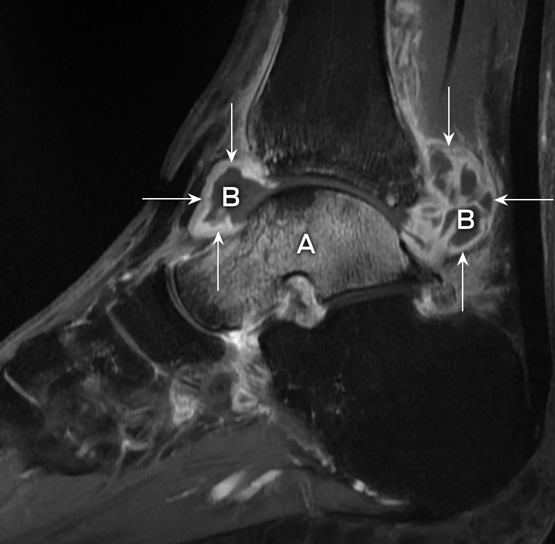
A. Bone marrow oedema of the talus. B. Ankle joint effusion. Arrows. Synovitis with synovial thickening and enhancement.
Competing interests
No relevant disclosures.
References
- Steffen CM, Smith M, McBride WJ. Mycobacterium ulcerans infection in North Queensland: the “Daintree ulcer”. ANZ J Surg 2010; 80: 732-736.
- O’Brien DP, Walton A, Hughes AJ, et al. Risk factors for recurrent Mycobacterium ulcerans disease after exclusive surgical treatment in an Australian cohort. Med J Aust 2013; 198: 436-439.
- Kanga JM, Kacou DE, Sangaré A, et al. Recurrence after surgical treatment of Buruli ulcer in Cote d’Ivoire. Bull Soc Pathol Exot 2003; 96: 406-409.
- Pommelet V, Vincent QB, Ardant MF, et al. Findings in patients from Benin with osteomyelitis and polymerase chain reaction-confirmed Mycobacterium ulcerans infection. Clin Infect Dis 2014; 59: 1256-1264.
- O’Brien DP, Jenkin G, Buntine J, et al. Treatment and prevention of Mycobacterium ulcerans infection (Buruli ulcer) in Australia: guideline update. Med J Aust 2014; 200: 267-270.
- Jenkin GA, Smith M, Fairley M, Johnson PD. Acute, oedematous Mycobacterium ulcerans infection in a farmer from far north Queensland. Med J Aust 2002; 176: 180-181.
- O’Brien DP, Robson M, Friedman ND, et al. Incidence, clinical spectrum, diagnostic features, treatment and predictors of paradoxical reactions during antibiotic treatment of Mycobacterium ulcerans infections. BMC Infect Dis 2013; 13: 416.
- Nienhuis WA, Stienstra Y, Abass KM, et al. Paradoxical responses after start of antimicrobial treatment in Mycobacterium ulcerans infection. Clin Infect Dis 2012; 54: 519-526.
Linked content
-
MJA Perspective: Tackling the worsening epidemic of Buruli ulcer in Australia in an information void: time for an urgent scientific response
-
MJA InSight: Buruli epidemic steps up; urgent funding needed
Provenance: Not commissioned; externally peer reviewed.
Costs of Delivering the Hep B PAST Model of Care in Remote Australia: A Retrospective Pre–Post Study
Hoa Nguyen, Anh Le Tuan Nguyen, Paula Binks, Melita McKinnon, Emily Vintour-Cesar, Karen Wills, Nicola Stephens, Benjamin Cowie, Joshua S. Davis, Yuejen Zhao, Peter Nihill, Julie A. Campbell, Andrew J. Palmer, Jane Davies, Barbara de Graaff
Buruli Ulcer Acquired From Ringtail Possum Rescue
Daniel P. O'Brien, Naomi Clarke, Sara Bonnici, Mohammad A. Hussain
Q Fever Visceral Abscesses After Contact With Native Australian Marsupials
Samantha J. Laws, Enzo Binotto, James Stewart, Jennifer Ho, Trent Yarwood, Josh Hanson, Simon Smith
Hospital-Admitted Injection-Related Infections Among Incarcerated People Who Inject Drugs in Australia: A Retrospective Cohort Study
Andrew Palmer, Matthew Carter, Jeremy Yeo, Cecilia Shim, Jason Connor, Jeremy Hayllar, Gerald Holtmann, Naomi Moy, Elliott G. Playford, Naomi Runnegar, Paul J. Clark
The Live Attenuated Influenza Vaccine in Australia: An Additional Tool for Influenza Prevention
Cyra Patel, Alexis Pillsbury, Tran Nguyen, Xia Wang, Helen E. Quinn, Clayton K. Chiu, Allen C. Cheng, Katie L. Flanagan, Zhicheng Wang
Reflex Testing for Hepatitis D Infection: A Unique Opportunity to Reduce Hepatitis D-Related Chronic Liver Disease Deaths in Australia
Jessica Howell, Lauren Andersson, Miriam T. Levy, James O'Beirne, Leon Adams, Katharine Irvine, Avik Majumdar, Golo Ahlenstiel, Kathy Jackson, Krispin Hajkowicz, Joseph Doyle, Jane Davies, Sarah Cherian, Wayne Dimech, Alexander J. Thompson