




Medical education
Lessons from practice
Volume 207 - Issue 7
Hyponatraemia and hypopituitarism: an easily missed entity
Authors: Melissa H Lee, Genevieve L Calder, Richard J MacIsaac and Nirupa Sachithanandan
Med J Aust 2017; 207 (7): 282-283. || doi: 10.5694/mja16.00921
Published online: 2 October 2017
Published online: 2 October 2017
A 53-year-old woman presented with 2 weeks of lethargy, vomiting, dizziness and confusion following a respiratory tract infection
Clinical record
A 53-year-old woman presented with 2 weeks of lethargy, vomiting, dizziness and confusion following a respiratory tract infection. She was previously well, with no regular medication. On admission, she was confused, hypotensive (systolic blood pressure level, 90 mmHg) and hyponatraemic (serum sodium, 109 mmol/L).
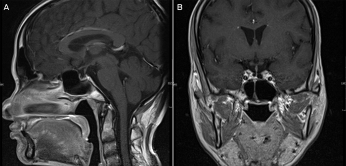
A comprehensive work-up for hyponatraemia was performed (Box 1). Her low serum cortisol level of 86 nmol/L, inappropriately low adrenocorticotropic hormone level and normokalaemia (potassium, 4.1 mmol/L) were suggestive of secondary adrenal insufficiency. She had evidence of hypopituitarism with secondary hypothyroidism and hypogonadism (low gonadotrophins for post-menopausal state). Her serum osmolality was low, with an inappropriately elevated urinary sodium and osmolality, suggestive of syndrome of inappropriate antidiuretic hormone secretion. Magnetic resonance imaging showed a partial empty sella with a thin rind of residual pituitary tissue (Box 2).
She was commenced on intravenous hydrocortisone, 50 mg three times daily, due to severe hyponatraemia and haemodynamic instability; followed by thyroxine 24 hours later. She was weaned to oral hydrocortisone 40 mg/20 mg after 48 hours, discharged on 20 mg twice daily and currently remains on 20 mg/10 mg. Her serum sodium level incremented to 130 mmol/L over 5 days, with symptom resolution.
This is a post-menopausal woman, who underwent early menopause at age 45 years, presenting with severe symptomatic hyponatraemia due to secondary adrenal insufficiency, triggered by an intercurrent illness. The presumed aetiology of her hypopituitarism was primary empty sella syndrome. She had no predisposing factors for secondary empty sella and, importantly, no pituitary adenoma; no history of Sheehan’s syndrome, with two uncomplicated pregnancies; no infiltrative, infective or autoimmune conditions; and no risk factors for drug-induced hypopituitarism.
Hyponatraemia is the most common electrolyte abnormality encountered in clinical practice,1 but the underlying aetiology is often overlooked. Recognition of hyponatraemia due to hypopituitarism (secondary adrenal insufficiency) or primary adrenal insufficiency is crucial, as it may be life-threatening if left untreated. Hypopituitarism is often longstanding and manifests during periods of illness or stress.2
The incidence of primary empty sella syndrome ranges from 5.5% to 35%3 and may present with hypopituitarism. Pathogenic factors include a congenital incomplete formation or defect of the sella diaphragm, in association with chronic cerebrospinal fluid hypertension.3
Hyponatraemia may be the initial manifestation of hypopituitarism or primary adrenal insufficiency, particularly in older individuals.4 Clinical manifestations are often non-specific, with fatigue, vomiting or altered mentation, and thus the diagnosis is often missed.
Hypopituitarism and adrenal insufficiency should be considered in all patients with hyponatraemia with a clinical picture mimicking the syndrome of inappropriate antidiuretic hormone secretion without an alternative cause.1 A basal morning serum cortisol is a sufficient screening test for adrenal insufficiency when the serum cortisol level is < 140 nmol/L.5 Dynamic testing using a short synacthen test or insulin tolerance test should be employed for equivocal cortisol levels (140–365 nmol/L).5
Distinguishing between primary or secondary adrenal insufficiency is important to determine the underlying aetiology and guide treatment. Primary adrenal insufficiency is associated with an elevated adrenocorticotropic hormone level (resulting in hyperpigmentation), while hyperkalaemia (due to aldosterone deficiency) is not present in secondary adrenal insufficiency.5
Cortisol is a physiological tonic inhibitor of arginine vasopressin secretion.4 Without cortisol, there is an inappropriate increase in arginine vasopressin secretion, resulting in impaired free water excretion and hyponatraemia. Glucocorticoids are the mainstay of treatment for hyponatraemia due to adrenal insufficiency by suppressing arginine vasopressin release and allowing excretion of excess solute-free water. The recommended glucocorticoid replacement regimen in adults is hydrocortisone 15–25 mg or cortisone acetate 20–35 mg in 2–3 divided doses per day.5 Glucocorticoid administration will usually normalise sodium levels within a few days. Patients on chronic glucocorticoid treatment must be educated about stress dosing and sick day management (Box 3).5,6 Mineralocorticoid replacement is not required with secondary adrenal insufficiency, as aldosterone secretion is unaffected.
There are no specific guidelines on how to manage acute symptomatic hyponatraemia in the setting of adrenal crisis; however, most accept that stress dosing — using maximal doses of hydrocortisone 200 mg per day5 — is necessary. Clinicians should be aware of the risk of osmotic demyelination syndrome with rapid correction of hyponatraemia. The recommended safe levels of correction for serum sodium is no more than 10 mmol/L per day or 18 mmol/L during the first 48 hours.1
Other concomitant hormonal deficiencies should also be treated. Glucocorticoid replacement must be commenced before thyroxine replacement if concurrent deficiency exists. The increase in cortisol metabolism after thyroxine administration increases the demand for cortisol, which if not replaced, may lead to adrenal crisis.
Determining the underlying cause of hyponatraemia can be a difficult diagnostic challenge. Clinicians should exclude hypopituitarism and adrenal insufficiency as a cause of hyponatraemia by measuring basal serum cortisol and adrenocorticotropic hormone levels. Early diagnosis and treatment of adrenal insufficiency is important to avoid potentially fatal consequences.
Lessons from practice
-
Consider evaluating for adrenal insufficiency when faced with a patient with unexplained euvolaemic hyponatraemia.
-
Severe hyponatraemia associated with non-specific symptoms may be the initial manifestation of secondary adrenal insufficiency, especially in older individuals.
-
Timely diagnosis and early glucocorticoid replacement are essential to correct the hyponatraemia in a patient with adrenal insufficiency.
-
Assess for glucocorticoid deficiency before commencing thyroxine replacement to avoid precipitating adrenal crisis.
Box 1 – Laboratory evaluation
|
Biochemical parameter |
Value |
Reference range* |
|||||||||||||
|
|
|||||||||||||||
|
Serum sodium |
109 |
135–145 mmol/L |
|||||||||||||
|
Serum osmolality |
264 |
280–300 mmol/kg |
|||||||||||||
|
Urine sodium |
214 |
20–150 mmol/L |
|||||||||||||
|
Urine osmolality |
587 |
50–1200 mmol/kg |
|||||||||||||
|
Cortisol |
86 |
100–545 nmol/L |
|||||||||||||
|
ACTH |
10 |
5–46 ng/L |
|||||||||||||
|
TSH |
6.86 |
0.35–4.94 μU/mL |
|||||||||||||
|
fT4 |
4.4 |
9–19 pmol/L |
|||||||||||||
|
LH |
2.6 |
> 5.2 IU/L† |
|||||||||||||
|
FSH |
8.9 |
> 27.0 IU/L† |
|||||||||||||
|
Oestradiol |
< 50 |
50–100 pmol/L† |
|||||||||||||
|
Prolactin |
231 |
110–560 mIU/L |
|||||||||||||
|
|
|||||||||||||||
|
ACTH = adrenocorticotropic hormone. FSH = follicle-stimulating hormone. fT4 = free thyroxine. LH = luteinising hormone. TSH = thyroxine-stimulating hormone. * As supplied by St Vincent’s Hospital, Melbourne. † Values specific for a post-menopausal state. |
|||||||||||||||
Box 2 – Sagittal view pituitary magnetic resonance image (MRI) with gadolinium contrast (A), and coronal view pituitary MRI with gadolinium contrast (B)
Box 3 – Guidelines for the management of adrenal insufficiency in adults during stress or acute illness*
|
Condition |
Suggested action |
||||||||||||||
|
|
|||||||||||||||
|
Minor medical illness with fever |
Glucocorticoid dose doubled (> 38°C) or tripled (> 39°C) until recovery |
||||||||||||||
|
Unable to tolerate oral medication |
IM or SC hydrocortisone, 100 mg |
||||||||||||||
|
Minor to moderate surgical stress |
Hydrocortisone, 25–75 mg/24 h |
||||||||||||||
|
Major surgery with general anaesthesia, trauma, delivery or requiring intensive care |
IV hydrocortisone 100 mg followed by 50 mg qid or continuous infusion of 200 mg/24 h |
||||||||||||||
|
|
|||||||||||||||
|
IM = intramuscular. qid = four times a day. SC = subcutaneous. * Adapted from Bornstein et al, Diagnosis and treatment of primary adrenal insufficiency: an Endocrine Society clinical practice guideline,5 and Jung and Inder, Management of adrenal insufficiency during the stress of medical illness and surgery.6 |
|||||||||||||||
Competing interests
No relevant disclosures.
Acknowledgements
We thank Xavier Yu of St Vincent’s Hospital, Melbourne, for his assistance in reviewing and providing the magnetic resonance images for this article.
References
- Spasovski G, Vanholder R, Allolio B, et al. Clinical practice guideline on diagnosis and treatment of hyponatraemia. Eur J Endocrinol 2014; 170: G1-G47.
- Smith JC. Hormone replacement therapy in hypopituitarism. Expert Opin Pharmacother 2004; 5: 1023-1031.
- Guitelman M, Garcia Basavilbaso N, Vitale M, et al. Primary empty sella (PES): a review of 175 cases. Pituitary 2013; 16: 270-274.
- Diederich S, Franzen NF, Bähr V, Oelkers W. Severe hyponatremia due to hypopituitarism with adrenal insufficiency: report on 28 cases. Eur J Endocrinol 2003; 148: 609-617.
- Bornstein SR, Allolio B, Arlt W, et al. Diagnosis and treatment of primary adrenal insufficiency: an Endocrine Society clinical practice guideline. J Clin Endocrinol Metab 2016; 101: 364-389.
- Jung C, Inder WJ. Management of adrenal insufficiency during the stress of medical illness and surgery. Med J Aust 2008; 188: 409-413.
Provenance: Not commissioned; externally peer reviewed.
When ‘Liver Enzymes’ Are Not Hepatic: Late-Onset Pompe Disease
Shauna Madigan, Georgina England, Wayne Rankin
Primary Hyperparathyroidism in Adults: Recent Developments in Diagnosis and Management
Elizabeth Wootton, Sunita M. C. De Sousa, Richard L. Prince, Donald S. A. McLeod, David A. Pattison, Mathis Grossmann
Severe Hypoglycaemia Secondary to Chronic Opioid-Induced Hypothalamic–Pituitary–Adrenal Axis Suppression: An Under-Recognised Phenomenon
Michael Do, Annabelle Hayes, Malgorzata Brzozowska
Rethinking diabetes care for Indigenous Australians: the need for Indigenous‐codesigned and led diabetes models of care
Natalie Nanayakkara, Sharon Atkinson‐Briggs, Alicia J Jenkins, Neale D Cohen
The first Australian evidence‐based guidelines on male infertility
Darren J Katz, Liza O’Donnell, Robert I McLachlan, Tim J Moss, Clare V Boothroyd, Veena Jayadev, Sarah R Catford