





Volume 207 - Issue 7
Diagnosing, monitoring and managing behavioural variant frontotemporal dementia
Authors: Olivier Piguet, Fiona Kumfor and John Hodges
Med J Aust 2017; 207 (7): 303-308. || doi: 10.5694/mja16.01458
Published online: 2 October 2017
Published online: 2 October 2017
Detailed medical history, carer information, targeted neuropsychological testing and brain neuroimaging help to differentiate bvFTD from other dementia syndromes
Summary
- Behavioural variant frontotemporal dementia is characterised by insidious changes in personality and interpersonal conduct that reflect progressive disintegration of the neural circuits involved in social cognition, emotion regulation, motivation and decision making.
- The underlying pathology is heterogeneous and classified according to the presence of intraneuronal inclusions of tau, TDP-43 or, occasionally, fused in sarcoma proteins. Biomarkers to detect these histopathological changes in life are increasingly important with the development of disease-modifying drugs.
- A number of gene abnormalities have been identified, the most common being an expansion in the C9orf72 gene, which together account for most familial cases.
- The 2011 international consensus criteria propose three levels of diagnostic certainty: possible, probable and definite. Detailed history taking from family members to elicit behavioural features underpins the diagnostic process, with support from neuropsychological testing designed to detect impairment in decision making, emotion processing and social cognition. Brain imaging is important for increasing the level of diagnosis certainty over time. Carer education and support remain of paramount importance.
Frontotemporal dementias (FTDs) are progressive neurodegenerative brain conditions characterised by brain atrophy in the prefrontal cortices or the anterior portions of the temporal lobes caused by various intraneuronal inclusions and abnormal protein depositions. FTD has a prevalence of 10–15/100 000 population in individuals aged 45–65 years, and is a common cause of younger onset dementia, although with large variability across studies.1,2 Recent evidence indicates that the occurrence of FTD beyond 65 years of age appears to be more common than previously assumed.2
Unlike Alzheimer disease (AD), the clinical profile and pathology of FTD are heterogeneous and characterised by two main phenotypes: a progressive deterioration in behaviour and personality, known as behavioural variant FTD (bvFTD); and a decline in language skills, known as primary progressive aphasia, which is further subdivided according to the main pattern of language breakdown into progressive non-fluent aphasia and semantic dementia.3,4 This review focuses on bvFTD. Although bvFTD is recognised as a potential cause of both major and mild neurocognitive disorder in the fifth edition of the Diagnostic and statistical manual of mental disorders,5 the international consensus criteria published in 20113 are usually preferred in the clinic.
Substantial clinical and pathological overlap exists between FTD and motor neuron disease (MND) as well as other extrapyramidal motor disorders. About 10% of patients with FTD have features of MND.6,7 Similarly, about 40% of patients with MND will develop behavioural or language deficits. In some instances, these deficits are severe enough to meet the FTD diagnostic criteria.8 FTD can also overlap with two other movement disorders — corticobasal degeneration and progressive supranuclear palsy — with which it shares abnormal tau pathology.9
Clinical presentation
The clinical presentation of bvFTD is that of progressive changes in personality, interpersonal conduct and emotion processing, which reflect the pathological changes in the brain networks supporting social cognition, emotion regulation, motivation and decision making (Box 1). This presentation contrasts with that of AD, where patients and their carers tend to report cognitive deficits, most commonly regarding episodic memory, while social graces remain relatively intact. As insight is commonly affected, it is crucial to interview a family member or a close friend to document the nature, severity, and progression of these early signs and symptoms. Additional information can be gathered from questionnaires aimed at carers.10-12 These questionnaires systematically probe for the presence and severity of various symptoms found in different types of dementia and are therefore helpful with differential diagnosis.10-12 What differentiates bvFTD from other dementias is not so much the presence of particular features, which are also found in other dementias, but rather their early presence and severity.
The presence of socially inappropriate behaviours (eg, disinhibition, socially inappropriate comments), stereotypical motor behaviour, and changes in eating habits (eg, increased food intake, hyperorality) are features that most clearly help distinguish bvFTD from AD in the early stages of the disease.13,14 As the condition advances, agitation and general irritability (ie, shortness of temper) seem to become more frequent, generally mixed with periods of apathy,15,16 while restlessness and hyperorality remain common throughout.17 Importantly, psychiatric elements such as delusions, paranoid ideation and hallucinations are relatively rare in bvFTD, except in patients harbouring the hexanucleotide repeat expansion in the C9orf72 gene, where psychosis has been documented in up to 40% of patients.18,19
Behavioural assessment is a crucial aspect of the examination when bvFTD is suspected. This investigation is sensitive and is a useful adjunct to the standard cognitive testing in helping to distinguish between bvFTD and AD. Although knowledge of the behavioural changes in bvFTD has increased markedly in recent years, the neural basis of these changes and their relation to underlying pathology remain poorly understood.
Cognitive profile of patients with bvFTD
In the early stages, patients with bvFTD may perform relatively well on formal tests of cognition, despite their personality and behavioural changes. The Mini-Mental State Examination is insensitive, but general screening measures that assess the major domains of cognition, such as the Addenbrooke’s Cognitive Examination20 offer improved detection.21 Performance on tasks assessing executive function may be variable despite being a central diagnostic feature.4 This variability may, in part, reflect the breadth of executive functions (eg, planning versus inhibition versus reasoning), and associated divergent neuroanatomical correlates. Tasks assessing disinhibition (eg, Hayling test) or generativity (eg, verbal fluency) appear particularly sensitive to bvFTD,22 and recent attempts to develop short executive function batteries have also shown promising results.23,24 Experimental tasks adopted from neuroeconomic theory, which focuses on behaviours such as social decision making, small short term gain versus larger long term gain, and aversion to monetary loss, have also shown promise. These tasks, which directly tap decision-making capacity, have helped further characterise the cognitive deficits seen in this syndrome.25,26
Relative sparing of episodic memory has long been considered a defining feature, and is recognised in the current diagnostic criteria;4 however, episodic memory capacity is variable, with some patients showing a similar amnestic profile to that seen in AD.27 Indeed, a small proportion (10–15%) of patients with bvFTD present with severe memory deficits, which affect all aspects of episodic memory such as novel information learning, personal (autobiographical) memory, and tests of future thinking.28 Nevertheless, assessment of topographical memory (eg, supermarket orientation task) shows promise in differentiating bvFTD from AD.29 Box 2 summarises the main features of bvFTD and AD with regard to the levels of clinical diagnostic certainty.
Disturbance of emotion processing and emotion regulation is another diagnostic criterion for the disease (ie, early emotional blunting, early decline in social interpersonal conduct).4 This clinical profile, together with recognition of overlap between the pattern of atrophy in bvFTD and the “social brain”30 has led to a rapid emergence of research on social cognition, including emotion recognition, theory of mind and empathy, in bvFTD over the past decade.
In contrast to patients with AD, those with bvFTD show widespread early deficits in social cognition. Early research has found a striking facial emotion recognition deficit early on.31 Difficulties in recognising emotions are observed across a range of modalities (eg, photos, films, voices) and extend to complex emotions, such as embarrassment.32 The extent that a decline in face processing capacity may compound emotion recognition impairments has recently been explored, with some evidence that face and emotion processing interact.33 Performance on some emotion recognition tasks appears to be compounded in part by the presence of additional cognitive deficits.34 These cognitive deficits may be amenable to retraining, which would potentially improve emotion processing as well as have a positive impact on interpersonal interactions.
Complex social cognition is also reduced in bvFTD, affecting varied skills such as theory of mind (ie, ability to infer intention and mental states in others), detection of social faux pas, discrimination of sincere from sarcastic exchanges, and moral judgement.35 The loss of empathy and coldness commonly reported by carers36 has further been demonstrated on objective measures, such as the Empathy for Pain task37 and the Multifaceted Empathy Task.38
Recently, deficits of social cognition have been interpreted as an inability to process and integrate relevant contextual information.39 This was in part driven by the observation that despite carers reporting profound changes in behaviour and social recognition, evidence of these behaviours can be difficult to elicit in the clinic. This apparent contradiction seems to reflect the fact that clinicians often become the patient’s “frontal lobes” during testing (eg, providing a focused, distraction-free environment). Hence, tests that incorporate contextual information and reflect ecological situations will be important for future research to examine. Clinically suitable tests of emotion and sarcasm detection exist,40,41 which also track disease progression.42 Thus, integration of social cognition tests is essential to complement the typical cognitive assessment in individuals where a differential diagnosis of bvFTD is considered.
Structural and functional neuroimaging
Early bilateral anterior brain atrophy (orbitofrontal, anterior cingulate, frontal insula) is observed on neuroimaging in bvFTD.43 Changes are not limited to the grey matter and the cortical surface, with additional involvement of the frontal white matter tracts, in particular those connecting subregions within the frontal lobe (eg, genu of the corpus callosum) or those connecting frontal and temporal brain regions (eg, uncinate fasciculus).44
The region comprising the anterior cingulate and frontal insula cortices contains von Economo cells. This unique population of neurons, which is involved in the development and maintenance of social cognition, is severely reduced in patients with bvFTD at autopsy.45 Patients with bvFTD experience additional changes in structural and functional connectivity in these regions sensitive to early atrophy, and more so than patients with other forms of dementia (Box 3, A).46,47 Although the location and severity of grey matter atrophy has been suggested to be predictive of the underlying pathology in bvFTD, the location and severity of the brain atrophy appear to relate more closely to clinical features than to specific pathologies.48 Reduction in functional and structural connectivity in subcortical structures (eg, amygdala, striatum, thalamus) have also been documented.49,50 Over time, grey and white matter changes become increasingly widespread and include posterior brain regions.51,52 This pattern is in contrast to the progression of atrophy in AD, which tends to follow a reverse posterior-to-anterior gradient (Box 3, B and C).47
Differential patterns of brain atrophy have been found in the genetic forms of bvFTD, with a more bilateral anterior temporal profile in patients carrying a MAPT mutation, compared with an increased parietal involvement in patients carrying a GRN mutation. Studies of patients with the C9orf72 expansion suggest a mild cortical atrophy with cerebellar involvement,53 with some patients exhibiting very little, if any, atrophy at first presentation.54 Recent investigations of asymptomatic carriers of genetic abnormalities have demonstrated that changes in structural brain integrity may be present up to 15 years before the onset of clinical signs.55
In most cases, atrophy in the frontal regions (medial, orbitofrontal and anterior insula) can easily be observed visually on magnetic resonance imaging (MRI) acquired in the coronal plane. Importantly, however, a seemingly normal MRI on visual inspection does not entirely rule out a diagnosis of bvFTD, as early changes may be subtle.
Functional neuroimaging is a helpful adjunct to diagnosis. Frontal hypoperfusion on hexamethylpropyleneamine oxime single-photon emission computed tomography is commonly seen in bvFTD. This pattern of hypoperfusion contrasts with that found in AD, which predominantly involves the temporoparietal and posterior cingulate brain regions.56 A consistent and reliable similar anterior–posterior contrasting profile of hypometabolism is found on fluorodeoxyglucose positron emission tomography (PET) between bvFTD (frontal) and AD (posterior) early in the disease process.57 This metabolic reduction on fluorodeoxyglucose PET is detected before any abnormalities are visible on structural MRI images, making it the most sensitive diagnostic tool currently available. Because focal frontal atrophy is a positive marker of FTD, little additional diagnostic benefit may be gained by conducting a PET scan in patients with clear brain atrophy on structural MRI.
In recent years, several ß-amyloid-detecting PET tracers have been developed. These show promising results in discriminating between cases with and without underlying AD pathology (eg, [11C]-Pittsburgh compound B, florbetapir, florbetaben),58 particularly in patients presenting with aphasia rather than behavioural changes. Its use as a routine test remains to be established, as about 30% of cognitively intact individuals over the age of 70 years will return a positive scan. Some radiotracers targeting tau protein aggregations are available (eg, THK5351, AV1451) but currently lack the specificity required for broad clinical applicability.59 No radiotracer targeting transactive response DNA-binding protein 43 kDa (TDP-43) is currently available. In summary, neuroimaging investigations are useful diagnostic tests that can help differentiate bvFTD from other FTD subtypes and from other dementia syndromes.
FTD pathology
The neuropathologies of FTD are classified according to the distribution of protein accumulation in the brain, referred to as frontotemporal lobar degeneration (FTLD).60 In FTD, the brain exhibits frontotemporal atrophy in both hemispheres with neuronal loss, microvacuolation and variable astrocytic gliosis, as well as different types of protein accumulations.61
The abnormal intracellular accumulation of the hyperphosphorylated protein tau is found in about 40% of FTD cases (FTLD-tau). Tau is involved in cell stability and plays a central role in neuronal integrity. Tau-positive cases are further classified based on the types of inclusions, with either three or four microtubule-binding repeat tau inclusions. Most other cases are tau-negative and ubiquitin-positive, with inclusions comprising TDP-43 (FTLD-TDP). TDP-43 is involved in multiple aspects of RNA processing. About 5–10% of cases are tau- and TDP-43-negative and show fused in sarcoma (FUS) protein inclusions (FTLD-FUS). Finally, a small proportion of cases have either no inclusions or show ubiquitin inclusions which are TDP-43- and FUS-negative, suggesting that other protein abnormalities exist in FTLD. In bvFTD, all neuropathological types are found; FTLD-tau and FTLD-TDP are detected in equal proportions,62 and there are a small number of FTLD-FUS cases.63 Currently, there are no reliable methods that enable the detection of pathology in life. With the development disease-modifying therapies, however, accurate determination of pathology in vivo is becoming increasingly important.
FTD genetics
Up to 40% of patients with FTD have a positive family history of dementia64 with an autosomal dominant pattern of inheritance found in 10–20% of cases.65 Mutations on two genes found on chromosome 17 (MAPT and GRN) account for 5–12% of total FTD cases each.66 In 2011, a hexamino acid expansion on the C9orf72 gene was identified,67,68 which is now the most common gene abnormality in patients with FTD.69 This abnormality is very commonly associated with families presenting with a combination of either FTD, MND or a combination of both, as well as some apparently sporadic cases.20 Mutations in the VCP, CHMP2B, and TBK1 genes have been found to cause FTD, but these are rare.70
From a clinical viewpoint, a detailed family history is recommended whenever FTD or MND is suspected, given the overlap between these two disorders. Clinical variability within families with the same gene mutation should also be considered. Indeed, one family member may present with bvFTD while others may have a progressive aphasia phenotype or a corticobasal syndrome. Screening for the presence of abnormality in the MAPT, GRN and C9orf72 genes is recommended in all patients with one or more first-degree relatives with a disease within the FTD spectrum, including MND. In the context of a combined presentation of FTD with MND, a family history of MND or features of psychosis, screening for C9orf72 mutation should be conducted first. Involvement of a clinician trained in genetic counselling or referral to an appropriate clinical genetics service is paramount.
Management of patients
There are currently no interventions or drugs that are specific for FTD. As such, treatment is mostly symptomatic, combining pharmacological and non-pharmacological measures, aimed at reducing the impact of debilitating symptoms.71 Evidence in support of pharmacological interventions in FTD is mixed. To date, only small treatment trials have been conducted, many of which have not considered the impact of the intervention on carer stress as a main outcome. These include trials of selective serotonin reuptake inhibitors to treat disinhibition and challenging behaviours,72,73 or atypical antipsychotics (eg, olanzapine) for agitation, aggressive behaviour or psychosis.74 Acetylcholinesterase inhibitors, commonly used in AD, do not show benefits in the treatment of FTD.75,76 Similarly, trials of memantine, a non-competitive inhibitor of N-methyl-D-aspartate receptors, have not shown any significant symptom improvement but a worsening in cognition.77 Recently completed double-blind, placebo-controlled trials of leuco-methylthioninium bis(hydromethanesulfonate), a drug targeting tau protein aggregation, failed to show any benefits in bvFTD or in AD.78
In the absence of disease-modifying medications, non-pharmacological interventions remain the most appropriate action. Because of their reduced insight, difficult behaviours in bvFTD patients are best addressed using redirection or distractions and a predictable environment. Nevertheless, applications of novel technologies such as virtual reality to manage behaviour appear promising.79 In the later stages of the disease, direct interventions that target a particular symptom (eg, repetitive behaviour) have had positive outcomes.80 Although such interventions are highly individualised, they can be applied to any condition in which such symptoms are present.
Overall, the burden is much greater for carers looking after patients with FTD compared with other dementias,81 predominantly due to the behavioural changes present in bvFTD.82 In addition, a younger age at disease onset and increased disease severity also appear to contribute to the severity of burden in carers.83 Direct carer interventions (eg, carer support groups, behaviour management techniques and education about the disease) are promising avenues with evidence of success in reducing distress and improving coping strategies,84 as well as in understanding the disease.80 Importantly, aspects of carer health (eg, depression, psychological wellbeing) also contribute to carer stress.81
Final remarks and recommendations
Our understanding of the phenomenology and pathology of bvFTD has improved considerably in the past 10–15 years. Prognosis, however, remains difficult and while the disease course is on average 5 years from the time of diagnosis, it is highly variable.85 The major challenge is now to improve the prediction of bvFTD neuropathology during life, which will also help to predict disease progression and duration. Research into disease biomarkers for the disease (eg, blood, cerebrospinal fluid) is promising but in its early days. Such research will become increasingly pertinent as novel pharmacological interventions are developed.
From a clinical viewpoint, clinicians (general practitioners, neurologists, psychiatrists) are encouraged to consider bvFTD as a possible diagnosis in the presence of progressive behavioural changes in midlife, particularly when combined with changes in eating habits or language disturbance. The combination of a detailed medical history, carer information, targeted neuropsychological testing (including investigations of social cognition) and brain neuroimaging will help to differentiate bvFTD from other dementia syndromes, or from psychiatric disorders (eg, major depressive disorder or bipolar disorder). Given the progressive nature of bvFTD, GPs have an important role to play throughout the disease course, as new symptoms emerge and others dissipate over time.42,86 We also recommend referral to dementia clinics that specialise in younger onset dementia, and support programs and services for patients with younger onset dementia. Organisations such as the Australian Frontotemporal Dementia Association and Dementia Australia, and international bodies such as the Association for Frontotemporal Degeneration in the United States and Dementia UK, provide valuable support for patients and carers.
Box 1 – Symptoms characteristic of behavioural variant frontotemporal dementia
|
Symptom |
Clinical characteristics |
||||||||||||||
|
|
|||||||||||||||
|
Apathy |
Very common; manifests as inertia, reduced motivation, lack of interest in previous hobbies, and progressive social isolation |
||||||||||||||
|
Disinhibition |
Often coexists with apathy; produces impulsive actions leading to overspending, tactless or sexually inappropriate remarks, and a range of socially embarrassing behaviours |
||||||||||||||
|
Repetitive or stereotypic behaviour |
May be apparent with perseveration and a tendency to repeat phrases, stories or jokes |
||||||||||||||
|
Hoarding |
When severe can result in squalor |
||||||||||||||
|
Mental rigidity |
Common; patients may have difficulty adapting to new situations or routines |
||||||||||||||
|
Blunting of affect |
Frequent reduction in range of emotional expression; elevation of mood resembling hypomania may also be seen |
||||||||||||||
|
Changes in eating behaviour |
Impaired satiety; change in preferences towards sweet food; common dysregulation of food intake |
||||||||||||||
|
Loss of empathy |
Common early symptom; lack of empathy towards others; inappropriate or subdued grief reaction |
||||||||||||||
|
Other symptoms |
New onset pathological gambling; hyper-religiosity (rare) |
||||||||||||||
|
|
|||||||||||||||
|
Reproduced with permission from Piguet O, Hodges JR. Behavioural-variant frontotemporal dementia: an update. Dement Neuropsychol 2013; 7: 10-18. |
|||||||||||||||
Box 2 – Diagnostic classification of behavioural variant frontotemporal dementia and Alzheimer disease
|
Behavioural variant frontotemporal dementia* |
Alzheimer disease† |
||||||||||||||
|
|
|||||||||||||||
|
Core clinical description |
|||||||||||||||
|
Progressive deterioration of behaviour and/or cognition by observation or history |
Progressive decline in functional ability with cognitive and/or behavioural changes involving at least two of the Pleft domains: episodic memory, reasoning skills, visuospatial skills, language, personality and behaviour |
||||||||||||||
|
Possible |
|||||||||||||||
|
Three of the following behavioural/cognitive symptoms must be present; symptoms must be persistent or recurrent:
|
Atypical course that meets the clinical/cognitive profile of probable AD (see below) but with sudden onset or lacking history of progression |
||||||||||||||
|
Probable |
|||||||||||||||
|
Must meet all the following criteria:
|
Meets criteria for dementia with additional features:
|
||||||||||||||
|
Definite |
Probable or possible with evidence of pathophysiology‡ |
||||||||||||||
|
Meets the criteria for either possible or probable bvFTD and either one of the following:
|
Meets criteria for probable AD with either of the following:
|
||||||||||||||
|
|
|||||||||||||||
|
AD = Alzheimer disease; bvFTD = behavioural variant frontotemporal dementia; CSF = cerebrospinal fluid; CT = computed tomography; FDG = fluorodeoxyglucose; MRI = magnetic resonance imaging; PET = positron emission tomography; PiB = Pittsburgh compound B. * Rascovsky K et al.3 † McKhann GM, Knopman DS, Chertkow H, et al. The diagnosis of dementia due to Alzheimer’s disease: recommendations from the National Institute of Aging-Alzheimer’s Association workgroups on diagnostic guidelines for Alzheimer’s disease. Alzheimer Dement 2011; 7: 263-269. ‡ This category is currently intended for research purposes. |
|||||||||||||||
Box 3 – Patterns of brain atrophy in behavioural variant frontotemporal dementia and Alzheimer disease and progression over time
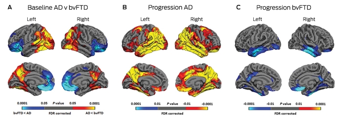
AD = Alzheimer’s disease. bvFTD = behavioural variant frontotemporal dementia. FDR = false discovery rate. A: Differences in the patterns of cortical thinning at baseline in bvFTD (blue) and AD (yellow-red). B: Annual progression in AD. C: Annual progression in bvFTD. Reproduced with permission from Landin-Romero et al.47
Competing interests
No relevant disclosures.
Acknowledgements
Olivier Piguet is supported by a National Health and Medical Research Council (NHMRC) Senior Research Fellowship (APP1103258). Fiona Kumfor is supported by an NHMRC–Australian Research Council Dementia Research Development Fellowship (APP1097026).
References
- Ratnavalli E, Brayne C, Dawson K, et al. The prevalence of frontotemporal dementia. Neurology 2002; 58: 1615-1621.
- Coyle-Gilchrist IT, Dick KM, Patterson K, et al. Prevalence, characteristics, and survival of frontotemporal lobar degeneration syndromes. Neurology 2016; 86: 1736-1743.
- Rascovsky K, Hodges JR, Knopman D, et al. Sensitivity of revised diagnostic criteria for the behavioural variant of frontotemporal dementia. Brain 2011; 134: 2456-2477.
- Gorno-Tempini ML, Hillis AE, Weintraub S, et al. Classification of primary progressive aphasia and its variants. Neurology 2011; 76: 1006-1014.
- American Psychiatric Association. Diagnostic and statistical manual of mental disorders. 5th ed. Washington, DC: APA, 2013.
- Lillo P, Garcin B, Hornberger M, et al. Neurobehavioral features in frontotemporal dementia with amyotrophic lateral sclerosis. Arch Neurol 2010; 67: 826-830.
- Lomen-Hoerth C, Anderson T, Miller B. The overlap of amyotrophic lateral sclerosis and frontotemporal dementia. Neurology 2002; 59: 1077-1079.
- Lillo P, Mioshi E, Zoing MC, et al. How common are behavioural changes in amyotrophic lateral sclerosis? Amyotroph Lateral Scler 2011; 12: 45-51.
- Kertesz A, McMonagle P, Blair M, et al. The evolution and pathology of frontotemporal dementia. Brain 2005; 128: 1996-2005.
- Cummings JL, Mega M, Gray K, et al. The Neuropsychiatric Inventory: comprehensive assessment of psychopathology in dementia. Neurology 1994; 44: 2308-2314.
- Wedderburn C, Wear H, Brown J, et al. The utility of the Cambridge Behavioural Inventory in neurodegenerative disease. J Neurol Neurosurg Psychiatry 2008; 79: 500-503.
- Kertesz A, Davidson W, Fox H. Frontal behavioral inventory: diagnostic criteria for frontal lobe dementia. Can J Neurol Sci 1997; 24: 29-36.
- Ahmed RM, Irish M, Kam J, et al. Quantifying the eating abnormalities in frontotemporal dementia. JAMA Neurol 2014; 71: 1540-1546.
- Liu W, Miller BL, Kramer JH, et al. Behavioral disorders in the frontal and temporal variants of frontotemporal dementia. Neurology 2004; 62: 742-748.
- Diehl-Schmid J, Pohl C, Perneczky R, et al. Behavioral disturbances in the course of frontotemporal dementia. Dement Geriatr Cogn Disord 2006; 22: 352-357.
- Srikanth S, Nagaraja AV, Ratnavalli E. Neuropsychiatric symptoms in dementia-frequency, relationship to dementia severity and comparison in Alzheimer’s disease, vascular dementia and frontotemporal dementia. J Neurol Sci 2005; 236: 43-48.
- Pasquier F, Lebert F, Lavenu I, et al. The clinical picture of frontotemporal dementia: diagnosis and follow-up. Dement Geriatr Cogn Disord 1999; 10 Suppl 1: 10-14.
- Snowden JS, Rollinson S, Thompson JC, et al. Distinct clinical and pathological characteristics of frontotemporal dementia associated with C9ORF72 mutations. Brain 2012; 135: 693-708.
- Devenney E, Hornberger M, Irish M, et al. Frontotemporal dementia associated with the C9ORF72 mutation: a unique clinical profile. JAMA Neurol 2014; 71: 331-339.
- Hsieh S, Schubert S, Hoon C, et al. Validation of the Addenbrooke’s Cognitive Examination III in frontotemporal dementia and Alzheimer’s disease. Dement Geriatr Cogn Disord 2013; 36: 242-250.
- Kipps CM, Nestor PJ, Dawson CE, et al. Measuring progression in frontotemporal dementia: Implications for therapeutic interventions. Neurology 2008; 70: 2046-2052.
- Hornberger M, Savage S, Hsieh S, et al. Orbitofrontal dysfunction discriminates behavioral variant frontotemporal dementia from Alzheimer’s disease. Dement Geriatr Cogn Disord 2010; 30: 547-552.
- Leslie F, Foxe D, Daveson N, et al. FRONTIER Executive Screen: a brief executive battery to differentiate frontotemporal dementia and Alzheimer’s disease. J Neurol Neurosurg Psychiatry 2016; 87: 831-835.
- Torralva T, Roca M, Gleichgerrcht E, et al. INECO Frontal Screening (IFS): a brief, sensitive, and specific tool to assess executive functions in dementia. J Int Neuropsychol Soc 2009; 15: 777-786.
- Chiong W, Wood KA, Beagle AJ, et al. Neuroeconomic dissociation of semantic dementia and behavioural variant frontotemporal dementia. Brain 2016; 139: 578-587.
- O’Callaghan C, Bertoux M, Irish M, et al. Fair play: social norm compliance failures in behavioural variant frontotemporal dementia. Brain 2016; 139: 204-216.
- Hornberger M, Piguet O. Episodic memory in frontotemporal dementia: a critical review. Brain 2012; 135: 678-692.
- Irish M, Hornberger M, Lah S, et al. Profiles of recent autobiographical memory retrieval in semantic dementia, behavioural-variant frontotemporal dementia, and Alzheimer’s disease. Neuropsychologia 2011; 49: 2694-2702.
- Tu S, Wong S, Hodges JR, et al. Lost in spatial translation - a novel tool to objectively assess spatial disorientation in Alzheimer’s disease and frontotemporal dementia. Cortex 2015; 67: 83-94.
- Kennedy DP, Adolphs R. The social brain in psychiatric and neurological disorders. Trends Cogn Sci 2012; 16: 559-572.
- Kumfor F, Piguet O. Disturbance of emotion processing in frontotemporal dementia: a synthesis of cognitive and neuroimaging findings. Neuropsychol Rev 2012; 22: 280-297.
- Sturm VE, Sollberger M, Seeley WW, et al. Role of right pregenual anterior cingulate cortex in self-conscious emotional reactivity. Soc Cogn Affect Neurosci 2013; 8: 468-474.
- Hutchings R, Palermo R, Piguet O, et al. Disrupted face processing in frontotemporal dementia: a review of the clinical and neuroanatomical evidence. Neuropsychol Rev 2017; 27: 18-30.
- Miller LA, Hsieh S, Lah S, et al. One size does not fit all: face emotion processing impairments in semantic dementia, behavioural-variant frontotemporal dementia and Alzheimer’s disease are mediated by distinct cognitive deficits. Behav Neurol 2012; 25: 53-60.
- Bora E, Walterfang M, Velakoulis D. Theory of mind in behavioural-variant frontotemporal dementia and Alzheimer’s disease: a meta-analysis. J Neurol Neurosurg Psychiatry 2015; 86: 714-719.
- Fernandez-Duque D, Hodges SD, Baird JA, et al. Empathy in frontotemporal dementia and Alzheimer’s disease. J Clin Exp Neuropsychol 2010; 32: 289-298.
- Baez S, Manes F, Huepe D, et al. Primary empathy deficits in frontotemporal dementia. Frontiers Aging Neurosci 2014; 6: 262.
- Oliver LD, Mitchell DG, Dziobek I, et al. Parsing cognitive and emotional empathy deficits for negative and positive stimuli in frontotemporal dementia. Neuropsychologia 2015; 67: 14-26.
- Ibanez A, Manes F. Contextual social cognition and the behavioral variant of frontotemporal dementia. Neurology 2012; 78: 1354-1362.
- McDonald S, Flanagan S, Rollins J, et al. TASIT: a new clinical tool for assessing social perception after traumatic brain injury. J Head Trauma Rehabil 2003; 18: 219-238.
- Bertoux M, Volle E, Funkiewiez A, et al. Social Cognition and Emotional Assessment (SEA) is a marker of medial and orbital frontal functions: a voxel-based morphometry study in behavioral variant of frontotemporal degeneration. J Int Neuropsychol Soc 2012; 18: 972-985.
- Kumfor F, Irish M, Leyton CE, et al. Tracking the progression of social cognition in neurodegenerative disorders. J Neurol Neurosurg Psychiatry 2014; 85: 1076-1083.
- Boccardi M, Sabattoli F, Laakso MP, et al. Frontotemporal dementia as a neural system disease. Neurobiol Aging 2005; 26: 37-44.
- Agosta F, Scola E, Canu E, et al. White matter damage in frontotemporal lobar degeneration spectrum. Cereb Cortex 2012; 22: 2705-2714.
- Seeley WW. Selective functional, regional, and neuronal vulnerability in frontotemporal dementia. Curr Opin Neurol 2008; 21: 701-707.
- Seeley WW, Crawford RK, Zhou J, et al. Neurodegenerative diseases target large-scale human brain networks. Neuron 2009; 62: 42-52.
- Landin-Romero R, Kumfor F, Leyton CE, et al. Disease-specific patterns of cortical and subcortical degeneration in a longitudinal study of Alzheimer’s disease and behavioural-variant frontotemporal dementia. Neuroimage 2017; 151: 72-80.
- Pereira JM, Williams GB, Acosta-Cabronero J, et al. Atrophy patterns in histologic vs clinical groupings of frontotemporal lobar degeneration. Neurology 2009; 72: 1653-1660.
- Gordon E, Rohrer JD, Fox NC. Advances in neuroimaging in frontotemporal dementia. J Neurochem 2016; 138: 193-210.
- Filippi M, Agosta F, Scola E, et al. Functional network connectivity in the behavioral variant of frontotemporal dementia. Cortex 2013; 49: 2389-2401.
- Lam BY, Halliday GM, Irish M, et al. Longitudinal white matter changes in frontotemporal dementia subtypes. Hum Brain Map 2014; 35: 3547-3557.
- Whitwell JL, Boeve BF, Weigand SD, et al. Brain atrophy over time in genetic and sporadic frontotemporal dementia: a study of 198 serial magnetic resonance images. Eur J Neurol 2015; 22: 745-752.
- Whitwell JL, Weigand SD, Boeve BF, et al. Neuroimaging signatures of frontotemporal dementia genetics: C9ORF72, tau, progranulin and sporadics. Brain 2012; 135: 794-806.
- Devenney E, Bartley L, Hoon C, et al. Progression in behavioral variant frontotemporal dementia: a longitudinal study. JAMA Neurol 2015; 72: 1501-1509.
- Rohrer JD, Nicholas JM, Cash DM, et al. Presymptomatic cognitive and neuroanatomical changes in genetic frontotemporal dementia in the Genetic Frontotemporal dementia Initiative (GENFI) study: a cross-sectional analysis. Lancet Neurol 2015; 14: 253-262.
- Varma AR, Adams W, Lloyd JJ, et al. Diagnostic patterns of regional atrophy on MRI and regional cerebral blood flow change on SPECT in young onset patients with Alzheimer’s disease, frontotemporal dementia and vascular dementia. Acta Neurol Scand 2002; 105: 261-269.
- Kanda T, Ishii K, Uemura T, et al. Comparison of grey matter and metabolic reductions in frontotemporal dementia using FDG-PET and voxel-based morphometric MR studies. Eur J Nucl Med Mol Imaging 2008; 35: 2227-2234.
- Morris E, Chalkidou A, Hammers A, et al. Diagnostic accuracy of 18 F amyloid PET tracers for the diagnosis of Alzheimer’s disease: a systematic review and meta-analysis. Eur J Nucl Med Mol Imaging 2016; 43: 374-385.
- Villemagne VL, Fodero-Tavoletti MT, Masters CL, et al. Tau imaging: early progress and future directions. Lancet Neurol 2015; 14: 114-124.
- Cairns NJ, Bigio EH, Mackenzie IR, et al. Neuropathologic diagnostic and nosologic criteria for frontotemporal lobar degeneration: consensus of the Consortium for Frontotemporal Lobar Degeneration. Acta Neuropathol (Berl) 2007; 114: 5-22.
- Mackenzie IR, Neumann M. Molecular neuropathology of frontotemporal dementia: insights into disease mechanisms from postmortem studies. J Neurochem 2016; 138 Suppl 1: 54-70.
- Shi J, Shaw CL, Du Plessis D, et al. Histopathological changes underlying frontotemporal lobar degeneration with clinicopathological correlation. Acta Neuropathol 2005; 110: 501-512.
- Seelaar H, Klijnsma KY, de Koning I, et al. Frequency of ubiquitin and FUS-positive, TDP-43-negative frontotemporal lobar degeneration. J Neurol 2010; 257: 747-753.
- Wood EM, Falcone D, Suh E, et al. Development and validation of pedigree classification criteria for frontotemporal lobar degeneration. JAMA Neurol 2013; 70: 1411-1417.
- Po K, Leslie FV, Gracia N, et al. Heritability in frontotemporal dementia: more missing pieces? J Neurol 2014; 261: 2170-2177.
- Rohrer JD, Guerreiro R, Vandrovcova J, et al. The heritability and genetics of frontotemporal lobar degeneration. Neurology 2009; 73: 1451-1456.
- DeJesus-Hernandez M, Mackenzie IR, Boeve BF, et al. Expanded GGGGCC hexanucleotide repeat in noncoding region of C9ORF72 causes chromosome 9p-linked FTD and ALS. Neuron 2011; 72: 245-256.
- Renton AE, Majounie E, Waite A, et al. A hexanucleotide repeat expansion in C9ORF72 is the cause of chromosome 9p21-linked ALS-FTD. Neuron 2011; 72: 257-268.
- Hodges J. Familial frontotemporal dementia and amyotrophic lateral sclerosis associated with the C9ORF72 hexanucleotide repeat. Brain 2012; 135: 652-655.
- Pottier C, Ravenscroft TA, Sanchez-Contreras M, et al. Genetics of FTLD: overview and what else we can expect from genetic studies. J Neurochem 2016; 138 Suppl 1: 32-53.
- Boxer AL, Boeve BF. Frontotemporal dementia treatment: current symptomatic therapies and implications of recent genetic, biochemical, and neuroimaging studies. Alzheimer Dis Assoc Disord 2007; 21: S79-S87.
- Moretti R, Torre P, Antonello RM, et al. Frontotemporal dementia: paroxetine as a possible treatment of behavior symptoms. A randomized, controlled, open 14-month study. Eur Neurol 2003; 49: 13-19.
- Deakin JB, Rahman S, Nestor PJ, et al. Paroxetine does not improve symptoms and impairs cognition in frontotemporal dementia: a double-blind randomized controlled trial. Psychopharmacology (Berl) 2004; 172: 400-408.
- Moretti R, Torre P, Antonello RM, et al. Olanzapine as a treatment of neuropsychiatric disorders of Alzheimer’s disease and other dementias: a 24-month follow-up of 68 patients. Am J Alzheimers Dis Other Demen 2003; 18: 205-214.
- Moretti R, Torre P, Antonello RM, et al. Rivastigmine in frontotemporal dementia: an open-label study. Drugs Aging 2004; 21: 931-937.
- Mendez MF, Shapira JS, McMurtray A, et al. Preliminary findings: behavioral worsening on donepezil in patients with frontotemporal dementia. Am J Geriatr Psychiatry 2007; 15: 84-87.
- Boxer AL, Knopman DS, Kaufer DI, et al. Memantine in patients with frontotemporal lobar degeneration: a multicentre, randomised, double-blind, placebo-controlled trial. Lancet Neurol 2013; 12: 149-156.
- Gauthier S, Feldman HH, Schneider LS, et al. Efficacy and safety of tau-aggregation inhibitor therapy in patients with mild or moderate Alzheimer’s disease: a randomised, controlled, double-blind, parallel-arm, phase 3 trial. Lancet 2016; 388: 2873-2884.
- Mendez MF, Joshi A, Jimenez E. Virtual reality for the assessment of frontotemporal dementia, a feasibility study. Disabil Rehabil Assist Technol 2015; 10: 160-164.
- O’Connor CM, Clemson L, Brodaty H, et al. Enhancing caregivers’ understanding of dementia and tailoring activities in frontotemporal dementia: two case studies. Disabil Rehabil 2016; 38: 704-714.
- Mioshi E, Bristow M, Cook R, et al. Factors underlying caregiver stress in frontotemporal dementia and Alzheimer’s disease. Dement Geriatr Cogn Disord 2009; 27: 76-81.
- Boutoleau-Bretonniere C, Vercelletto M, Volteau C, et al. Zarit burden inventory and activities of daily living in the behavioral variant of frontotemporal dementia. Dement Geriatr Cogn Disord 2008; 25: 272-277.
- Miller LA, Mioshi E, Savage S, et al. Identifying cognitive and demographic variables that contribute to carer burden in dementia. Dement Ger Cogn Disord 2013; 36: 43-49.
- Mioshi E, McKinnon C, Savage S, et al. Improving burden and coping skills in frontotemporal dementia caregivers : a pilot study. Alzheimer Dis Assoc Disord 2013; 27: 84-86.
- Garcin B, Lilo P, Hornberger M, et al. Determinants of survival in behavioral variant frontotemporal dementia. Neurology 2009; 73: 1656-1661.
- Chow TW, Fridhandler JD, Binns MA, et al. Trajectories of behavioural disturbance in dementia. J Alzheimers Dis 2012; 31: 143-149.
Linked content
-
MJA Editorial: Dementia of the personality
-
MJA InSight: Frontotemporal dementias: treat the symptoms, spare the carers
-
MJA Podcast: Professor Olivier Piguet
Provenance: Not commissioned; externally peer reviewed.
Motor Neuron Disease Mortality Trends in Australia From 1986 to 2023: A Population-Based Study
Carol M. Y. Lee, Rupendra N. Shrestha, Julian Gold, Mark Stevenson, Kelly L. Williams, Lyndal Henden, Sandrine K. K. Chan Moi Fat, Zoe Zussa, Amanda Wright, Martin Jude, Mark A. S. Laidlaw, Willem J. R. Fokkink, Dominic B. Rowe
Mild traumatic brain injury and concussion and persisting post‐concussion symptoms: new guidelines to support evidence‐based assessment and management in Australia and Aotearoa New Zealand
Karen M Barlow, Jennie L Ponsford, Alice Theodom, Gill Cowen, Gavin A Davis, Vicki Anderson, Franz E Babl, David Cole, Jennifer Cullen, Stuart R Dalziel, Melinda Fitzgerald, Howard Flavell, Caroline Yates, Rebecca Kimble, John H Olver, Rhonda Orr, Mark Ralfe, Michael Rose, Nick Rushworth, Julia Treleaven, Gary Browne, Nathan Delang, Sarah Harris, Gary Mitchell, Sean Tweedy
The prevalence of and variation in indicators of the quality and safety of long term aged care in Australia, 2019: a cross‐sectional population‐based study
Tesfahun C Eshetie, Gillian E Caughey, Catherine Lang, Olivia Ryan, Renuka Visvanathan, Craig Whitehead, Keith Evans, Janet K Sluggett, Jyoti Khadka, Carolyn Dawkins, Helena Williams, Miranda Starke, Sara Blunt, Anne Liddell, Megan Corlis, Anna Sheppeard, Penelope Lello, Marilyn Thien, Steven L Wesselingh, Maria C Inacio
Health and economic benefits of improving pre‐hospital identification of stroke in Australian women: a modelling study
Thomas Gadsden, Lei Si, Emily R Atkins, Cheryl Carcel, Xia Wang, Stephen Jan, Mark Woodward, Laura E Downey
Symptom burden, quality of life, and diagnostic journey of people with postural orthostatic tachycardia syndrome, Australia, 2021–24: a descriptive patient registry data study
Marie‐Claire Seeley, Gemma Wilson, Eric Ong, Amy Langdon, Jonathan Chieng, Danielle Bailey, Kristina Comacchio, Amanda Page, Dennis H Lau, Celine Gallagher
The intersection of rurality and dementia prevalence in Australia for Aboriginal and Torres Strait Islander and non‐Indigenous peoples
Antonia J Clarke, Marwan Ahmed, Judith M Katzenellenbogen, John Towney (Wiradjuri), Anna H Balabanski, Adrienne Withall (Dharawal Yuin), Kylie Radford, Amy Brodtmann