




Volume 207 - Issue 6
Invasive Neisseria gonorrhoeae producing pre-septal cellulitis and keratoconjunctivitis: diagnosis and management
Authors: Shivesh Varma, Nathan Wong and Jwu Jin Khong
Med J Aust 2017; 207 (6): 269. || doi: 10.5694/mja17.00223
Published online: 18 September 2017
Published online: 18 September 2017
To the Editor:
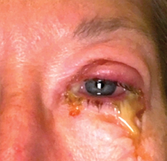
A 53-year-old woman experienced rapid onset of left eyelid pain, swelling and purulent discharge (Box 1). She presented to our eye hospital 3 days later and, on assessment, the lids were exquisitely tender, inflamed and difficult to open. There was chemosis, microcystic corneal oedema with Descemet membrane folds and moderate inflammation in the anterior chamber. Visual acuity was 6/24 in the left eye and 6/9 in the right eye. Ocular motility appeared normal.
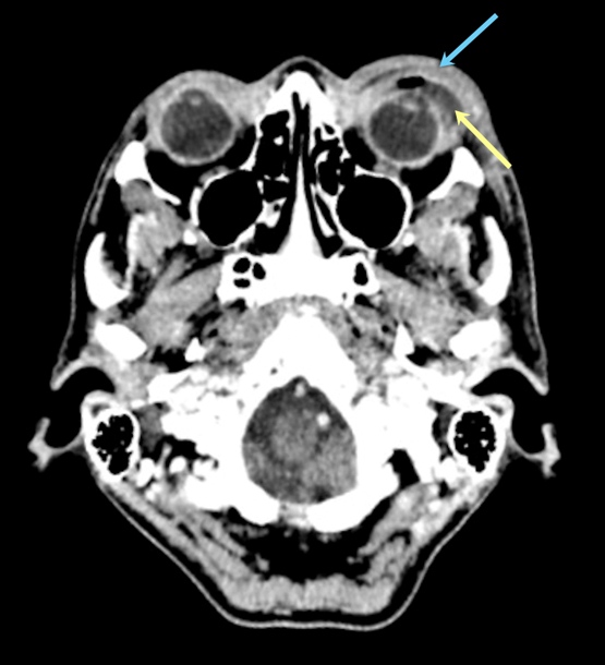
The severe inflammation and visual impairment raised concerns for post-septal orbital cellulitis. A computed tomography scan showed severe pre-septal stranding only (Box 2). A diagnosis of left pre-septal cellulitis and keratoconjunctivitis was made. She was given intravenous ceftriaxone 2 g daily, intravenous flucloxacillin 2 g four times a day, topical chloramphenicol 0.5% four times a day and regular eye washings.
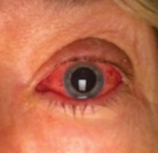
Microscopy found gram-negative diplococci, and polymerase chain reaction testing was positive for Neisseria gonorrhoeae. The patient denied history of sexually transmissible infection. A urine and serum sexually transmissible infection screen for N. gonorrhoeae, Chlamydia trachomatis and blood-borne viruses was negative. Treatment was rationalised to intravenous ceftriaxone 1 g daily, with a single 1 g dose of oral azithromycin. The patient had dramatic improvement over 4 days, and visual acuity improved to 6/7.5 at discharge (Box 3). One week later, the infection had predominately resolved.
N. gonorrhoeae typically produces a hyperacute unilateral conjunctivitis. While the source of infection in this patient is unclear, in most cases infection is acquired via sexual transmission and reaches the conjunctiva through hand–eye auto-inoculation.1,2 The bacteria can invade the corneal epithelium, producing keratitis and corneal melting necessitating therapeutic keratoplasty.1,2 Gonococcal periorbital infection is rare, with only three cases of pre-septal cellulitis and two cases of post-septal cellulitis reported.3 All patients were successfully treated with parenteral antibiotics.
N. gonorrhoeae has increasing antimicrobial resistance, creating a therapeutic challenge. The Australian Gonococcal Surveillance Programme has shown an emerging resistance to ceftriaxone — the antibiotic of choice — from 0.6% in 2006 to 5.4% in 2014.4 Azithromycin resistance occurs in 2.4% of strains.4 The Australian Therapeutic Guidelines recommend treatment of N. gonorrhoeae with parenteral ceftriaxone and oral azithromycin, for bactericidal synergy and cotreatment for C. trachomatis.
When presented with pre-septal cellulitis and purulent keratoconjunctivitis, the clinician should be vigilant for a sight-threatening N. gonorrhoeae infection.
Competing interests
No relevant disclosures.
References
- McAnena L, Knowles SJ, Curry A, Cassidy L. Prevalence of gonococcal conjunctivitis in adults and neonates. Eye (Lond) 2015; 29: 875-880.
- Kawashima M, Kawakita T, Den S, et al. Surgical management of corneal perforation secondary to gonococcal keratoconjunctivitis. Eye (Lond) 2009; 23: 339-344.
- Yao HY, Wang CH. Preseptal cellulitis caused by Neisseria gonorrhoeae: a rare disease need to be vigilant. J Microbiol Immunol Infect 2017; 50: 397-398.
- Lahra MM, Australian Gonococcal Surveillance Programme. Australian Gonococcal Surveillance Programme annual report, 2014. Commun Dis Intell Q Rep 2015; 39: E347-E354.
A Diagnostic Headache
Thomas Glynn, Michelle T. Leech, Stacy K. Goergen, Emily Lin, Nadeem Toodayan, Ralph Junckerstorff
Neisseria meningitidis: an uncommon cause of conjunctivitis
Chloe Story, Tina Marinelli, Simeon Crawford, Amrita Ronnachit
Dismantling barriers to research and clinical care for individuals with a vision impairment
Eden G Robertson, Kate Hetherington, Meredith Prain, Julia Hall, Leighton Boyd AM, Rosemary Boyd OAM, Emily Shepard, Hollie Feller, Sally Karandrews, Fleur O'Hare, Kanae Yamamoto, Matthew P Simunovic, Robyn V Jamieson, Alan Ma, Lauren Ayton AM, Anai Gonzalez‐Cordero
The New South Wales Pharmacy Trial for herpes zoster: on the nose?
Christian P Pappas, Timothy R Holmes, Minas T Coroneo
Skin fragility disorder misdiagnosed as child abuse: a cautionary tale
Heather G Mack, Fred K Chen, John R Grigg
Health economic aspects of inherited retinal diseases: looking for cost‐effective treatments
Benjamin Kamien, Rachael Heath Jeffery, Fred K Chen