




Volume 207 - Issue 5
A stitch in time: stitching errors in digital radiology
Authors: Clare Faurie, Nicole Williams and Peter J Cundy
Med J Aust 2017; 207 (5): 224. || doi: 10.5694/mja17.00098
Published online: 4 September 2017
Published online: 4 September 2017
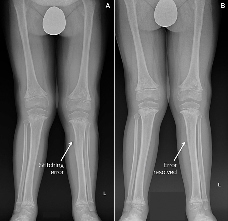
Modern digital radiological techniques include “stitching” together multiple x-ray images. This provides one overall image of the area of interest, such as the entire spine or lower limbs, for deformity assessment. Dedicated software automatically combines separate exposures and allows for overlap. Image acquisition is rapid, minimising patient motion artefact and reducing distortion.1 However, if images are inappropriately put together (digitally stitched), stitching errors may occur with this computer-driven process. If not manually corrected by the radiology technician, the supplied image may hide pathology or give the false impression of abnormality. Four cases of digital stitching error have recently occurred in our tertiary referral paediatric hospital.
Digitally stitched x-rays from the lower limbs were obtained for routine assessment of a child with skeletal dysplasia, and fractures of the left tibia and fibula were apparent (Box, A). This did not correlate with the clinical findings, including the child dancing in the waiting room. The radiology department was contacted and they re-issued corrected images (Box, B). Spinal x-rays in two patients after scoliosis surgery incorrectly showed broken surgical rods. In the context of ongoing symptoms, the option of revision surgery was discussed with the family in the first case. Repeat x-rays, obtained as part of the pre-operative planning, showed the rod was in fact not broken. A stitching error was immediately recognised by the spinal surgeon when a second similar case occurred. In a fourth case, comparison of the anteroposterior and lateral views of a child’s spine showed different numbers of vertebrae in the two views (11 vertebrae on the frontal view and 12 on the lateral).
Digital radiology is commonplace and allows rapid image acquisition and ease of digital measurement.2 However, technical errors — digital stitching errors — may occur. Although rarely reported, these may be relatively common, with one study identifying stitching errors in 14 out of 86 reviewed scoliosis studies.1 Despite ongoing technological refinements,3 clinicians should always carefully check digitally stitched images: look for soft tissue mismatch, correlate with source images and clinical presentation, and avoid digital stitching in certain patients, such as people with movement disorders.
Competing interests
No relevant disclosures.
References
- Supakul N, Newbrough K, Cohen MD, Jennings SG. Diagnostic errors from digital stitching of scoliosis images — the importance of evaluating the source images prior to making a final diagnosis. Pediatr Radiol 2012; 42: 584-598.
- Kuklo TR, Potter BK, Schroeder TM, O’Brien MF. Comparison of manual and digital measurements in adolescent idiopathic scoliosis. Spine (Phila Pa 1976) 2006; 31: 1240-1246.
- Yang F, He Y, Deng ZS, Yan A. Improvement of automated image stitching system for DR X-ray images. Comput Biol Med 2016; 71: 108-114.
Reducing Nitrous Oxide Emissions Across the Melbourne Biomedical Precinct
Ross Robertson, Andrew Downey, Daryl Williams, Bjorn Makein, Ben Dunne, Tugce Ozturk, Ying Gu, Rebecca McIntyre
Multimorbidity Clusters Among People Aged 65 Years and Over in Australia: A Nationwide Cross-Sectional Data Linkage Study
Weisi Chen, Christine Y. Lu, Sarah N. Hilmer, Alice A. Gibson, Edwin C. K. Tan
When ‘Liver Enzymes’ Are Not Hepatic: Late-Onset Pompe Disease
Shauna Madigan, Georgina England, Wayne Rankin
West Nile virus Kunjin subtype in rural NSW
Emily Gibson, Megan Whitley, Peter Murray, Linda Hueston, Jane Bennett, Raguharan Kathiresu, David N Durrheim
The impact of the Breast Screen NSW transition from film to digital mammography, 2002–2016: a linked population health data analysis
Rachel Farber, Nehmat Houssami, Katy J L Bell