




Volume 207 - Issue 5
Radiation therapy and early breast cancer: current controversies
Author: John Boyages
Med J Aust 2017; 207 (5): 216-222. || doi: 10.5694/mja16.01020
Published online: 4 September 2017
Published online: 4 September 2017
Recent advances are likely to further improve the incremental benefit of radiation over and above surgery and systemic therapy and thus increase survival rates
Summary
- Radiation therapy (RT) is an important component of breast cancer treatment.
- RT reduces local recurrence and breast cancer mortality after breast conservation for all patients and for node-positive patients after a mastectomy.
- Short courses of RT over 3–4 weeks are generally as effective as longer courses.
- A patient subgroup where RT can be avoided after conservative surgery has not been consistently identified.
- A radiation boost reduces the risk of a recurrence in the breast but may be omitted for older patients with good prognosis tumours with clear margins.
- Axillary recurrences can take a long time to appear, with 35% occurring after 5 years.
- Leaving disease untreated in regional nodes is associated with reduced survival.
- Not all patients require radiation after neoadjuvant chemotherapy and a subsequent mastectomy.
- Modern RT equipment and techniques will further improve survival rates.
A year after Wilhelm Röntgen discovered x-rays in 1895, radiation was used by Emil Grubbé in Chicago to treat a patient with inoperable breast cancer, applying radiation energies that could penetrate at most a superficial skin cancer today.1 In the 1920s, radium was used following conservative surgery by Geoffrey Keynes at St Bartholomew’s Hospital, London, producing equivalent survival rates to radical mastectomy, which was introduced by William Halsted in the late 19th century.2,3
After World War 1, deep x-ray therapy was introduced, with energies up to 200 kV. Megavoltage linear accelerators and cobalt beam were largely introduced in the 1960s.4 However, radiation therapy (RT) remained imprecise, with lower energies causing severe skin reactions, and planning involving surface anatomy, gentian violet marks and tracing paper, rather than the sophisticated imaging and 3D computing techniques of today.
With more effective chemotherapies and hormonal and targeted therapies, controversy exists as to whether RT can be reduced in extent or avoided altogether after surgery for early breast cancer given that RT after breast conservation or a mastectomy reduces recurrence rates and improves survival (Box 1).5,6 After conservative surgery, RT to the breast halves the overall recurrence rate and reduces breast cancer mortality by about one-sixth.5 Cosmetic results are excellent if limited surgery is combined with moderate dose radiation.
Substantial changes in practice have occurred since these clinical trials were completed and their results may not be totally applicable today. Many women now have smaller screen-detected cancers and radiotherapy techniques better target the cancer while protecting normal structures such as the heart. Serial sectioning of sentinel nodes and more frequent recognition of micrometastases has meant that some women classified as node-negative in earlier studies would have been identified as node-positive and given better systemic therapy if treated today. Therefore, the challenge is to integrate historical data with our better understanding of disease biology and treatments to formulate a personalised treatment strategy.
Radiation after conservative surgery
Several trials firstly attempted conservative surgery with or without RT and later with or without hormonal treatment, usually tamoxifen. Although recent trials have reported lower recurrence rates, this is partly due to shorter follow-up and better patient selection and may not generally apply to a community setting. A meta-analysis found that older patients with small, low grade, oestrogen-receptor (ER)-positive tumours have a low recurrence rate and are possible candidates for omitting RT in some clinical circumstances,5 and other researchers have tested accelerated partial breast irradiation in this low risk group.7
Box 2 shows recent trials of lower risk older patients with small, ER-positive, node-negative tumours randomised between conservative surgery with or without RT and/or hormonal treatment.8-14 Adjuvant hormonal treatment reduces the risk of an in-breast recurrence when compared with surgery alone, but the combination of RT with or without hormonal treatment is more effective and more sustainable.
The question remains as to the lesser of two evils. For an 80-year-old with 10.2 years average life expectancy15 with a small ER-positive tumour, is it best to advise 5 years of tamoxifen with potential side effects including a small risk of mortality from thromboembolic disease, or a 3-week course of RT with a small risk of recurrence?16-18 This is the art of oncology — working out a personalised plan based on listening to the patient to find the right solution for their time of life. For a frailer patient, starting with hormonal treatment such as an aromatase inhibitor (with less thrombosis risk) and avoiding surgery or RT is another option, but 5-year local recurrence rates are as high as 70%.19,20 Therefore, this known development of tumour resistance remains a concern for fitter patients treated with conservative surgery and tamoxifen without radiation.
Shorter courses of radiation
Hypofractionation means a shorter radiation course with higher daily doses for the same biological effect. This saves the patient time and money and reduces pressure on public radiotherapy units. The United Kingdom Standardisation of Breast Radiotherapy (START) trial A compared a standard regimen of 50 Gy in 25 fractions with 41.6 Gy or 39 Gy in 13 fractions all over 5 weeks. In START trial B, 50 Gy in 25 fractions over 5 weeks was compared with 40 Gy in 15 fractions over 3 weeks.21 A Canadian trial for node- and margin-negative disease compared 42.5 Gy in 16 fractions over 3 weeks to 50 Gy in 25 fractions over 5 weeks.22 Boost was optional in the UK trials and omitted in the Canadian trial. Shorter schedules were not inferior for local recurrence or survival except for high grade tumours (15.6%, 10-year local recurrence) compared with the control group (4.7%; P = 0.01), perhaps due to the lack of a radiation boost in the Canadian trial.22 More detailed analysis and longer follow-up of the Canadian study found that grade on its own did not influence local recurrence, which was lower for low grade ER-positive tumours (4.5%) and higher grade triple-negative breast cancer (4.5%), but significantly higher (P < 0.001) for higher grade ER-positive tumours (7.9%) and human epidermal growth factor receptor 2 (HER2)-positive tumours (6.9%).23 The START trials did not show a higher local recurrence rate in higher grade tumours, perhaps because 61% received an additional RT boost.
In practice, standard fractionation appears to be better for larger breasted women for whom increased breast oedema can be a problem. It remains to be seen if this can be reduced with more sophisticated radiation techniques. The Canadian trial excluded women with large breasts, few women received chemotherapy and nodal radiation was not given.22 In the START trials, cosmetic result was not inferior for larger breasted women, but only a small fraction were in this group (15.6%).21
In START trial A, one patient (0.1%) who received 41.6 Gy in 13 fractions developed brachial plexopathy;21 a concern for faster schedules involving nodal irradiation, particularly if neurotoxic chemotherapeutic agents such as the taxanes are also used. The Australian and United States guidelines recommend hypofractionated treatment only for patients aged 50 years or older, with node-negative tumours up to 5 cm and no chemotherapy and where breast size and treatment technique minimise dose variation across the volume.24,25 The Australian guidelines also advise caution when recommending this approach to other patients. The UK NICE guidelines recommend that all patients with early invasive breast cancer after breast-conserving surgery receive 40 Gy in 15 fractions.26
The utility of accelerated partial breast irradiation is not yet entirely clear. This type of radiation can be delivered using intra-operative electron beam, placement of radioactive guide wires that can be loaded post-operatively, or targeted external beam radiation. A review of these techniques has been addressed elsewhere.27 Recent trials add to the debate by using intracavitary brachytherapy, or 3D conformal accelerated partial breast irradiation to deliver radiation in shorter periods to the at-risk primary tumour area.28,29
Boost to the primary tumour site
Detailed pathological studies done in the 1980s found that cancer cells were often located as far as 4 cm away from the edge of the primary tumour and this was often ductal carcinoma in situ.30 Further, early studies of the pattern of recurrence after conservative surgery and RT found that 60–70% occurred at the primary site,31,32 identifying this as an important focus for clinical examination, additional mammographic views, ultrasound and non-surgical biopsy after treatment. Predictive factors for local recurrence included young age at diagnosis, lymphatic vessel invasion, an extensive intraductal component and a positive margin, leading to studies of surgical extent, margin width and radiation boost dose31-34 in an attempt to improve local control rates. A European trial of over 5000 patients reported a 20-year breast tumour recurrence rate of 16.4% in the no-boost group compared with 12.0% in the boost group (P < 0.0001).35 This study found that the boost significantly reduced the risk of a recurrence for young patients or those with grade 3 tumours or an extensive intraductal component. For the patient subgroup with grade 3 ER-negative tumours, the boost reduced the risk of breast tumour recurrence from 31% to 5% (P = 0.01), highlighting the need to strongly consider a boost for this scenario irrespective of fractionation schedule.36
Local recurrence rates have fallen and not all patients require a boost.18,37,38 Recent US guidelines on surgical margins recommended “no ink at the margin” to mean a negative margin, rather than a specific margin width of 1 or 2 mm. This should reduce unnecessary re-excisions for many patients.37,39,40 Early results from a large Danish study of 11 900 patients found no evidence of improved local control by margin depth.41 A previous study found that patients treated without a boost (usually because of a large excision specimen or a negative re-excision) had no increase in the risk of local recurrence compared with patients who did receive a boost.31
In practice, the surgeon can assist boost planning by placing titanium clips at the excision borders. The radiation oncologist must correlate margin depth with pre-operative imaging and pathological predictors for local recurrence and sometimes consider post-operative mammography or magnetic resonance imaging. It is insufficient simply to boost the scar, because with modern oncoplastic techniques, the scar often has no relationship to the actual primary tumour site where the risk of recurrence is highest.42 Different boost techniques include electron beam, which creates more skin reaction, or an external photon boost, which may involve treating more normal tissue or more localised synchronous boost techniques.
Radiation therapy after neoadjuvant chemotherapy
The use of post-mastectomy RT is widely accepted for patients with four or more positive nodes but there is still controversy about its use in patients with one to three positive nodes43 or after neoadjuvant chemotherapy. Today, chemotherapy is used for patients with larger tumours not only to reduce tumour size to facilitate breast conservation but also to assess in vivo tumour response to the chosen chemotherapy. Patients with HER2-positive disease may receive trastuzumab in addition to chemotherapy.
A 2012 study used National Surgical Adjuvant Breast and Bowel Project (NSABP) B-18 and B-27 trial data to identify patient subgroups with a low rate of loco-regional recurrence after neoadjuvant chemotherapy and mastectomy and no RT.44 Cohorts with a less than 10–12% risk of loco-regional recurrence had pathologically negative nodes after neoadjuvant chemotherapy irrespective of whether or not they presented with clinically node-positive or -negative disease or had larger tumours.44 Chemotherapy can reduce the likelihood of pathological nodal disease, but the reductions are not large, particularly for lower grade tumours. In the NSABP B-18 trial, patients randomised to four cycles of doxorubicin and cyclophosphamide after an axillary clearance had a 57% node-positivity rate, compared with 41% for patients who had a clearance after neoadjuvant doxorubicin and cyclophosphamide.45 The addition of a taxane to doxorubicin and cyclophosphamide in the NSABP B27 study increased the overall pathological complete response rate from 14% to 26% (P < 0.001).46 Pathological complete response rate was significantly higher for patients with triple-negative (38.2%) or HER2-positive (45.4%) tumours than for patients with ER-receptor-positive, HER2-negative disease (11.4%) (P < 0.0001).47 One review suggested that clinical stage II (T1-2 N0–N1) patients aged over 40 years, with ER-positive disease and no lymphatic vessel invasion who have a pathological complete response rate or 0–3 positive axillary nodes without cancer extending beyond the lymph node capsule, gain little or no benefit from post-mastectomy RT after neoadjuvant chemotherapy.48
The pre-operative classification of breast cancer by molecular subtypes has influenced not only the response to chemotherapy, but also the choice of systemic agent. It also helps to predict the risk of recurrence. It is well known, for example, that invasive lobular carcinomas have a low pathological complete response rate (4.9%) to neoadjuvant chemotherapy compared with invasive ductal carcinoma (20.2%) (P < 0.0001).49 Residual lobular carcinoma can be hard for the pathologist to detect and can infiltrate the skin. This makes clinical detection difficult, so post-mastectomy RT is usually indicated. It is less well known that patients with triple-negative disease have better loco-regional control rates with conservative surgery and RT than with mastectomy alone.50-52 Further, not all node-negative patients with a tumour over 5 cm (T3 N0) require post-mastectomy radiation.53 Ongoing trials are investigating the role of post-mastectomy RT in patients receiving neoadjuvant chemotherapy.54 Pre-operative assessment by a radiation oncologist can help with decision making and subsequent field placements.
Regional node irradiation after positive axillary sentinel node
There is increasing long term data showing that any cells in a sentinel node reduce survival, including isolated tumour cells, micrometastases or larger deposits, although this remains an area of debate in the era of more effective systemic therapy.55 The risk of a non-sentinel node metastasis largely depends on the thoroughness of the subsequent axillary dissection. In a consecutive validation study at Westmead Hospital, Sydney,56 140 patients underwent a complete axillary dissection (mean of 22 nodes dissected) after a sentinel node biopsy, including nodes in level 3 inferior to the medial end of the clavicle and involving three surgeons; other validation studies usually had less complete axillary dissections and multiple surgeons, potentially underestimating the true rate of non-sentinel node metastasis.57 Of 51 patients with a positive sentinel node, 47% had non-sentinel node metastases. Primary tumour size was the only significant predictor for non-sentinel node involvement; 25% of patients with a primary tumour size = 20 mm and 67% with a primary tumour size > 20 mm had additional disease in the axilla (P = 0.005).56
The American College of Surgeons Oncology Group Z0011 trial randomised older patients with predominantly small (69% T1), grade 1 and 2 (72%) ER-positive tumours who underwent conservative surgery and RT to an axillary dissection or observation after a positive sentinel lymph node biopsy.57,58 Patients were treated with RT to the breast alone. Nodal positivity was micrometastatic in 45% of the sentinel lymph node biopsy group and 38% in the dissection group. After an axillary dissection, 27% had additional positive axillary nodes. Nearly all patients received systemic therapy. Regional nodal failure rate at 10 years was 0.5% in the nodal dissection group and 1.5% in the sentinel lymph node biopsy group (P = 0.28).
Unfortunately, clinicians have applied the Z0011 trial results in clinical scenarios outside the strict study entry criteria, including the post-mastectomy setting. Micrometastases have been observed in a small subgroup of patients after a mastectomy,59 but current guidelines caution against not dissecting the axilla if radiation is not given.60 Of concern, 35% of axillary recurrences occur after 5 years and late recurrences cause significant morbidity.5 Further, a retrospective audit found 69% of patients in the Z0011 trial received radiation that treated the lymph nodes.61 In many ways, the Z0011 study compared axillary radiation with axillary surgery, which is known to be equivalent.62
A randomised trial compared axillary and supraclavicular fossa radiation after a positive sentinel node to axillary dissection and found no difference in outcome except for less arm oedema (11% v 23%; P < 0.0001) in the RT group.63 The 5-year axillary recurrence rate was 0.4% in the axillary dissection group and 1.2% in the nodal radiation group, with no survival difference. This approach would avoid a second operation to the axilla for selected patients with limited isolated tumour cells or micrometastases after a sentinel node biopsy.63
However, treatment to the full axilla can also be personalised based on the risk of non-sentinel node involvement and prognostic features of the tumour. High-tangent RT encompassing levels 1 and 2 of the axilla is an option if the risk is low (eg, ER-positive tumour, primary tumour = 20 mm, no lymphatic vessel invasion, single micrometastasis), whereas comprehensive regional node irradiation is preferred if the patient has grade 3 tumours, extensive lymphatic vessel invasion and more than one sentinel node involved.64 In some patients with high grade, triple-negative or HER2-positive disease, a post-operative positron emission tomography scan (Box 3, A and B) may help determine treatment strategy. Various calculators are available to determine the probability of non-sentinel node involvement to guide therapy (eg, http://nomograms.mskcc.org/breast/BreastAdditionalNonSLNMetastasesPage.aspx).
In summary, leaving a small burden of axillary disease after a sentinel node biopsy is not unreasonable in most situations, provided radiation to the regional nodes is added.
The internal mammary chain
In 1918, Stibbe documented that the first to sixth intercostal mammary spaces contained lymph nodes in 97%, 98%, 83%, 9%, 13% and 63% of cases respectively;65 other studies have found lymph nodes to be as infrequent as 10% in the sixth space.66 The nodes lie medial to the internal mammary chain (IMC) vessels in 88%, 76% and 21% respectively in the first three interspaces and laterally thereafter.65 Coombs and colleagues highlighted the dilemma of a lymphoscintigram draining to the IMC.67 Involvement of cancer in biopsied IMC sentinel lymph nodes was 22.2% but increased significantly to over 35% for risk factors such as high grade, lymphatic vessel invasion or medial tumours with a positive axillary sentinel node. IMC recurrence is uncommon but morbid, and causes destruction of the sternum and adjacent ribs (Box 3, C).
IMC RT has been shown to reduce distant metastases and potentially improve survival (Box 1 and Box 4).5,6,68-73 For example, the MA.20 trial randomised patients with node-positive or high risk, node-negative disease treated with conservative surgery and RT to additional RT to the regional nodes including the IMC.69 Distant metastasis rates were 12.9% in the nodal irradiation group and 16.5% in the control group (P = 0.03), with no difference in breast cancer mortality. Patients with ER-negative disease who received IMC RT had a lower 10-year mortality rate than patients who did not receive IMC RT (18.7% v 26.1%; P = 0.05). Other trials show similar findings and support the notion that leaving IMC disease behind is probably detrimental to survival and that this area should be selectively treated using RT (Box 4).43
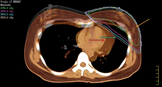
IMC radiation is problematical because it potentially increases heart dose, morbidity and mortality. There have been significant advances in RT technology, with sophisticated imaging integrated into planning systems using techniques that protect the heart with shielding or deep inspiration breath holding (Box 5), preferably using volumetric modulated arc therapy (Box 6). Volumetric modulated arc therapy can achieve highly conformal dose distributions by rotating the linear accelerator gantry at varying speeds through one or more arcs while simultaneously changing the field shape. This allows shaping or sculpting radiation doses to complex cancer volumes, while using modern equipment with on-board computed tomography scans with treatment times of about 5 minutes to reduce the dose to normal structures such as the heart. These advances are likely to further improve the incremental benefit of radiation over and above surgery and systemic therapy and thus increase survival rates.
Box 1 – Meta-analyses of randomised trials of breast-conserving surgery or mastectomy with or without radiation therapy (RT)5,6
|
|
No. of patients |
Treatment |
Any first recurrence |
Loco-regional recurrence at 10 years |
Breast cancer mortality at 15–20 years* |
Loco-regional recurrence reduction with RT |
Breast cancer mortality reduction with RT |
||||||||
|
|
|||||||||||||||
|
Conservative surgery ± RT |
|
|
|
|
|
|
|
||||||||
|
All |
10 801 |
No RT |
35.0% |
25.1% |
25.2% |
|
|
||||||||
|
|
|
RT |
19.3% |
7.7% |
21.4% |
17.4%† |
3.8%‡ |
||||||||
|
pN0 |
7287 |
No RT |
31.0% |
22.8% |
20.5% |
|
|
||||||||
|
|
|
RT |
15.6% |
7.3% |
17.2% |
15.5%† |
3.3%‡ |
||||||||
|
pN+ |
1050 |
No RT |
63.7% |
43.0% |
51.3% |
|
|
||||||||
|
|
|
RT |
42.5% |
12.4% |
42.8% |
30.6%† |
8.5%‡ |
||||||||
|
Mastectomy ± RT |
|
|
|
|
|
|
|
||||||||
|
pN+ |
3131 |
No RT |
62.5% |
26.0% |
66.4% |
|
|
||||||||
|
|
|
RT |
51.9% |
8.1% |
58.3% |
17.9%† |
8.1%‡ |
||||||||
|
pN0 |
700 |
No RT |
21.1% |
1.6% |
26.6% |
|
|
||||||||
|
|
|
RT |
22.4% |
3.0% |
28.8% |
- 1.4%§ |
- 2.2%§ |
||||||||
|
1–3 pN+ |
1314 |
No RT |
45.7% |
20.3% |
50.2% |
|
|
||||||||
|
|
|
RT |
34.2% |
3.8% |
42.3% |
16.5%† |
7.9%‡ |
||||||||
|
> 4 pN+ |
1772 |
No RT |
75.1% |
32.1% |
80.0% |
|
|
||||||||
|
|
|
RT |
66.3 |
13.0% |
70.7% |
19.1%† |
9.3%‡ |
||||||||
|
|
|||||||||||||||
|
pN0 = pathologically node-negative. pN+ = pathologically node-positive. * Breast cancer mortality was at 15 years for conservative surgery and 20 years for mastectomy. † * P < 0.00001. ‡ P < 0.05. § Not significant. |
|||||||||||||||
Box 2 – Recent trials of older patients with small node-negative cancers treated with surgery with or without radiation therapy (RT) and hormone therapy (HT)*
|
Study |
First year |
No. of patients |
Age = 50 years |
Tumour = 20 mm |
Hormonal treatment† |
Follow-up (years) |
Ipsilateral breast tumour recurrence after conservative surgery |
||||||||
|
Nil |
HT |
RT |
RT + HT |
||||||||||||
|
|
|||||||||||||||
|
Fisher 20028 |
1989 |
1000 |
80% |
98% |
67% |
7.2 |
|
16.5% |
9.3% |
2.8% |
|||||
|
Winzer 20049 |
1991 |
361 |
91% |
99% |
50% |
5.9 |
15.7% |
2.8% |
3.7% |
3.8% |
|||||
|
Fyles 200410 |
1992 |
769 |
100% |
83% |
50% |
5.6 |
|
7.7% |
|
0.6% |
|||||
|
Hughes 200411 |
1994 |
636 |
100% |
100% |
50% |
12.6 |
|
4.4% |
|
0.6% |
|||||
|
Potter 200712 |
1996 |
869 |
97% |
93% |
100% |
4.5 |
|
5.1% |
|
0.4% |
|||||
|
Blamey 201313 |
1992 |
1135 |
ns |
100% |
50% |
13.9 |
|
10.2% |
|
3.9% |
|||||
|
Kunkler 201514 |
2003 |
1326 |
100% |
88% |
50% |
5 |
|
4.1% |
|
1.3% |
|||||
|
|
|||||||||||||||
|
ns = not specified. * None of the studies used chemotherapy and patients were pathologically (and a few clinically) node-negative. † Tamoxifen (20 mg daily for 5 years) was the standard adjuvant endocrine treatment, but some studies (eg, Kunkler 2015)14 allowed other forms of adjuvant or neoadjuvant endocrine treatment. About 50% of patients in Potter 200712 received anastrozole. |
|||||||||||||||
Box 3 – Positron emission tomography (PET) scans
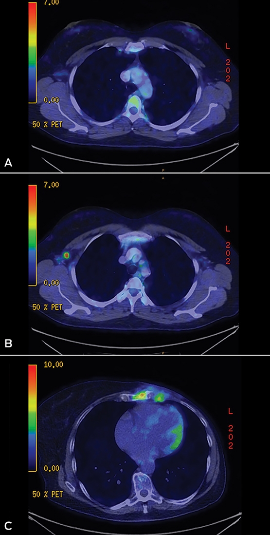
A: Hot internal mammary chain (IMC) node on post-operative PET scan. B: Persistent hot node in axilla level 1 in post-operative PET scan after sentinel node showed micrometastasis. C: Left-sided IMC recurrence 10 years after mastectomy and 2 years after stopping hormonal treatment for a 55 mm invasive lobular carcinoma of the upper inner quadrant. Images courtesy of Macquarie Medical Imaging.
Box 4 – Trials of patients treated with breast conservation or mastectomy with or without internal mammary chain (IMC) radiation therapy (RT)
|
Study |
First year |
No. of patients |
Median follow-up (years) |
= 4 pN+ |
pN0 |
RT location |
Distant metastases |
Benefit from RT |
|||||||
|
No IMC RT |
IMC RT |
||||||||||||||
|
|
|||||||||||||||
|
Thorsen 201668* |
2003 |
3089 |
8.9 |
41.1% |
0 |
Breast/chest wall, SCF, ICF, axilla II + III (axilla I if > 6 N+) + IMC (I-IV) if right-sided |
29.7% |
27.4% |
2.3% |
||||||
|
Whelan 201569† |
2000 |
1832 |
9.5 |
5.3% |
9.7% |
Breast ± SCF, axilla III, axilla I + II if < 10 nodes dissected or > 3N+ and IMC (I–III) |
16.5% |
12.9% |
3.6% |
||||||
|
Bartelink 201535† |
1996 |
4004 |
10.9 |
12.5% |
44.4% |
Breast/chest wall ± SCF and IMC (I–III or I–IV if lower inner quadrant) |
19.6% |
15.9% |
3.7% |
||||||
|
Hennequin 201370† |
1991 |
1334 |
11.3 |
31.1% |
24.8% |
Chest wall and SCF ± IMC (I–V) |
39.9% |
35.2% |
4.7% |
||||||
|
Chang 201371‡ |
1994 |
396 |
12.4 |
84.0% |
4.3% |
Chest wall + SCF ± IMC (I–VI) |
42.7% |
35% |
7.7% |
||||||
|
Courdi 201372‡ |
1975 |
2558 |
12.8 |
0 |
100% |
Breast ± SCF, ± IMC (I–IV) |
18.4% |
8.1% |
10.3% |
||||||
|
|
|||||||||||||||
|
ICF = intercondylar fossa. pN+ = pathologically node-positive. pN0 = pathologically node-negative. SCF = supraclavicular fossa. * Prospective non-randomised trial. † Randomised controlled trial. ‡ Case series. |
|||||||||||||||
Box 5 – Fused image showing heart covered by radiation beam (normal breathing, green arrow) and missed after deep inspiration (orange arrow); primary tumour site marked at surgery by titanium clips (yellow arrow)
Reproduced with permission from the Royal Australian College of General Practitioners from Tailby E, Boyages J. Conservation surgery and radiation therapy in early breast cancer – an update. Aust Fam Physician 2017; 46: 214-219. www.racgp.org.au/afp/2017/april/conservation-surgery-and-radiation-therapy-in-early-breast-cancer-an-update. Image courtesy of Genesis Cancer Care.
Box 6 – Volumetric modulated arc therapy plan of a patient who underwent a left nipple-sparing mastectomy for a triple-negative breast cancer and prophylactic right mastectomy
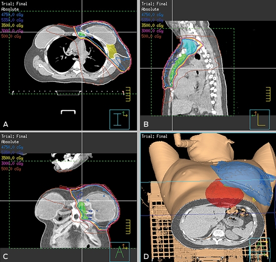
A: Axial slice. B: Sagittal slice. C: Coronal slice. D: 3D volume render. Shading: green, internal mammary chain volume; yellow, axilla level 1; light blue, supraclavicular fossa; red, heart; blue, 47.5 Gy dose cloud. Reproduced with permission from the Royal Australian College of General Practitioners from Tailby E, Boyages J. Conservation surgery and radiation therapy in early breast cancer – an update. Aust Fam Physician 2017; 46: 214-219. www.racgp.org.au/afp/2017/april/conservation-surgery-and-radiation-therapy-in-early-breast-cancer-an-update. Images courtesy of Genesis Cancer Care.
Competing interests
No relevant disclosures.
Acknowledgements
I thank Ellen Tailby for research assistance, Philippa Sutton for editing and managing earlier versions of the manuscript, and Sergio Duque and Lesley Baker for the volumetric modulated arc therapy plan of the patient shown in Box 5.
References
- Grubbé EH. Priority in the therapeutic use of X-rays. Radiology 1933; 21: 156-162.
- Keynes G. The Radium Treatment of Primary Carcinoma of the Breast. Can Med Assoc J 1934; 30: 24-30.
- Halsted WS. I. The results of radical operations for the cure of carcinoma of the breast. Ann Surg 1907; 46: 1-19.
- Ginzton EL, Nunan CS. History of microwave electron linear accelerators for radiotherapy. Int J Radiat Oncol Biol Phys 1985; 11: 205-216.
- Darby S, McGale P, Correa C, et al. Effect of radiotherapy after breast-conserving surgery on 10-year recurrence and 15-year breast cancer death: meta-analysis of individual patient data for 10,801 women in 17 randomised trials. Lancet 2011; 378: 1707-1716.
- McGale P, Taylor C, Correa C, et al. Effect of radiotherapy after mastectomy and axillary surgery on 10-year recurrence and 20-year breast cancer mortality: meta-analysis of individual patient data for 8135 women in 22 randomised trials. Lancet 2014; 383: 2127-2135.
- Esposito E, Anninga B, Harris S, et al. Intraoperative radiotherapy in early breast cancer. Br J Surg 2015; 102: 599-610.
- Fisher B, Anderson S, Bryant J, et al. Twenty-year follow-up of a randomized trial comparing total mastectomy, lumpectomy, and lumpectomy plus irradiation for the treatment of invasive breast cancer. N Engl J Med 2002; 347: 1233-1241.
- Winzer KJ, Sauer R, Sauerbrei W, et al. Radiation therapy after breast-conserving surgery; first results of a randomised clinical trial in patients with low risk of recurrence. Eur J Cancer 2004; 40: 998-1005.
- Fyles AW, McCready DR, Manchul LA, et al. Tamoxifen with or without breast irradiation in women 50 years of age or older with early breast cancer. N Engl J Med 2004; 351: 963-970.
- Hughes KS, Schnaper LA, Berry D, et al. Lumpectomy plus tamoxifen with or without irradiation in women 70 years of age or older with early breast cancer. N Engl J Med 2004; 351: 971-977.
- Potter R, Gnant M, Kwasny W, et al. Lumpectomy plus tamoxifen or anastrozole with or without whole breast irradiation in women with favorable early breast cancer. Int J Radiat Oncol Biol Phys 2007; 68: 334-340.
- Blamey RW, Bates T, Chetty U, et al. Radiotherapy or tamoxifen after conserving surgery for breast cancers of excellent prognosis: British Association of Surgical Oncology (BASO) II trial. Eur J Cancer 2013; 49: 2294-2302.
- Kunkler IH, Williams LJ, Jack WJ, et al. Breast-conserving surgery with or without irradiation in women aged 65 years or older with early breast cancer (PRIME II): a randomised controlled trial. Lancet Oncol 2015; 16: 266-273.
- Australian Government Actuary. Australian life tables 2010–12: females. http://www.aga.gov.au/publications/life_table_2010-12/05-ALT-Females.asp (accessed July 2017).
- Early Breast Cancer Trialists' Collaborative Group (EBCTCG). Effects of chemotherapy and hormonal therapy for early breast cancer on recurrence and 15-year survival: an overview of the randomised trials. Lancet 2005; 365: 1687-1717.
- Boyages J, Taylor R, Chua B, et al. A risk index for early node-negative breast cancer. Br J Surg 2006; 93: 564-571.
- Mannino M, Yarnold JR. Local relapse rates are falling after breast conserving surgery and systemic therapy for early breast cancer: can radiotherapy ever be safely withheld? Radiother Oncol 2009; 90: 14-22.
- Hernandez RK, Sorensen HT, Pedersen L, et al. Tamoxifen treatment and risk of deep venous thrombosis and pulmonary embolism: a Danish population-based cohort study. Cancer 2009; 115: 4442-4449.
- Chakrabarti J, Kenny FS, Syed BM, et al. A randomised trial of mastectomy only versus tamoxifen for treating elderly patients with operable primary breast cancer-final results at 20-year follow-up. Crit Rev Oncol Hematol 2011; 78: 260-264.
- Haviland JS, Owen JR, Dewar JA, et al. The UK Standardisation of Breast Radiotherapy (START) trials of radiotherapy hypofractionation for treatment of early breast cancer: 10-year follow-up results of two randomised controlled trials. Lancet Oncol 2013; 14: 1086-1094.
- Whelan TJ, Pignol JP, Levine MN, et al. Long-term results of hypofractionated radiation therapy for breast cancer. N Engl J Med 2010; 362: 513-520.
- Bane AL, Whelan TJ, Pond GR, et al. Tumor factors predictive of response to hypofractionated radiotherapy in a randomized trial following breast conserving therapy. Ann Oncol 2014; 25: 992-998.
- Australian Government Cancer Australia. Clinical practice recommendations and practice points. https://canceraustralia.gov.au/publications-and-resources/clinical-practice-guidelines/hypofractionated-radiotherapy-early-operable-breast-cancer/clinical-practice-recommendations-and-practice-points (accessed July 2017).
- Smith BD, Bentzen SM, Correa CR, et al. Fractionation for whole breast irradiation: an American Society for Radiation Oncology (ASTRO) evidence-based guideline. Int J Radiat Oncol Biol Phys 2011; 81: 59-68.
- Harnett A, Smallwood J, Titshall V, et al. Diagnosis and treatment of early breast cancer, including locally advanced disease–summary of NICE guidance. BMJ 2009; 338: 598-600.
- Shah C, Parsai S, Kotecha R, et al. Overview of outcomes with accelerated partial breast irradiation. In: Arthur DW, Vicini FA, Wazer DE, et al., editors. Short course breast radiotherapy. Cham: Springer, 2016; pp 229-244.
- Coles CE, Yarnold JR. Accelerated partial breast irradiation: the new standard? Lancet 2016; 387: 201-202.
- Strnad V, Ott OJ, Hildebrandt G, et al. 5-year results of accelerated partial breast irradiation using sole interstitial multicatheter brachytherapy versus whole-breast irradiation with boost after breast-conserving surgery for low-risk invasive and in-situ carcinoma of the female breast: a randomised, phase 3, non-inferiority trial. Lancet 2016; 387: 229-238.
- Holland R, Connolly JL, Gelman R, et al. The presence of an extensive intraductal component following a limited excision correlates with prominent residual disease in the remainder of the breast. J Clin Oncol 1990; 8: 113-118.
- Boyages J, Bosch C, Langlands AO, et al. Breast conservation: long-term Australian data. Int J Radiat Oncol Biol Phys 1992; 24: 253-260.
- Boyages J, Recht A, Connolly JL, et al. Early breast cancer: predictors of breast recurrence for patients treated with conservative surgery and radiation therapy. Radiother Oncol 1990; 19: 29-41.
- Connolly JL, Boyages J, Nixon AJ, et al. Predictors of breast recurrence after conservative surgery and radiation therapy for invasive breast cancer. Mod Pathol 1998; 11: 134-139.
- Leong C, Boyages J, Jayasinghe UW, et al. Effect of margins on ipsilateral breast tumor recurrence after breast conservation therapy for lymph node-negative breast carcinoma. Cancer 2004; 100: 1823-1832.
- Bartelink H, Maingon P, Poortmans P, et al. Whole-breast irradiation with or without a boost for patients treated with breast-conserving surgery for early breast cancer: 20-year follow-up of a randomised phase 3 trial. Lancet Oncol 2015; 16: 47-56.
- Vrieling C, van Werkhoven E, Maingon P, et al. Prognostic factors for local control in breast cancer after long-term follow-up in the EORTC boost vs no boost trial: a randomized clinical trial. JAMA Oncol 2017; 3: 42-48.
- Houssami N, Macaskill P, Marinovich ML, et al. Meta-analysis of the impact of surgical margins on local recurrence in women with early-stage invasive breast cancer treated with breast-conserving therapy. Eur J Cancer 2010; 46: 3219-3232.
- Hunt KK, Ballman KV, McCall LM, et al. Factors associated with local-regional recurrence after a negative sentinel node dissection: results of the ACOSOG Z0010 trial. Ann Surg 2012; 256: 428-436.
- Houssami N, Marinovich ML. Margins in breast-conserving surgery for early breast cancer: how much is good enough? Curr Breast Cancer Rep 2016; 8: 127-134.
- Moran MS, Schnitt SJ, Giuliano AE, et al. Society of Surgical Oncology-American Society for Radiation Oncology consensus guideline on margins for breast-conserving surgery with whole-breast irradiation in stages I and II invasive breast cancer. Int J Radiat Oncol Biol Phys 2014; 88: 553-564.
- Bodilsen A, Bjerre K, Offersen BV, et al. Importance of margin width in breast-conserving treatment of early breast cancer. J Surg Oncol 2016; 113: 609-615.
- Wang W, French J, Boyages J. Put the felt pen away: time to move on from a clinical mark-up for a breast boost. J Med Imaging Radiat Oncol 2012; 56: 375-378.
- Recht A, Comen EA, Fine RE, et al. Postmastectomy radiotherapy: an American Society of Clinical Oncology, American Society for Radiation Oncology, and Society of Surgical Oncology focused guideline update. Pract Radiat Oncol 2016; 6: e219-e234.
- Mamounas EP, Anderson SJ, Dignam JJ, et al. Predictors of locoregional recurrence after neoadjuvant chemotherapy: results from combined analysis of National Surgical Adjuvant Breast and Bowel Project B-18 and B-27. J Clin Oncol 2012; 30: 3960-3966.
- Fisher B, Brown A, Mamounas E, et al. Effect of preoperative chemotherapy on local-regional disease in women with operable breast cancer: findings from National Surgical Adjuvant Breast and Bowel Project B-18. J Clin Oncol 1997; 15: 2483-2493.
- Bear HD, Anderson S, Smith RE, et al. Sequential preoperative or postoperative docetaxel added to preoperative doxorubicin plus cyclophosphamide for operable breast cancer: National Surgical Adjuvant Breast and Bowel Project Protocol B-27. J Clin Oncol 2006; 24: 2019-2027.
- Boughey JC, McCall LM, Ballman KV, et al. Tumor biology correlates with rates of breast-conserving surgery and pathologic complete response after neoadjuvant chemotherapy for breast cancer: findings from the ACOSOG Z1071 (Alliance) Prospective Multicenter Clinical Trial. Ann Surg 2014; 260: 608-614; discussion 614-606.
- Fowble BL, Einck JP, Kim DN, et al. Role of postmastectomy radiation after neoadjuvant chemotherapy in stage II-III breast cancer. Int J Radiat Oncol Biol Phys 2012; 83: 494-503.
- Truin W, Vugts G, Roumen RM, et al. Differences in response and surgical management with neoadjuvant chemotherapy in invasive lobular versus ductal breast cancer. Ann Surg Oncol 2016; 23: 51-57.
- Abdulkarim BS, Cuartero J, Hanson J, et al. Increased risk of locoregional recurrence for women with T1-2N0 triple-negative breast cancer treated with modified radical mastectomy without adjuvant radiation therapy compared with breast-conserving therapy. J Clin Oncol 2011; 29: 2852-2858.
- Lowery AJ, Kell MR, Glynn RW, et al. Locoregional recurrence after breast cancer surgery: a systematic review by receptor phenotype. Breast Cancer Res Treat 2012; 133: 831-841.
- Voduc KD, Cheang MC, Tyldesley S, et al. Breast cancer subtypes and the risk of local and regional relapse. J Clin Oncol 2010; 28: 1684-1691.
- Taghian AG, Jeong JH, Mamounas EP, et al. Low locoregional recurrence rate among node-negative breast cancer patients with tumors 5 cm or larger treated by mastectomy, with or without adjuvant systemic therapy and without radiotherapy: results from five National Surgical Adjuvant Breast and Bowel Project randomized clinical trials. J Clin Oncol 2006; 24: 3927-3932.
- Bazan JG, White JR. The role of postmastectomy radiation therapy in patients with breast cancer responding to neoadjuvant chemotherapy. Semin Radiat Oncol 2016; 26: 51-58.
- de Boer M, van Dijck JA, Bult P, et al. Breast cancer prognosis and occult lymph node metastases, isolated tumor cells, and micrometastases. J Natl Cancer Inst 2010; 102: 410-425.
- Chua B, Ung O, Taylor R, et al. Treatment implications of a positive sentinel lymph node biopsy for patients with early-stage breast carcinoma. Cancer 2001; 92: 1769-1774.
- Giuliano AE, McCall L, Beitsch P, et al. Locoregional recurrence after sentinel lymph node dissection with or without axillary dissection in patients with sentinel lymph node metastases: the American College of Surgeons Oncology Group Z0011 randomized trial. Ann Surg 2010; 252: 426-432; discussion 432-423.
- Giuliano AE, Ballman K, McCall L, et al. Locoregional recurrence after sentinel lymph node dissection with or without axillary dissection in patients with sentinel lymph node metastases: long-term follow-up from the American College of Surgeons Oncology Group (Alliance) ACOSOG Z0011 randomized trial. Ann Surg 2016; 264: 413-420.
- Boyages J, Winch C. Axillary versus sentinel-lymph-node dissection for micrometastatic breast cancer. Lancet Oncol 2013; 14: e250-e251.
- Coates AS, Winer EP, Goldhirsch A, et al. Tailoring therapies–improving the management of early breast cancer: St Gallen International Expert Consensus on the Primary Therapy of Early Breast Cancer 2015. Ann Oncol 2015; 26: 1533-1546.
- Jagsi R, Chadha M, Moni J, et al. Radiation field design in the ACOSOG Z0011 (Alliance) Trial. J Clin Oncol 2014; 32: 3600-3606.
- Fisher B, Jeong JH, Anderson S, et al. Twenty-five-year follow-up of a randomized trial comparing radical mastectomy, total mastectomy, and total mastectomy followed by irradiation. N Engl J Med 2002; 347: 567-575.
- Donker M, van Tienhoven G, Straver ME, et al. Radiotherapy or surgery of the axilla after a positive sentinel node in breast cancer (EORTC 10981–22023 AMAROS): a randomised, multicentre, open-label, phase 3 non-inferiority trial. Lancet Oncol 2014; 15: 1303-1310.
- Haffty BG, Hunt KK, Harris JR, et al. Positive sentinel nodes without axillary dissection: implications for the radiation oncologist. J Clin Oncol 2011; 29: 4479-4481.
- Stibbe EP. The Internal Mammary Lymphatic Glands. J Anat 1918; 52: 257-264.
- Putti F. Ricerche anatomiche sui linfonodi mammari interni. Chir Ital 1953; 7: 161-172.
- Coombs NJ, Boyages J, French JR, et al. Internal mammary sentinel nodes: ignore, irradiate or operate? Eur J Cancer 2009; 45: 789-794.
- Thorsen LB, Offersen BV, Dano H, et al. DBCG-IMN: a population-based cohort study on the effect of internal mammary node irradiation in early node-positive breast cancer. J Clin Oncol 2016; 34: 314-320.
- Whelan TJ, Olivotto IA, Parulekar WR, et al. Regional nodal irradiation in early-stage breast cancer. N Engl J Med 2015; 373: 307-316.
- Hennequin C, Bossard N, Servagi-Vernat S, et al. Ten-year survival results of a randomized trial of irradiation of internal mammary nodes after mastectomy. Int J Radiat Oncol Biol Phys 2013; 86: 860-866.
- Chang JS, Park W, Kim YB, et al. Long-term survival outcomes following internal mammary node irradiation in stage II-III breast cancer: results of a large retrospective study with 12-year follow-up. Int J Radiat Oncol Biol Phys 2013; 86: 867-872.
- Courdi A, Chamorey E, Ferrero JM, et al. Influence of internal mammary node irradiation on long-term outcome and contralateral breast cancer incidence in node-negative breast cancer patients. Radiother Oncol 2013; 108: 259-265.
- Verma V, Vicini F, Tendulkar RD, et al. Role of internal mammary node radiation as a part of modern breast cancer radiation therapy: a systematic review. Int J Radiat Oncol Biol Phys 2016; 95: 617-631.
Linked content
-
MJA InSight: Radiotherapy for breast cancer: a fast-evolving field
-
MJA Podcast: Prof John Boyages
Provenance: Commissioned; externally peer reviewed.
From Ambition to Action: Closing Policy, Equity and Implementation Gaps in the Australian Cancer Plan's ‘Enhanced Consumer Experience’
Carla Thamm, Elise Button, Oluwaseyifunmi A. Agbejule, Reegan Knowles, Karen van Gorp, Mark B. Pinkham, Meinir Krishnasamy, Alexandra L. McCarthy, Raymond J. Chan
Sovereignty and Survival: Understanding Lung Cancer Outcomes for Aboriginal and Torres Strait Islander People in Australia, a Case–Control Study
Alice R. T. Bergin, Luc te Marvelde, Roger L. Milne, Benjamin J. Solomon, Kris Ivanova, Peter Savas, Stephen J. Luen, Jay Hamann, Andrea Casey, Nicole Watt, Sue M. Evans, Gail Garvey, Sherene Loi
The Changing Indications for Adult Liver Transplantation in Australia: A Narrative Review
Fadak Mohammadi, Alan J. Wigg, Ken Liu, Simone I. Strasser
Uneven Ground: Survival Differences Among Victorian Lung Cancer Patients by Location of Residence (2011–2023): A Retrospective Cohort Study
Evangeline Samuel, Eldho Paul, Mike Lloyd, Sanuki Tissera, Craig Underhill, Sagun Parakh, Phillip Parente, Inger Olesen, Javier Torres, Katharine See, Gavin M. Wright, David Langton, Thomas John, Matthew Conron, James Bartlett, Nicola Atkin, Nikolajs Zeps, Susan V. Harden, Wasek Faisal, John R. Zalcberg, Rob G. Stirling
Early-Onset Colorectal Cancer With Liver-Only Metastases: A Retrospective Cohort Study Integrating Prospectively Collected Real-World Clinical and Molecular Data From an Australian National Database (2009–2024) to Guide Treatment Planning
Savio G. Barreto, Christos S. Karapetis, Shahid Ullah, Matthew Burge, Susan Caird, Angus Campbell, Azim Jalali, Ross Jennens, Muhammad A. Khattak, Belinda Lee, Stephanie H. Lim, Shehara Mendis, Louise Nott, Timothy J. Price, Jeremy D. Shapiro, Jeanne Tie, Javier Torres, Colin Williams, Rachel Wong, Vanessa Wong, Peter Gibbs
Australian Pathways for Specialist Pain Management and Early Palliative Care for People With Pancreatic Cancer: Developed Using a Community Consensus Approach
Jennifer Philip, Melanie R. Lovell, Kylee Bellingham, Gail Garvey, Gregory B. Crawford, Nicole M. Rankin, Kara Burns, Isabel Young, Vivienne Milch, Dorothy Keefe, Katrina Anderson, James Lawson, Meinir Krishnasamy