




Medical education
Clinical skills
Volume 206 - Issue 5
Detecting the gallop: the third heart sound and its significance
Authors: Subha Ramani and Brittany N Weber
Med J Aust 2017; 206 (5): 198-199. || doi: 10.5694/mja16.00613
Published online: 20 March 2017
Published online: 20 March 2017
Good technique and a reflective approach assist clinicians to identify an easily missed indicator of ventricular dysfunction
Good technique and a reflective approach assist clinicians to identify an easily missed indicator of ventricular dysfunction
The term “gallop rhythm” was first coined in 1847 by Jean-Baptiste Bouillaud to describe the cadence of three heart sounds occurring in rapid succession. It was further described by his pupil Pierre Potain as follows:
One distinguishes therein three sounds, namely: two normal sounds of the heart and a superadded sound… It is a shock, a perceptible elevation; it is hardly a sound. If one applies the ear to the chest it is affected by a tactile sensation, perhaps more so than an auditory one… In addition to the two normal sounds, this bruit completes the triple rhythm of the heart… This is the bruit de gallop.1
Third heart sound (S3) and fourth heart sound gallops are indicators of underlying ventricular dysfunction; however, the term gallop is only used when the sounds are pathological.
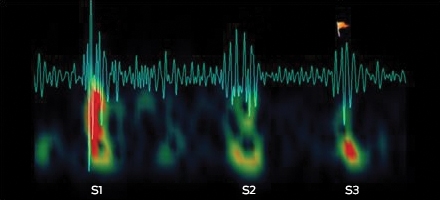
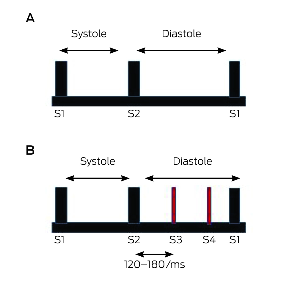
The S3 is a mid-diastolic, low pitched sound (Box 1). In early diastole, the ventricular pressure falls below atrial pressure, and the atrioventricular valves open initiating rapid ventricular filling. Filling slows as the ventricles reach the limit of their elasticity. It is thought that the S3 occurs in the presence of volume overload or ventricular dysfunction when the rapid filling phase ends abruptly.2 It is not completely clear whether the sound arises from vibrations in the valve cusps or tautening of the papillary muscles. Factors that affect S3 intensity include age, atrial pressure, unobstructed flow across the atrioventricular valve, rapidity of early diastolic filling, compliance of the ventricle, blood volume, ventricular cavity size, and patient positioning.
The sound is easily missed, as the level of clinical experience correlates with the ability to detect it.3 The S3 is a low frequency sound in the range of 20 to 70 Hz, with a thudding quality, easily masked by respiratory or environmental sounds.2 It is localised to a small area of the precordium, does not radiate and can be completely missed unless the bell or its equivalent is used during auscultation. An S3 can originate from the left or right ventricle. The differential diagnosis includes splitting of the second heart sound, opening snap of the mitral or tricuspid valve, tumour “plop” from atrial myxoma, or pericardial knock. An S3 is best detected when it is anticipated based on valuable clinical clues of heart failure or valvular pathology (Box 2).
While the sound challenges many clinicians, there are several strategies that can be effective in its detection. It is essential to tune out all other sounds, including systole, and focus on diastole alone (Box 3). As the sound itself may not be distinct or sharp like the first or second heart sounds, clinicians should become accustomed to recognising the cadence which could indicate its presence. An S3 originating in the left ventricle is best heard in the left lateral decubitus position (a position that brings the apex closest to the stethoscope), in held end expiration, and by using the bell or light pressure. It can be augmented by mild exertion which worsens left ventricular dysfunction. An S3 originating in the right ventricle is best heard along the left lower sternal border, sometimes in the epigastrium, and augmented during inspiration and manoeuvres that increase venous return.
The S3 is often physiological in asymptomatic children and young adults, but usually pathological in people over 40 years of age. It correlates with ventricular dysfunction or volume overload. Typical causes of ventricular dysfunction include ischaemic heart disease, cardiomyopathy, myocarditis and cor pulmonale. The S3 can also be heard in aortic and mitral regurgitation, and high output heart failure associated with anaemia, pregnancy, arteriovenous fistulae, thyrotoxicosis and left-to-right shunts.
The S3 can serve as a vital clue in the detection of patients with congestive cardiac failure as well as their risk stratification. The acute decompensated heart failure syndromes (ATTEND) registry study, a multivariate analysis performed in 4107 patients hospitalised with acute heart failure, suggested that the presence of the S3 was independently associated with increased in-hospital all-cause death (adjusted odds ratio [OR], 1.69; 95% CI, 1.19–2.41) and cardiac death (adjusted OR, 1.66; 95% CI, 1.08–2.54).4 Box 4 lists the haemodynamic significance of the presence of the S3.3,5,6
As elusive as the S3 might seem, good technique and reflective practice can help clinicians in eliciting the sound — a finding that provides clues to important cardiac pathology as well as severity of disease.
Box 2 – Symptoms of heart failure and associated physical examination findings
Symptoms of left-sided heart failure |
Dyspnoea on exertion, orthopnoea, paroxysmal nocturnal dyspnoea, cough, wheeze |
||||||||||||||
Symptoms of right-sided heart failure |
Poor appetite, abdominal distension, right hypochondrial discomfort, oedema |
||||||||||||||
Associated examination findings |
Increased jugular venous pressure, oedema, resting tachycardia, displaced apical impulse, parasternal heave, murmurs of valvular dysfunction |
||||||||||||||
Box 3 – Normal heart sounds (A) and abnormal diastolic heart sounds showing the third and fourth sounds (B)
Box 4 – Significance of the third heart sound: cardiovascular pathology
The third heart sound can be predictive of:
- Depressed left ventricular ejection fraction of < 50% (likelihood ratio [LR], 4)
- Depressed left ventricular ejection fraction of < 30% (sensitivity [Sn], 68–78%; specificity [Sp], 80–88%; LR, 4.1)
- Post-operative pulmonary oedema (Sn, 17%; Sp, 99%; LR, 14.6)
- Myocardial infarction in patients with chest pain (LR, 3.2)
- Post-myocardial infarction mortality (LR, 8.0)
Competing interests
No relevant disclosures.
Series editors
Balakrishnan (Kichu) Nair
Simon O’Connor
Series editors
Balakrishnan (Kichu) Nair
Simon O’Connor
References
- Silverman ME. The third heart sound. In: Walker HK, Hall WD, Hurst JW, editors. Clinical Methods: The History, Physical, and Laboratory Examinations. 3rd ed. Boston: Butterworths, 1990.
- Talley NJ, O’Connor S. Clinical examination: a systematic guide to physical diagnosis. 7th ed. Sydney: Elsevier, 2013.
- Marcus GM, Vessey J, Jordan MV, et al. Relationship between accurate auscultation of a clinically useful third heart sound and level of experience. Arch Intern Med 2006; 166: 617-622.
- Sato N, Kajimoto K, Asai K, et al. Acute decompensated heart failure syndromes (ATTEND) registry. A prospective observational multicenter cohort study: rationale, design, and preliminary data. Am Heart J 2010; 159: 949-955 e1.
- McGee SR. Evidence-based physical diagnosis. 3rd ed. Philadelphia: Elsevier/Saunders: 2012.
- Wang CS, FitzGerald JM, Schulzer M, et al. Does this dyspneic patient in the emergency department have congestive heart failure? JAMA 2005; 294: 1944-1956.
Provenance: Commissioned; externally peer reviewed.
Shifting focus to adolescent wellbeing and inclusive participation in the digital age
Allyson R Todd, Elena Wang, Stephanie R Partridge
The clinical presentation, investigation, and management of women diagnosed with endometriosis in Australian general practices, 2011–2021: an open cohort study
Danielle Mazza, Kailash Thapaliya, Sharinne B Crawford, Alissia Hui, Maryam Moradi, Luke E Grzeskowiak
The impact of the Breast Screen NSW transition from film to digital mammography, 2002–2016: a linked population health data analysis
Rachel Farber, Nehmat Houssami, Katy J L Bell
Non‐technical errors associated with deaths in surgical care, Australia, 2012–2019, by surgical specialty (Australian and New Zealand Audit of Surgical Mortality): a retrospective cohort study
Jesse Ey, Victoria Kollias, Octavia Lee, Kelly Hou, Matheesha Herath, John B North, Ellie Treloar, Suzanne Edwards, Martin Bruening, Adam J Wells, Guy J Maddern
The CURE Asthma roadmap
Gary P Anderson, Anthony Flynn, Phil G Bardin, John D Blakey, Shyamali C Dharmage, Paul Foster, Peter G Gibson, Adam Jaffe, Alan James, Christine R Jenkins, Sundram Sivamalai, Peter D Sly, Guy B Marks, Vanessa M McDonald, Judy Wetttenhall
Evidence from Australian cohort studies about asthma trajectories and transitions across the life course: a narrative review
Caroline Lodge, Xin Dai, Ingrid A. Laing, Michael P Menden, Anthony Flynn, Gary P Anderson, Sarath Ranganathan, Shyamali C Dharmage