




Lessons from practice
Volume 204 - Issue 3
The early bird and the worm: a case of cercarial dermatitis
Authors: John W Frew, Christopher JA Henderson and Ian D McCrossin
Med J Aust 2016; 204 (3): 122-123. || doi: 10.5694/mja15.00802
Published online: 15 February 2016
Published online: 15 February 2016
A common but under-recognised condition
Clinical record
A 36-year-old man presented to our dermatology department with a 5-day history of an intensely pruritic papular eruption on the dorsum of both feet, associated with significant swelling and discomfort. This began the morning after a night-time fishing trip on the central coast of New South Wales at the mouth of the Lake Tuggerah estuary, a system of brackish tidal estuaries.
The patient reported standing ankle-deep in water for 6 hours from dusk (9 pm) until early morning (3 am). He noted that the water was particularly warm after a daytime temperature of 33°C with seaward tidal flows. He awoke the following morning with multiple discrete nodules, oedema and intense pruritus to the dorsa of the feet. He reported previous episodes of “bites” after fishing trips that spontaneously resolved after 24–48 hours.
The next 12 hours saw an increase in pruritus and the size of lesions. The patient presented to a general practitioner, who diagnosed pelican itch with secondary infection and prescribed cephalexin 500 mg four times a day for 5 days. Pain and pruritus continued to increase, resulting in the patient presenting to a metropolitan emergency department where he was diagnosed with bilateral foot cellulitis and administered intravenous cefazolin 1 g twice daily for 5 days. Investigations showed leucocytosis (white cell count, 10.1 × 10⁹/L; reference interval [RI], 4.0–10.0 × 10⁹/L) with eosinophilia (0.8 × 10⁹/L; RI, 0–0.5 × 10⁹/L) and negative blood cultures. On Day 3 of intravenous antibiotics, he was referred to our dermatology department for reassessment of diagnosis.
On presentation, the patient was in significant discomfort with swelling, difficulty ambulating and inability to wear shoes. He did not describe any fever, vomiting, diarrhoea, abdominal cramping or haematuria. Multiple violaceous papules were noted over the dorsum of each foot, with sparing of the plantar surfaces, web spaces of the toes and distal third of the lower limb (Figure, A). Dusky oedema was evident surrounding the papules, with no evidence of lymphangitis. Popliteal and inguinal lymph nodes were palpable.
Histopathological examination (Figure, C and D) revealed epidermal spongiosis and prominent dermal oedema. A heavy inflammatory infiltrate predominately of eosinophils involving all layers of skin down to the subcutis with scattered flame figures was also noted. No parasites were identified. Bacterial, fungal and mycobacterial tissue cultures were negative. This histology supported a markedly pronounced hypersensitivity reaction in a patient clinically suspected to have cercarial dermatitis, and was not compatible with other clinical differential diagnoses such as hypertrophic lichen planus, Sweet syndrome and reticulohistiocytosis.
Our patient was successfully treated with 3 days of oral prednisolone therapy (25 mg daily) with complete resolution of lesions and residual post-inflammatory pigmentation by Day 7 (Figure, B).
Cercarial dermatitis (commonly known as swimmer’s itch, pelican itch or duck itch) conventionally refers to a cutaneous hypersensitivity reaction to non-human trematode larvae (cercariae) of schistasome flukes after penetration of human skin.1,2 In the classic avian form of the disease, the flukes undergo a two-host (digenetic) life cycle with egg development in freshwater snails and release of the larvae into open water (in temperatures above 23°C) to infest migratory aquatic birds (ducks and seagulls, the fluke’s definitive host).2 Human infections with avian cercariae represent a disruption of the natural schistasome life cycle. The fluke was previously thought to die within human skin within 48 hours; however, animal models have shown migration and survival of the trematodes in distant organs weeks after initial infection.1,2
Cercarial dermatitis is a common condition found worldwide1,2 but is under-recognised and under-reported in Australia owing to its self-limiting nature.1 High concentrations of cercariae occur during days of intense sunlight and higher air and sea surface temperatures, with coastal currents concentrating cercariae around shallow estuary outlets.2 Cases have been reported from Queensland,3,4 Terrigal on the central coast of NSW (eponymous for the schistasome Austrobilharzia terrigalensis5), and the Swan River estuary of Western Australia.1,6
The condition manifests as a monomorphic, papular, pruritic dermatitis confined to water-submerged areas that begins to spontaneously regress within 72 hours.1,2 Recurrent exposure can result in sensitisation phenomena precipitating a florid hypersensitivity reaction as seen in our case. Cercarial dermatitis can be differentiated from sea-bather’s eruption, as the latter condition affects areas of skin covered by swimwear, commonly as a swimmer exits the water or after swimwear has dried. It is caused by discharging nematocysts of sea anemones and jellyfish, and can be associated with systemic symptoms of fevers, vomiting and malaise.
Avian schistasomes are homologous to (but not to be confused with) human schistosomiasis, an endemic and potentially fatal tropical disease, manifesting in portal hypertension, bladder carcinoma, granulomatous central nervous system disease, pulmonary fibrosis and glomerulonephritis.7,8 In endemic tropical areas, cercarial dermatitis and schistosomiasis can be difficult to differentiate, although the early cercarial stage of human schistosomiasis is less inflammatory than that of avian schistosomiasis.2 The histopathology of cercarial dermatitis in humans is poorly documented owing to the acute self-limiting nature of the disease.2 Where descriptions are available, they are briefly described as a hypersensitivity reaction with oedema and eosinophils (plus or minus neutrophils). Specificity is provided by finding cercariae in the specimen (located within the epidermis) but they are usually not seen,7 presumably due to the limited life span of the cercariae within non-definitive hosts and the difficulty in finding isolated small (0.75–0.95 mm long) organisms within a disproportionately large area of inflammatory reaction. It is surmised that without identification of the organism, some cases could easily be misclassified as arthropod bite reactions in the absence of awareness of the specific clinical context.
Treatment for mild cercarial dermatitis includes reassurance and symptomatic management with potent topical corticosteroids (betamethasone dipropionate 0.05% ointment twice daily) and emollients. Oedema can be relieved with intermittent cold compress, rest and elevation. Exuberant reactions may require oral prednisolone (0.5 mg/kg up to a maximum dose of 25 mg daily for 3 consecutive days). Some authors recommend routine treatment with oral ivermectin (200 μg/kg up to a maximum of 12 mg per dose)9 until possible systemic complications arising from visceral migration of trematodes in humans are better understood.
It is important for physicians to be aware of this common and under-recognised entity, as well as the potential for schistosomiasis with similar presentations in returning travellers. Although the cutaneous manifestations of this condition spontaneously resolve, further research will clarify the potential for chronic infestation and the role of routine antihelminthic therapy.Lessons from practice
Cercarial dermatitis is a common, under-reported condition caused by a hypersensitivity reaction to the penetration of human skin by the trematode fluke of avian schistosomiasis.
It commonly occurs due to submergence of unprotected skin in warm waters of freshwater tidal estuaries.
Clinicians should be aware of similar presentations in returning travellers, which may represent the potentially fatal tropical disease of human schistosomiasis.
Although most cases spontaneously resolve, topical and systemic corticosteroids are treatment options for exuberant hypersensitivity reactions.
Figure
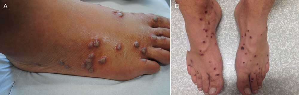
A: Grouped violaceous papules on the dorsolateral aspect of the foot with associated oedema. B: Significant improvement in foot oedema with healing blisters and erosions after 3 days of oral prednisolone (25 mg). C: Low-power image of upper dermis and epidermis with marked subepidermal oedema and spongiosis responsible for the bullous clinical appearance (pseudobullous change) (objective magnification, ×4; haematoxylin–eosin stain). D: High-power image showing prominent interstitial dermal infiltrate of eosinophils with flame figure (arrow) at top of image (objective magnification, ×20; haematoxylin–eosin stain).
Figure
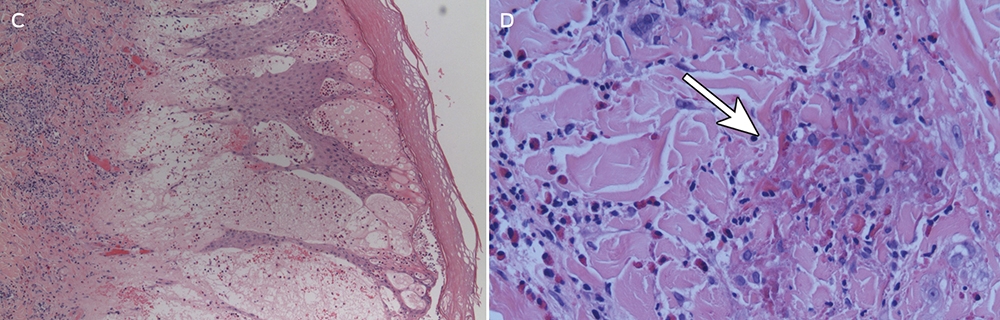
A: Grouped violaceous papules on the dorsolateral aspect of the foot with associated oedema. B: Significant improvement in foot oedema with healing blisters and erosions after 3 days of oral prednisolone (25 mg). C: Low-power image of upper dermis and epidermis with marked subepidermal oedema and spongiosis responsible for the bullous clinical appearance (pseudobullous change) (objective magnification, ×4; haematoxylin–eosin stain). D: High-power image showing prominent interstitial dermal infiltrate of eosinophils with flame figure (arrow) at top of image (objective magnification, ×20; haematoxylin–eosin stain).
Competing interests
No relevant disclosures.
References
- Appleton CC, Lethbridge RC. Schistosome dermatitis in the Swan estuary, Western Australia. Med J Aust 1979; 1: 141-144.
- Kolarova L, Horak P, Skirnisson K, et al. Cercarial dermatitis, a neglected allergic disease. Clin Rev Allerg Immunol 2013; 45: 63-74.
- Rohde K The bird schistosome Austrobilharzia terrigalensis from the Great Barrier Reef, Australia. Z Parasitenkd 1977; 52: 39-51.
- Freier K, Wheller L, Barrington G, Soyer P. A case of duck itch. Australas J Dermatol 2013; S3: 17.
- Bearup AJ. Life cycle of Austrobilharzia terrigalensis. Parasitology 1956; 46: 470-479.
- Appleton CC. Studies on Austrobilharzia terrigalensis (Trematoda: Schistosomatidae) in the Swan estuary, Western Australia: infection in the definitive host, Larus novaehollandiae. Int J Parasitol 13: 249-259.
- Weedon D. Weedon’s skin pathology. 3rd ed. London: Churchill Livingstone Elsevier, 2010: p 646.
- Colley DG, Secor WE. Immunology of human schistosomiasis. Parasite Immunol 2014; 36: 347-357.
- Burkhart CG, Burkhart CN. Swimmer’s itch: an assessment proposing possible treatment with ivermectin. Int J Dermatol 2003; 42: 917-918.
Diagnosing Acute Kava Dermopathy: A Case Report of a Characteristic Cutaneous Eruption
Ali Abid, Nicholas Allen, Abeer Hagelamin, Christopher Henderson, Artiene Tatian
Localised Herpes Simplex Following Midline Laparotomy
Jessica S. Bulluss, Paul Chee, Matthew J. Verheyden
No silver lining with health misinformation: argyria caused by intentional silver consumption
Luke Collins, Logesh Palanikumar, Stephen Bacchi
Lingual Raynaud phenomenon
Michael Taggart, Mina John
Melasma in the male: a less well recognised entity
Tim Aung, Rowland Noakes
Age group‐specific changes in keratinocyte cancer treatment rates in Australia, 2012–2021: a retrospective cohort study based on MBS claims data
Catherine M Olsen, Nirmala Pandeya, Rachel E Neale, David C Whiteman