




Medical education
Open access CC BY NC
Volume 221 - Issue 9
Melasma in the male: a less well recognised entity
Authors: Tim Aung and Rowland Noakes
Med J Aust 2024; 221 (9): 468-468. || doi: 10.5694/mja2.52478
Published online: 4 November 2024
Published online: 4 November 2024
An otherwise healthy man of Southeast-Asian descent aged in his mid-30s presented with non-pruritic hyperpigmented patches involving the face for over 12 months
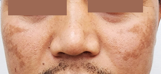
An otherwise healthy man of Southeast‐Asian descent aged in his mid‐30s presented with non‐pruritic hyperpigmented patches involving the face for over 12 months (Figure). Clinically, the findings were consistent with melasma, characterised by brown or dark brown hyperpigmented patches with demarcated or ill‐defined borders, involving the face (zygoma and cheeks) bilaterally. There was no history or clinical features suggestive of diagnoses such as post‐inflammatory hyperpigmentation, drug‐induced pigmentation or photosensitivity, or actinic lichen planus.
Melasma is a common acquired hyperpigmentary condition, described mostly in women of Asian, Middle Eastern or Latin American descent aged 30–50 years.1 Melasma in men is less well recognised,2 with a reported sex ratio of 1:9 (male:female).3 The precise aetiopathogenesis of melasma is incompletely understood. However, there are risk factors related to melasma development, such as sunlight exposure, genetic predisposition, hormonal exposure (contraceptive use and pregnancy), photosensitive medications and cosmetics use, and, rarely, thyroid disorder.2,3,4,5 Melasma can be diagnosed clinically based on distribution and characteristics of pigmentation and skin type or ethnicity; biopsy is rarely required.
Management includes sun protection with the use of a wide‐brim hat and sunscreen (with a sun protection factor of 30 or over), and avoidance of risk factors such as phototoxic drugs, cosmetics, and hormonal contraception (in women). The triple combined topical therapy (hydroquinone 4–5% with tretinoin 0.05–0.1% and low potency corticosteroid), daily for three months, is usually the first line treatment of choice.1,2,3 Adjunct oral tranexamic acid may be considered for extensive lesions, dermal involvement, or for patients refractory to topical therapy, although there are no established studies in men.2 There are also advanced treatment options, such as chemical peeling, microneedling, and light and laser‐based therapies, with unpredictable or mixed results.1,3
Competing interests
No relevant disclosures.
References
- Neagu N, Conforti C, Agozzino M, et al. Melasma treatment: a systematic review. J Dermatolog Treat 2022; 33: 1816‐1837.
- Sarkar R, Ailawadi P, Garg S. Melasma in men: a review of clinical, etiological, and management issues. J Clin Aesthet Dermatol 2018; 11: 53‐59.
- Basit H, Godse KV, Al Aboud AM. Melasma [updated 8 Aug 2023]. In: StatPearls [internet]. Treasure Island (FL): StatPearls Publishing, 2024.
- Majid I, Aleem S. Melasma: update on epidemiology, clinical presentation, assessment, and scoring. J Skin Stem Cell 2021; 8: e120283.
- Doolan BJ, Gupta M. Melasma. Aust J Gen Pract 2021; 50: 880‐885.
Diagnosing Acute Kava Dermopathy: A Case Report of a Characteristic Cutaneous Eruption
Ali Abid, Nicholas Allen, Abeer Hagelamin, Christopher Henderson, Artiene Tatian
Localised Herpes Simplex Following Midline Laparotomy
Jessica S. Bulluss, Paul Chee, Matthew J. Verheyden
No silver lining with health misinformation: argyria caused by intentional silver consumption
Luke Collins, Logesh Palanikumar, Stephen Bacchi
Lingual Raynaud phenomenon
Michael Taggart, Mina John
Age group‐specific changes in keratinocyte cancer treatment rates in Australia, 2012–2021: a retrospective cohort study based on MBS claims data
Catherine M Olsen, Nirmala Pandeya, Rachel E Neale, David C Whiteman
Good's syndrome associated with multiple basal cell carcinomas
Jacques Zimmer, Markus Ollert