




Volume 184 - Issue 12
Slowly progressive cranial nerve palsies
Authors: Giles C Warner, Mitesh Gandhi and Benidict Panizza
Med J Aust 2006; 184 (12): 641-643. || doi: 10.5694/j.1326-5377.2006.tb00423.x
Published online: 19 June 2006
Published online: 19 June 2006
Clinical records
The details of four patients treated at the Head and Neck Unit, Princess Alexandra Hospital, Brisbane, over a 1-year period are summarised in the table. All four patients presented with progressive trigeminal or facial nerve palsies following excision of cutaneous lesions from the head and neck.
3 years’ progressive paraesthesia of the left upper lip and cheek |
Nasal tip lesion removed with cryotherapy: no histology available |
Intracranial/skull base surgery with postoperative radiotherapy |
|||||||||||||
8 months’ progressive left cheek paraesthesia, jaw pain and trismus |
Excision of a left lower lip SCC 2 years previously: histology showed small nerve PNS, so postoperative radiotherapy given |
MRI (showed hyperintensity of the left masseter consistent with denervation changes) Masseteric muscle biopsy |
|||||||||||||
Extensive facial SCCs, including an aggressive recurrent right cheek SCC treated with radical excision and radiotherapy |
Intracranial/skull base surgery with postoperative radiotherapy |
||||||||||||||
5 months’ progressive left facial nerve palsy and left forehead and cheek paraesthesia |
Two skin lesions excised from the right (contralateral to nerve palsies) nasolabial sulcus 20 years previously: no histology available |
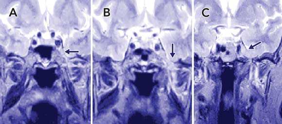
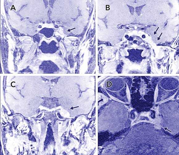
Initial MRI (Box 1) reported as “normal”, although on retrospective review PNS was seen. Subsequent MRI (Box 2) showed extension of the tumour to the pons. |
|||||||||||||
MRI = magnetic resonance imaging. PNS = perineural spread. SCC = squamous cell carcinoma. V2 = maxillary division of trigeminal nerve. V3 = mandibular division of trigeminal nerve. VII = facial nerve. |
|||||||||||||||
The incidence of non-melanotic head and neck skin cancers in Queensland is among the highest in the world.1 Perineural spread (PNS) from these lesions involves either small nerves, identified at pathological examination (incidental), or large nerves, presenting clinically as cranial nerve palsies. Basal cell carcinoma is the more common skin cancer, but incidental PNS is most frequently associated with squamous cell carcinoma.2 In 32 out of 34 patients over a 5-year period, isolated major nerve PNS was due to squamous cell carcinoma (unpublished data). PNS from melanoma and microcystic adnexal carcinoma has also been described.3,4
Because many clinicians are not familiar with PNS involving large cranial nerves, the diagnosis can easily be missed or delayed. The disease is associated with high treatment morbidity and poor prognosis once clinical or radiological evidence becomes apparent, with a 5-year survival rate of 20%–30%.5 Early detection of PNS in large cranial nerves is essential, as the condition is often unsalvageable once the tumour has spread through the skull base.
The facial and trigeminal nerves are most commonly affected,6 although forehead tumours can gain access to the orbit via the ophthalmic nerve.7 Symptoms of trigeminal nerve infiltration include formication, dysaethesia, paraesthesia, numbness and pain (often severe and “electric shock-like” in nature).8 Slowly progressive facial nerve palsy may represent seventh cranial nerve infiltration or infiltration within the parotid, whereas diplopia and visual impairment indicate advanced orbital disease. These symptoms can mimic other diagnoses, such as Bell’s palsy or trigeminal neuralgia, but almost always manifest as slowly progressive and irreversible palsies. Close follow-up of patients to ensure resolution of symptoms is mandatory.
The four patients we have described illustrate delays between clinical presentation and diagnosis. These delays could have been avoided by establishing the link between unresolving cranial nerve palsies and excision of cutaneous lesions from the head and neck. Patients 2 and 3 had a history of aggressive squamous cell carcinoma of the face treated with radical excision and postoperative radiotherapy. Advanced, recurrent skin squamous cell carcinomas have a higher incidence of PNS, particularly when located close to a cranial nerve, and should alert the clinician to the possibility of neural metastasis.9 Tumour size before excision, postoperative defect size, subclinical extension and Moh’s micrographic surgery levels are significantly larger in patients with PNS than in patients without PNS.10
PNS can be subtle and missed on initial pathology but, because of the propensity for local recurrence, most specialist multidisciplinary units that deal with skin cancer will recommend adjuvant radiotherapy, particularly if the lesion is excised from the trigeminal nerve distribution. In Patients 1 and 4, no histology was available. Small skin lesions are often treated with cryotherapy or curettage, resulting in no pathological report. However, these lesions may still have a propensity for PNS, so their excision should be elicited in the history. In Patient 4, the skin lesions were excised from the side of the face contralateral to the cranial nerve palsies and were unlikely to represent the primary disease. Nevertheless, this case illustrates that patients with a history of cutaneous lesions from any site on the head and neck are at risk of developing PNS. These cases also illustrate that the interval between excision of a lesion and presentation of PNS can be long.11 Clinicians should hold a high index of suspicion even if the interval is several years.
A further source of delay is that patients are seen by a variety of clinicians before diagnosis. Cross-referral for second opinions is common, resulting in further delay. Patients 1–3 illustrate how the unfamiliarity of clinicians with the disease process resulted in patients seeking opinions from several specialties before definitive diagnosis. In addition, investigations may be performed to exclude a different pathology or misinterpreted because the clinician is unaware of the phenomenon, resulting in further delay. In Patients 2 and 4, initial imaging revealed changes compatible with PNS, but these were not recognised, either because the radiologist was not familiar with the entity or because the referring clinician may not have raised the possibility of PNS. In Patient 2, the hyperintensity of the masseter muscle on magnetic resonance imaging (MRI) led the clinician to suspect a primary muscular disorder, instead of the correct interpretation of denervation changes from trigeminal nerve involvement. The patient underwent an unnecessary muscle biopsy. Earlier detection may have resulted in Patients 2 and 4 being treated with curative intent instead of palliative care.
High resolution MRI is the investigation of choice, and may identify the earliest changes of PNS. However, disease may not be radiologically apparent until it has reached the orbit, cranial fossa or the skull base foramina. Computed tomography (CT) identifies the disease at a late stage when the tumour has eroded adjacent bony margins. MRI has the added advantage of defining the relationship of the perineural tumour to important anatomical landmarks such as the cavernous internal carotid artery; this is important when planning a resection or biopsy. However, subtle changes on MRI may be missed even by experienced radiologists and, given a strong clinical suspicion with normal imaging, nerve biopsies should be performed. Presently, positron emission tomograph (PET) scanning has no role to play in the staging of PNS.
Delayed diagnosis of major cranial nerve PNS may result in devastating outcomes for patients, so early detection is crucial. Patients who present with slowly progressive cranial nerve palsies with a history of head and neck cutaneous malignancies should be investigated for PNS with a high index of suspicion. We recommend that all patients who undergo excision of high risk skin malignancies should be advised to seek a medical opinion if they develop facial numbness or weakness in subsequent years.
Competing interests
None identified.
Acknowledgements
We would like to acknowledge the contribution of the neurosurgical, plastic surgery and radiology departments of the Mater Private Hospital, Queensland X-ray and the Princess Alexandra Hospital, Brisbane, for their involvement in these complex cases.
References
- Staples MP, Elwood M, Burton RC, et al. Non-melanoma skin cancer in Australia: the 2002 national survey and trends since 1985. Med J Aust 2006; 184: 6-10. 0_i1091922
- Brown CI, Perry AE. Incidence of perineural invasion in histologically aggressive types of basal cell carcinoma. Am J Dermatopathol 2000; 22: 123-125. 0_i1091924
- Clement CI, Genge J, O’Donnell BA, Lochhead AG. Orbital and periorbital microcystic adnexal carcinoma. Ophthal Plast Reconstr Surg 2005; 21: 97-102. 0_i1091926
- Newlin HE, Morris CG, Amdur RJ, Mendenhall WM. Neurotropic melanoma of the head and neck with clinical perineural invasion. Am J Clin Oncol 2005; 28: 399-402. 0_i1091928
- Mendenhall WM, Parsons JT, Mendenhall NP, et al. Carcinoma of the skin of the head and neck with perineural invasion. Head Neck 1989; 11: 301-308. 0_i1091930
- Goepfert H, Dichtel WJ, Medina JE, et al. Perineural invasion in squamous cell skin carcinoma of the head and neck. Am J Surg 1984; 148: 542-547. 0_i1091932
- McNab AA, Francis IC, Benger R, Crompton JL. Perineural spread of cutaneous squamous cell carcinoma via the orbit. Clinical features and outcome in 21 cases. Ophthalmology 1997; 104: 1457-1462. 0_i1091934
- Veness MJ. Perineural spread in head and neck skin cancer. Aust J Dermatol 2000; 41: 117-119. 0_i1091936
- Mendenhall WM, Amdur RJ, Williams LS, et al. Carcinoma of the skin of the head and neck with perineural invasion. Head Neck 2002; 24: 78-83. 0_i1091938
- Leibovitch I, Huilgol SC, Selva D, et al. Cutaneous squamous cell carcinoma treated with Mohs micrographic surgery in Australia II. Perineural invasion. J Am Acad Dermatol 2005; 53: 261-266. 0_i1091940
- McCord MW, Mendenhall WM, Parsons JT, et al. Skin cancer of the head and neck with clinical perineural invasion. Int J Radiat Oncol Biol Phys 2000; 47: 89-93. 0_i1091942
Diagnosing Acute Kava Dermopathy: A Case Report of a Characteristic Cutaneous Eruption
Ali Abid, Nicholas Allen, Abeer Hagelamin, Christopher Henderson, Artiene Tatian
Localised Herpes Simplex Following Midline Laparotomy
Jessica S. Bulluss, Paul Chee, Matthew J. Verheyden
No silver lining with health misinformation: argyria caused by intentional silver consumption
Luke Collins, Logesh Palanikumar, Stephen Bacchi
Lingual Raynaud phenomenon
Michael Taggart, Mina John
Melasma in the male: a less well recognised entity
Tim Aung, Rowland Noakes
Age group‐specific changes in keratinocyte cancer treatment rates in Australia, 2012–2021: a retrospective cohort study based on MBS claims data
Catherine M Olsen, Nirmala Pandeya, Rachel E Neale, David C Whiteman