




Volume 183 - Issue 6
An unexpected cause of macroscopic haematuria
Authors: John S Lubel, Louise M Burrell and Vicki Levidiotis
Med J Aust 2005; 183 (6): 321-323. || doi: 10.5694/j.1326-5377.2005.tb07064.x
Published online: 19 September 2005
Published online: 19 September 2005
A 25-year-old man presented with macroscopic haematuria associated with a body mass index of 20 kg/m2 and a severe coagulopathy consistent with vitamin K deficiency. The diagnosis of a profound malabsorption syndrome secondary to coeliac disease was confirmed by small bowel histology and positive coeliac serology.
Clinical record
A 25-year-old man presented to our hospital’s emergency department with frank haematuria and bilateral loin pain. He had been unwell for 10 days with abdominal pain and vomiting. Two days before presentation, he sought medical attention and was prescribed naproxen for the abdominal pain (he only took two of these tablets); he was on no other medications. Two years previously, a diagnosis of irritable bowel syndrome had been made by his doctor, based on a history of loose bowel motions over 2 years and a normal colonoscopy.
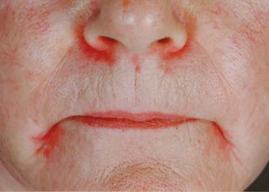
At the time of his hospital presentation, our patient had a body mass index (BMI) of 20 kg/m2 and appeared pale, but had no sign of bruising. He was apyrexial and well hydrated, with unremarkable cardiovascular and respiratory findings. Gastrointestinal examination revealed a red swollen tongue and angular cheilitis (Box 1). There was some mild left renal angle tenderness.
Urinalysis revealed > 500 ×106 non-glomerular red blood cells per litre (normal range < 13 ×106/L) but no pyuria. Abdominal ultrasound performed on the day of review was unremarkable. Peripheral blood tests revealed normal electrolyte levels and renal function, with a coagulopathy reflected by an international normalised ratio (INR) of > 10 (reference range, < 1.4) and activated partial thromboplastin time (APTT) of 115 seconds (reference range, 25–38 seconds) (Box 2). These results were verified with repeat testing. Clotting factor studies revealed severe deficiencies in factors II, VII, IX and X. There were also deficiencies in iron, vitamin B12, and serum folate, vitamin A and vitamin E concentrations (Box 2). Vitamin D levels were normal, but the serum alkaline phosphatase level was twice the upper limit of normal. Results of other liver function tests were normal. Endomysial antibody test results were positive, and tissue transglutaminase antibody (IgA) levels were more than five times the upper limit of normal.
A preliminary diagnosis of vitamin K deficiency, leading to a profound coagulopathy, secondary to a malabsorption syndrome from coeliac disease was made. Duodenal biopsies confirmed the diagnosis of coeliac disease (Box 3). Gastroscopy showed no gastritis or peptic ulcer disease. The duodenal mucosa appeared abnormal and was consistent with villous atrophy. No conclusion could be reached regarding the nonspecific abdominal pain the patient experienced on presentation. Inflammatory bowel disease was a differential diagnosis, but inflammatory markers were only marginally elevated (Box 2).
Parenteral vitamin K was administered, and the prothrombin time reduced to 15 seconds (INR, 1.3) and APTT became normal within 24 hours. The macroscopic haematuria resolved.
The patient was also given parenteral iron and vitamin B12 and oral multivitamin replacement. He was reviewed by the dietetic services and educated about coeliac disease. He also started a gluten-free diet. On clinical review 2 months later, he was feeling well and had put on 15 kg in weight (BMI, 26 kg/m2).
Duodenal biopsies 6 months after the initial presentation revealed variable villous abnormality, consistent with partially treated coeliac disease. The vitamin deficiencies had all resolved, and clotting factor studies were normal. Endomysial antibodies had become negative, and tissue transglutaminase antibody levels (IgA) were within the normal reference range. Bone mineral density at this time was normal.
Genotyping revealed the presence of the HLA DQ2 allele.
Discussion
The most common presentation of coeliac disease is diarrhoea (43%); other reasons for presentation include anaemia (8%), bone disease (6%), weight loss (6%), and abdominal pain (5%).1 The remaining 32% of patients are asymptomatic or present with vague symptoms. Delay in the diagnosis of coeliac disease is common (mean, 11 years).2 About 47% of patients will have been misdiagnosed, and of those with classical symptoms of coeliac disease, 59% have been misdiagnosed as having irritable bowel syndrome.3 The clinical diversity of coeliac disease is recognised, and disorders involving nearly every organ system have been described with this condition.4
Haematuria is an unusual presenting symptom of coeliac disease.5 The haematuria in our patient indicated a systemic bleeding diathesis due to malabsorption of vitamin K, with subsequent prolongation of the prothrombin time and APTT.6 In patients with untreated coeliac disease, the prothrombin time is prolonged (INR, ≥ 1.4) in about 20%,7 and the coagulopathy is usually asymptomatic,8,9 but corrects quickly with administration of vitamin K.10
In the long-term treatment of coeliac disease, a gluten-free diet is paramount and leads to resolution of the underlying small bowel villous abnormality. A partial histological response to a gluten-free diet may reflect poor compliance, accidental exposure to gluten-containing food products, or slow recovery. Our patient claimed close adherence to the diet and was provided with adequate education and dietetic review. Histological recovery can be delayed, with 35% of patients still showing features of coeliac disease up to 2 years after commencing a gluten-free diet.11,12 Failure to achieve histological recovery can occur in the absence of gluten.13 The use of serology tests as surrogate markers for histological resolution is controversial.14-16 Some coeliac patients with negative results for endomysial and tissue transglutaminase antibodies have persistent villous abnormalities, necessitating the use of biopsies to monitor the response to a gluten-free diet (as in our patient).
On HLA genotyping, our patient possessed the HLA DQ2 allele, which is present in more than 90% of patients with coeliac disease, but in only 20% of the general population.17 Endomysial IgA antibodies have a sensitivity of 90% and specificity of 100%, tissue transglutaminase IgA has a sensitivity and specificity of 98%. Sensitivities for the IgG class endomysial and tissue transglutaminase antibodies are around 40%.18 Deficiency of IgA occurs in 1.7%–2.6% of coeliac patients, and these patients have negative results on IgA antibody testing. Total serum IgA levels should be tested together with IgG (endomysial, tissue transglutaminase) to improve the overall sensitivity of antibody testing.19
Our patient demonstrates the need to be aware of the various presenting features of coeliac disease, and reminds us that coeliac disease is often misdiagnosed.
2 Results of laboratory tests
Investigation |
Result |
Reference range |
|||||||||||||
Haemoglobin |
123 |
130–180 g/L |
|||||||||||||
White cell count |
12.4 |
4.0–11.0 × 10⁹/L |
|||||||||||||
Platelets |
727 |
150–400 × 109/L |
|||||||||||||
Mean corpuscular volume |
73 |
82–95 fL |
|||||||||||||
Mean corpuscular haemoglobin |
23.3 |
24–32 pg |
|||||||||||||
Reticulocytes |
74 |
10–90 × 109/L |
|||||||||||||
Differential white cell count |
|||||||||||||||
Neutrophils |
9.52 |
2.0–7.5 × 10⁹/L |
|||||||||||||
Lymphocytes |
1.48 |
1.0–4.0 × 10⁹/L |
|||||||||||||
Monocytes |
1.25 |
0.1–0.8 × 10⁹/L |
|||||||||||||
Eosinophils |
0.12 |
< 0.4 × 10⁹/L |
|||||||||||||
Basophils |
0.02 |
< 0.2 × 10⁹/L |
|||||||||||||
Blood film |
Microcytosis with moderate hypochromasia, and small numbers of elongated cells. Neutrophilia with no left shift and thrombocytosis. |
||||||||||||||
CRP |
22.4 |
1.6–8.7 mg/L |
|||||||||||||
ESR |
14 |
5–12 mm/hour |
|||||||||||||
Alkaline phosphatase |
249 |
40–129 U/L |
|||||||||||||
Tissue transglutaminase antibody |
101 |
< 20 U/mL |
|||||||||||||
Serum folate |
4.7 |
> 6.8 nmol/L |
|||||||||||||
Red cell folate |
Not measured |
|
|||||||||||||
Vitamin B12 |
93 |
132–857 pmol/L |
|||||||||||||
Vitamin A |
0.1 |
0.8–3.1 μmol/L |
|||||||||||||
Vitamin D |
38 |
30–110 nmol/L |
|||||||||||||
Vitamin E |
4.9 |
> 18.6 μmol/L |
|||||||||||||
Ferritin |
10 |
24–336 μg/L |
|||||||||||||
International normalised ratio |
> 10 |
< 1.4 |
|||||||||||||
Activated partial thromboplastin time |
115 |
25–38 seconds |
|||||||||||||
Factor II |
15% |
50%–120% |
|||||||||||||
Factor VII |
3% |
50%–120% |
|||||||||||||
Factor IX |
17% |
50%–120% |
|||||||||||||
Factor X |
5% |
50%–120% |
|||||||||||||
3 Duodenal biopsies
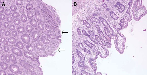
A At time of diagnosis. There is severe villous abnormality associated with marked enterocyte damage (black arrows), in keeping with untreated coeliac disease.
B After 6 months of gluten-free diet. There is partial improvement of the previous villous abnormality, but a moderate villous abnormality with associated crypt hyperplasia remains.
Competing interests
None identified.
Acknowledgements
We thank Dr P A Atkin, Consultant/Honorary Senior Lecturer in Oral Medicine, Cardiff Dental Hospital and School, Heath Park, Cardiff, UK, for his kind contribution of the photograph of angular cheilitis.
References
- Lo W, Sano K, Lebwohl B, et al. Changing presentation of adult celiac disease. Dig Dis Sci 2003; 48: 395-398.
- Green PHR, Stavropoulos SN, Panagi SG, et al. Characteristics of adult celiac disease in the USA: results of a national survey. Am J Gastroenterol 2001; 96: 126-131. 0_i1091809
- Corazza GR, Brusco G, Andreani ML, et al. Previous misdiagnosis and diagnostic delay in adult celiac sprue. J Clin Gastroenterol 1996; 22: 324-325. 0_i1091811
- Duggan JM. Coeliac disease: the great imitator. Med J Aust 2004; 180: 524-526. 0_i1091815
- Moussa AM, Cavestro GM, Coruzzi P, et al. Macrohematuria caused by a fall in prothrombin activity as a clinical presentation of celiac disease. J Clin Gastroenterol 2002; 35: 359-360. 0_i1091817
- Hussaini SH, Ahmed S, Heatley RV. Celiac disease and hypoprothrombinemia. Nutrition 1999; 15: 389-391. 0_i1091820
- Cavallaro R, Iovino P, Castiglione F, et al. Prevalence and clinical associations of prolonged prothrombin time in adult untreated coeliac disease. Eur J Gastroenterol Hepatol 2004; 16: 219-223. 0_i1091822
- Jones RB. Diarrhoea and vomiting with bleeding. An unusual presentation of coeliac disease. Clin Pediatr (Phila) 1973; 12: 24A-25A. 0_i1091824
- Laudage G, Schirp J. Life threatening epistaxis due to plasma coagulation disorder as a partial manifestation of gluten sensitive enteropathy Leber Magen Darm 1995; 25: 94-96. 0_i1091826
- Bottaro G, Fichera A, Ricca O, et al. Effect of the therapy with vitamin K on coagulation factors in celiac disease in children: Pediatr Med Chir 1986; 8: 551-554. 0_i1091829
- Wahab PJ, Meijer JW, Mulder CJ. Histologic follow-up of people with celiac disease on a gluten-free diet: slow and incomplete recovery. Am J Clin Pathol 2002; 118: 459-463. 0_i1091831
- Grefte JM, Bouman JG, Grond J, et al. Slow and incomplete histological and functional recovery in adult gluten sensitive enteropathy. J Clin Pathol 1988; 41: 886-891. 0_i1091833
- Selby WS, Painter D, Collins A, et al. Persistent mucosal abnormalities in coeliac disease are not related to the ingestion of trace amounts of gluten. Scand J Gastroenterol 1999; 34: 909-914. 0_i1091835
- Ghoshal UC, Ghoshal U, Misra A, Choudhuri G. Partially responsive celiac disease resulting from small intestinal bacterial overgrowth and lactose intolerance. BMC Gastroenterol 2004; 4: 10. 0_i1091837
- Tursi A, Brandimarte G, Giorgetti GM. Lack of usefulness of anti-transglutaminase antibodies in assessing histologic recovery after gluten-free diet in celiac disease. J Clin Gastroenterol 2003; 37: 387-391.
- Dickey W, Hughes DF, McMillan SA. Disappearance of endomysial antibodies in treated celiac disease does not indicate histological recovery. Am J Gastroenterol 2000; 95: 712-714. 0_i1091840
- Sollid LM, Markussen G, Ek J, et al. Evidence for a primary association of celiac disease to a particular HLA-DQ alpha/beta heterodimer. J Exp Med 1989; 169: 345-350. 0_i1091842
- Rostom A, Dube C, Cranney A, et al. The diagnostic accuracy of serologic tests for celiac disease: a systematic review. Gastroenterology 2005; 128: S38-S46. 0_i1091844
- Cataldo F, Lio D, Marino V, et al. IgG(1) antiendomysium and IgG antitissue transglutaminase (anti-tTG) antibodies in coeliac patients with selective IgA deficiency. Working Groups on Celiac Disease of SIGEP and Club del Tenue. Gut 2000; 47: 366-369. 0_i1091848
Early-Onset Colorectal Cancer With Liver-Only Metastases: A Retrospective Cohort Study Integrating Prospectively Collected Real-World Clinical and Molecular Data From an Australian National Database (2009–2024) to Guide Treatment Planning
Savio G. Barreto, Christos S. Karapetis, Shahid Ullah, Matthew Burge, Susan Caird, Angus Campbell, Azim Jalali, Ross Jennens, Muhammad A. Khattak, Belinda Lee, Stephanie H. Lim, Shehara Mendis, Louise Nott, Timothy J. Price, Jeremy D. Shapiro, Jeanne Tie, Javier Torres, Colin Williams, Rachel Wong, Vanessa Wong, Peter Gibbs
Australian Pathways for Specialist Pain Management and Early Palliative Care for People With Pancreatic Cancer: Developed Using a Community Consensus Approach
Jennifer Philip, Melanie R. Lovell, Kylee Bellingham, Gail Garvey, Gregory B. Crawford, Nicole M. Rankin, Kara Burns, Isabel Young, Vivienne Milch, Dorothy Keefe, Katrina Anderson, James Lawson, Meinir Krishnasamy
Cause‐specific mortality among Queensland people with cirrhosis, by cirrhosis aetiology and decompensation status, 2007–22: a retrospective cohort study
Vikas Bhasker, Jessica R Fong, Paul J Clark, Gunter F Hartel, Richard Skoien, James O’Beirne, Elizabeth E Powell, Patricia C Valery
Clinical practice guidelines for hepatocellular carcinoma surveillance for people at high risk in Australia: summary of recommendations
Jacob George, Nicole L Allard, Stuart K Roberts, Leon A Adams, Jane Davies, Behzad Hajarizadeh, Jennifer H MacLachlan, Suzanne E Mahady, Rosalie Altus, Catherine Brown, David C Fry, Belinda Greenwood‐Smith, Natali Smud, Patricia C Valery, Nafisa Yussf, Kate Broun, Denise Campbell, Karen Canfell, Chelsea Carle Harrison, Victoria Freeman, Paul Grogan, Catherine Holliday, Suzanne Hughes, Anna Kelly, Cathelijne Kemenade, Claire Latumahina, Amanda McAtamney, Megan Varlow, Joachim Worthington, Susan Yuill, Eleonora Feletto
Assessment of metabolic dysfunction‐associated fatty liver disease in primary care: a consensus statement summary
Leon A Adams, William W Kemp, Kate R Muller, Elizabeth E Powell, Stuart K Roberts, Luis Calzadilla Bertot, Stephanie Best, Gary Deed, Jon D Emery, Samantha L Hocking, Graham R Jones, John S Lubel, Sinead Sheils, Stephen M Twigg, Gerald F Watts, Jacob George
Treatment outcomes for people with hepatitis C referred to tertiary care in Victoria, 2021–22: a retrospective observational study
Elly Layton, Nicole Matthews, Brendan Quinn, Nasra Higgins, Gabrielle Lindeman, Mielle Abbott, Jennifer MacLachlan, Elizabeth Birbilis, Margaret E Hellard, Joseph Doyle, Benjamin C Cowie, Mark Stoové