




Volume 217 - Issue 8
Osteopoikilosis: benign bone islands in periarticular bones
Authors: Te‐Feng A Chou and Ching‐Hua Hsiao
Med J Aust 2022; 217 (8): 397-397. || doi: 10.5694/mja2.51719
Published online: 17 October 2022
Published online: 17 October 2022
A 41-year-old man presented with right knee pain for 3 months
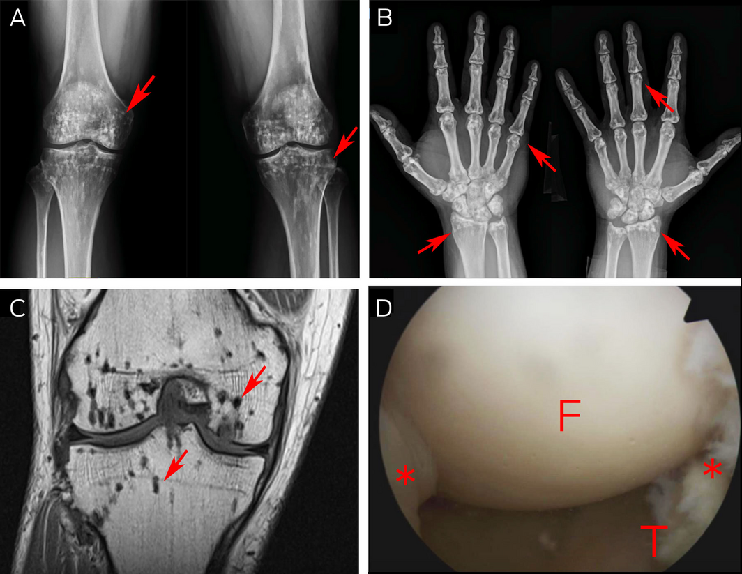
A 41‐year‐old man presented with right knee pain for 3 months. Physical exams noted a positive Hughston plica test (ie, the presence of pain or a popping sensation during flexion and extension of the knee while the tibia is internally rotated, indicative of an abnormal plica). An x‐ray revealed multiple radiopaque lesions in bilateral knees (Figure, A, arrows) and in all periarticular bones (Figure, B, arrows). A magnetic resonance imaging scan revealed benign, hypointense lesions on T1‐ and T2‐weighted images (Figure, C, arrows), compatible with osteopoikilosis. Due to persistent pain, he underwent an arthroscopy of the right knee, which showed normal articular cartilage with synovitis secondary to a medial plica (Figure, D [F = femur; T = tibia; * synovium]). Successful resection of the medial plica was performed; the pain disappeared and the patient was able to return to his usual daily activities without difficulty. Osteopoikilosis is an autosomal‐dominant disorder with a prevalence of one in 50000 population.1 This condition can be associated with a mutation to the LEMD3 gene and is characterised by sclerosing bone dysplasias.2 In addition, these patients may be associated with dermatofibrosis, scleroderma and dwarfism.3 The lesions are benign, and if asymptomatic, further workup is unnecessary.3 The asymptomatic presentation and relatively benign radiologic appearances differentiate this condition from metastatic cancer, tuberous sclerosis, and other diseases.3,4
Competing interests
No relevant disclosures.
References
- Woyciechowsky TG, Monticielo MR, Keiserman B, Monticielo OA. Osteopoikilosis: what does the rheumatologist must know about it? Clin Rheumatol 2012; 31: 745‐748.
- Hellemans J, Preobrazhenska O, Willaert A, et al. Loss‐of‐function mutations in LEMD3 result in osteopoikilosis, Buschke–Ollendorff syndrome and melorheostosis. Nat Genet 2004; 36: 1213‐1218.
- Negi RS, Manchanda KL, Sanga S, et al. Osteopoikilosis — spotted bone disease. Med J Armed Forces India 2013; 69: 196‐198.
- Mahbouba J, Mondher G, Amira M, et al. Osteopoikilosis: a rare cause of bone pain. Caspian J Intern Med 2015; 6: 177‐179.
Patterns of Osteoporosis Treatment Initiation Following Low-Trauma Fracture: A Population-Based Retrospective Cohort Study in New South Wales, Australia, Using Linked Administrative Health Data
Mike Lin, Huy Nguyen, Thach Tran, Robert D. Blank, Dana Bliuc, Jacqueline R. Center
Rapid Growth in Diagnostic CT Knee Imaging in Australia: Implications for Imaging Stewardship
Hanwen Chen, Rachelle Buchbinder, Sean Docking, Ian A Harris
Spinal cord stimulation patterns of care, re‐interventions, and costs for private health insurers, Australia, 2011–22: a retrospective observational study
Caitlin MP Jones, Christopher G Maher, Rachelle Buchbinder, Ian A Harris, Chung‐Wei Christine Lin, Christopher Hayes, Alexandra Gorelik
The incidence of and risk factors for hospitalisations and amputations for people with diabetes‐related foot ulcers in Queensland, 2011–19: an observational cohort study
Yuqi Zhang, Susanna M Cramb, Steven M McPhail, Rosana Pacella, Jaap J Netten, Ewan M Kinnear, Peter A Lazzarini
Bone health perspectives among Indigenous people: a qualitative study
Troy Walker (Yorta Yorta), Karan P Singh, Vanessa Gan, Brooke Conley (Ngiyampaa), Jessica Bravo, Nigel Smith (Weilwan), April Clarke (Eastern Maar, Kirrae Whurrung, Djap Wurrung), Jackson Baker, Louise J Maple‐Brown, Robin M Daly, Jennifer Browne, Jesse Zanker, Cat Shore‐Lorenti, David Scott, Peter R Ebeling, Ayse Zengin
I am a Civil War
Jennifer G Mowbray