




Medical education
Lessons from practice
Volume 217 - Issue 8
Mycobacterium haemophilum in a patient with inflammatory bowel disease
Authors: Fleur W Kong, David Wong, Kendall Sharpe, Louis Pool and James Muir
Med J Aust 2022; 217 (8): 394-396. || doi: 10.5694/mja2.51720
Published online: 17 October 2022
Published online: 17 October 2022
A 38-year-old woman with longstanding Crohn’s disease presented with 7 months of a nonhealing widespread rash
Clinical record
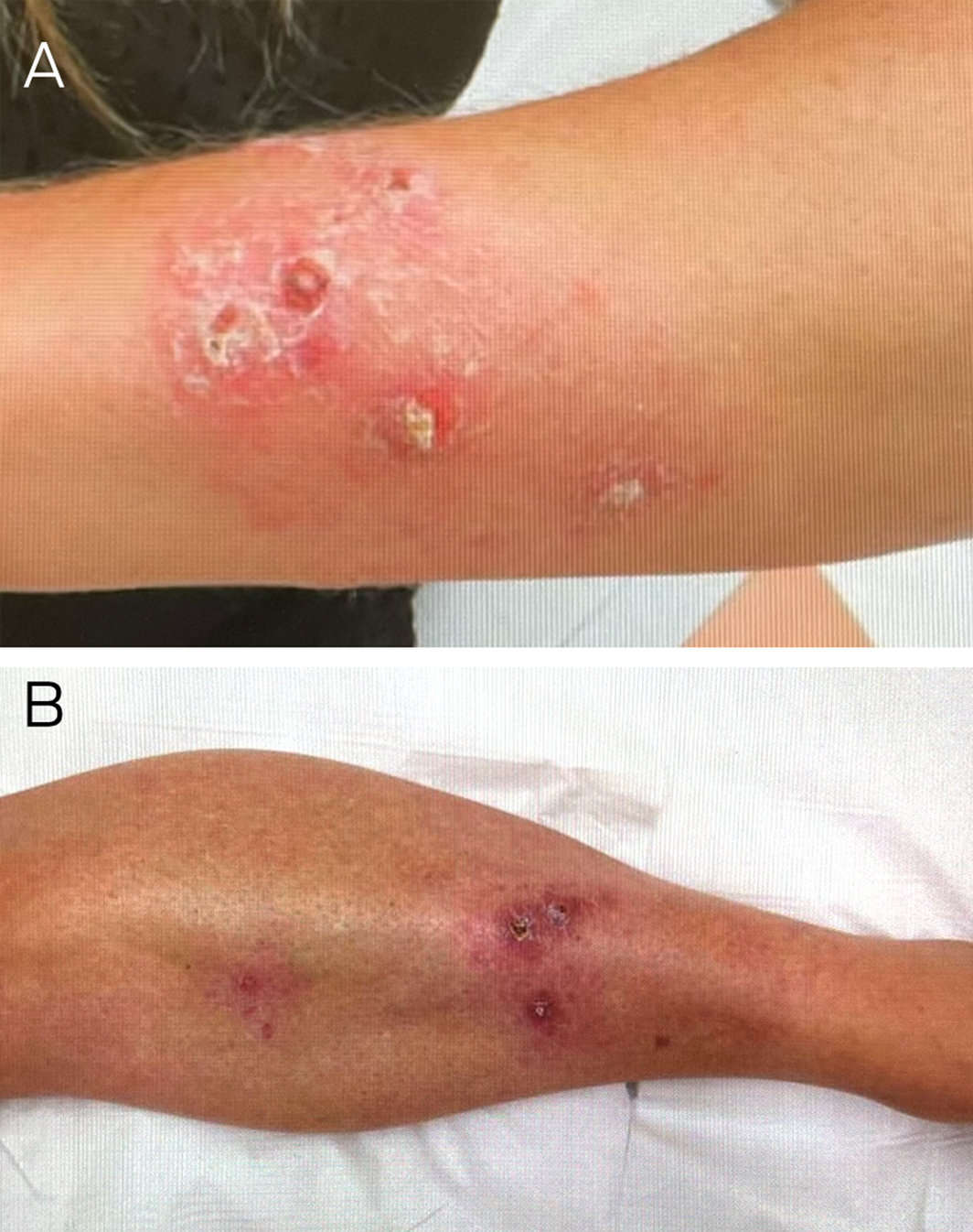
A 38‐year‐old woman with longstanding Crohn’s disease presented with 7 months of a non‐healing widespread rash. Examination revealed indurated and ulcerated papulonodular lesions on the abdomen and extremities (Box 1). Medications included adalimumab, 6‐mercaptopurine, allopurinol and an oral contraceptive pill. There were no other significant medical conditions. The main differentials considered were cutaneous/metastatic Crohn’s disease or a disseminated atypical infection such as atypical mycobacterium or deep fungal. The lesions were too numerous for a skin malignancy.
Incisional biopsy samples with her initial dermatologist revealed small non‐necrotising granulomas. Special stains such as periodic acid–Schiff, Wade–Fite and Ziehl–Neelsen sections were negative for fungi and acid‐fast bacilli. Biopsy culture was negative after one week incubating at 35°C in the standard media. All other investigations were normal or negative.
Given the clinical and histopathological features combined with negative special stains and cultures, the diagnosis of cutaneous Crohn’s disease was favoured. This resulted in the decision to increase the dose of adalimumab and institute both topical and intralesional steroids.
Treatment with topical betamethasone dipropionate 0.05% ointment and intralesional triamcinolone 10mg/mL with her initial dermatologist and subsequent doubling of the adalimumab dose from 40mg fortnightly to weekly under the guidance of her gastroenterologist had no benefit over 6weeks.
In view of the clinical picture and lack of response to treatment, the patient was urgently referred to our dermatology department where we reinvestigated for an infectious aetiology.
Haematoxylin–eosin stains showed a complex pattern of inflammation with superficial ulceration (Box 2). There were poorly formed granulomas which were not as small and discrete as typically seen in Crohn’s disease. Plasma cells were present but no eosinophils. Special stains (periodic acid–Schiff and Ziehl–Neelsen) did not find any fungi or acid‐fast bacilli. Culture swabs grew sensitive Staphylococcus aureus. Oral cephalexin provided no benefit. No further treatment was given pending the results of repeat cultures.
After 6 weeks of incubation at 32C° and 35°C, Mycobacterium haemophilum was isolated. Her Crohn’s disease was in remission with adalimumab, 6‐mercaptopurine and allopurinol, and all were ceased when the mycobacteria were cultured.
A computed tomography scan of the chest, abdomen and pelvis showed no systemic involvement. Treatment was instituted with 6 months of oral ciprofloxacin 500mg, rifampicin 300mg and clarithromycin 500mg under the guidance of the infectious diseases team.
After 8 weeks, the lesions improved and her Crohn’s disease remained in remission. The patient became pregnant 4 weeks later and, after discussion, she ceased all antibiotics. She remains systemically well. Antibiotics will be recommenced after delivery.
Discussion
Mycobacterium haemophilum is a non‐tuberculous, slow growing acid‐fast bacillus and an increasingly recognised pathogen in immunocompromised patients.1 The organism is found in the environment, especially water reservoirs, and has been associated with tattoo parlours and acupuncture needles.2 Cutaneous M. haemophilum typically presents as multiple tender violaceous nodules on extremities which develop into abscesses or ulcers.1 Systemic involvement includes arthritis, lymphadenitis, and disseminated and pulmonary infections. Its occurrence on extremities is likely due to its preference for lower temperatures of 30–32°C.2
M. haemophilum is a slow growing organism needing up to 8 weeks of incubation, making diagnosis difficult.2,3 Methods of rapid species identification include DNA sequencing or polymerase chain reaction (PCR).4 Although pan‐mycobacterial PCR panels are available in Australia, and can be diagnostically helpful, in the presence of a low mycobacterial load, false negative results are possible and therefore cannot be a substitute for culture.4
M. haemophilum infection can be difficult to differentiate from cutaneous Crohn’s disease clinically and histologically (Box 3).5,6
The likelihood of detection of M. haemophilum in biopsy samples is increased by shorter laboratory transit time, using liquid media and specific mediums, such as an iron source, and lower incubation temperature at 30°C.4
Tumour necrosis factor (TNF)‐α has a critical role against Mycobacterium infection through T helper cell 1 based cell‐mediated immune response.7 Adalimumab is a TNF‐α inhibitor used in the treatment of inflammatory bowel disease, hidradenitis suppuritiva and other conditions.8 Granulomatous infections in patients receiving adalimumab are well recognised complications, as reflected by the increasing number of cutaneous M. haemophilum infections in patients receiving adalimumab and other TNF‐α inhibitors.3,9,10
Repeat cultures were likely positive because they were performed after intralesional steroids and higher doses of adalimumab. Greater immunosuppression decreases local inhibition of mycobacterial growth. The diagnosis of atypical infections can be difficult with false negative results on histopathology and culture. Empirical treatment of granulomatous skin disease in immunocompromised patients should be avoided.
The consensus for treatment of M. haemophilum is a combination of clarithromycin, ciprofloxacin and rifampicin.2 Ciprofloxacin and clarithromycin are both pregnancy Category B drugs. Rifampicin is Category C but has been used widely to treat tuberculosis during pregnancy.11
Maternal–fetal complications in Crohn’s disease are related to the activity of the disease at time of conception.8 Most Crohn’s disease medications can be continued during pregnancy, except for methotrexate and thalidomide.8
Lessons from practice
- Persistently inflamed or ulcerated skin lesions should raise the possibility of atypical infection, especially in immunocompromised patients.
- Routine histopathology, special stains and tissue culture may not differentiate cutaneous Crohn’s disease from mycobacterial infection.
- Repeat investigations such as tissue culture should be considered, especially if treatment fails. It is important to alert the laboratory with the repeat biopsy if there is high suspicion for atypical mycobacteria, so that it can be incubated with the right media, for longer duration, and at the correct temperatures.
- Caution should be exercised when treating cutaneous granulomatous conditions in immunocompromised patients.
Box 2 – Biopsy sample from patient’s left wrist (haematoxylin–eosin stain)
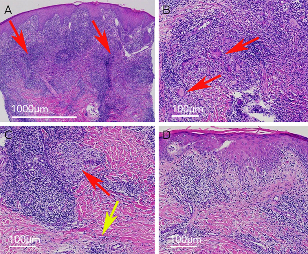
A: A superficial to deep inflammatory infiltrate is dense in the top dermal half (arrows) (magnification ×40). B: Poorly demarcated granulomatous focus with scattered Langerhan multinucleate giant cells (arrows) (magnification ×100). C: A small, better formed granuloma can be seen in the mid top third of the image (red arrow); plasma cells are shown in the bottom right field (yellow arrow) (magnification ×100). D: Inflammatory infiltrate at the dermo‐epidermal junction (magnification ×100).
Box 3 – Comparison of cutaneous Crohn’s disease and Mycobacterium haemophilum infection
|
|
Cutaneous Crohn’s disease |
M. haemophilum infection |
|||||||||||||
|
|
|||||||||||||||
|
Granulomas |
|
|
|||||||||||||
|
Other features |
|
|
|||||||||||||
|
|
|||||||||||||||
|
|
|||||||||||||||
Competing interests
No relevant disclosures.
References
- Burns T, Breathnach S, Cox N, Griffiths C. Rook’s textbook of dermatology. 8th ed. Wiley‐Blackwell, 2010.
- Lindeboom JA, Bruijnesteijn van Coppenraet LE, van Soolingen D, et al. Clinical manifestations, diagnosis, and treatment of Mycobacterium haemophilum infections. Clin Microbiol Rev 2011; 24: 701‐717.
- Bao F, Yu C, Pan Q, et al. Cutaneous Mycobacterium haemophilum infection in an immunocompetent patient. J Dtsch Dermatol Ges 2020; 18: 1186‐1188.
- Simon A, Onya O, Mazza‐Stalder J, et al. Added diagnostic value of 16S rRNA gene pan‐mycobacterial PCR for nontuberculous mycobacterial infections: a 10‐year retrospective study. Eur J Clin Microbiol Infect Dis 2019; 38: 1873‐1881.
- Calonje JE, Brenn T, Lazar AJ, Billings SD. Granulomatous, necrobiotic and perforating dermatoses. In: McKee’s pathology of the skin. 5th ed. Elsevier, 2018.
- Grayson W, Calonje E. Infectious diseases of the skin. In: McKee’s pathology of the skin. 5th ed. Elsevier, 2018.
- Winthrop KL, Chang E, Yamashita S, et al. Nontuberculous mycobacteria infections and anti‐tumor necrosis factor‐alpha therapy. Emerg Infect Dis 2009; 15: 1556‐1561.
- Hashash JG, Kane S. Pregnancy and inflammatory bowel disease. Gastroenterol Hepatol (N Y) 2015; 11: 96‐102.
- Chen CJ, Yen HH. Cutaneous Mycobacterium haemophilum infection: a rare cutaneous manifestation in a patient with Crohn’s disease. Dig Liver Dis 2020; 52: 1057‐1058.
- Collins CS, Terrell C, Mueller P. Disseminated Mycobacterium haemophilum infection in a 72‐year‐old patient with rheumatoid arthritis on infliximab. BMJ Case Rep 2013; 2013: bcr2012008034.
- Bothamley G. Drug treatment for tuberculosis during pregnancy: safety considerations. Drug Saf 2001; 24: 553‐565.
Provenance: Not commissioned; externally peer reviewed.
Costs of Delivering the Hep B PAST Model of Care in Remote Australia: A Retrospective Pre–Post Study
Hoa Nguyen, Anh Le Tuan Nguyen, Paula Binks, Melita McKinnon, Emily Vintour-Cesar, Karen Wills, Nicola Stephens, Benjamin Cowie, Joshua S. Davis, Yuejen Zhao, Peter Nihill, Julie A. Campbell, Andrew J. Palmer, Jane Davies, Barbara de Graaff
Buruli Ulcer Acquired From Ringtail Possum Rescue
Daniel P. O'Brien, Naomi Clarke, Sara Bonnici, Mohammad A. Hussain
Q Fever Visceral Abscesses After Contact With Native Australian Marsupials
Samantha J. Laws, Enzo Binotto, James Stewart, Jennifer Ho, Trent Yarwood, Josh Hanson, Simon Smith
Hospital-Admitted Injection-Related Infections Among Incarcerated People Who Inject Drugs in Australia: A Retrospective Cohort Study
Andrew Palmer, Matthew Carter, Jeremy Yeo, Cecilia Shim, Jason Connor, Jeremy Hayllar, Gerald Holtmann, Naomi Moy, Elliott G. Playford, Naomi Runnegar, Paul J. Clark
The Live Attenuated Influenza Vaccine in Australia: An Additional Tool for Influenza Prevention
Cyra Patel, Alexis Pillsbury, Tran Nguyen, Xia Wang, Helen E. Quinn, Clayton K. Chiu, Allen C. Cheng, Katie L. Flanagan, Zhicheng Wang
Reflex Testing for Hepatitis D Infection: A Unique Opportunity to Reduce Hepatitis D-Related Chronic Liver Disease Deaths in Australia
Jessica Howell, Lauren Andersson, Miriam T. Levy, James O'Beirne, Leon Adams, Katharine Irvine, Avik Majumdar, Golo Ahlenstiel, Kathy Jackson, Krispin Hajkowicz, Joseph Doyle, Jane Davies, Sarah Cherian, Wayne Dimech, Alexander J. Thompson