




Medical education
Lessons from practice
Volume 215 - Issue 9
Violaceous skin lesions on a returned traveller
Authors: David WJ Griffin, Jenny SJ Wong, Orla Morrissey and Cristina Mateevici
Med J Aust 2021; 215 (9): 398-400. || doi: 10.5694/mja2.51298
Published online: 1 November 2021
Published online: 1 November 2021
A 34-year-old Australian man, with no relevant past medical history, presented with 4 weeks of progressive, nodular skin lesions on his non-dominant hand and arm
Clinical record
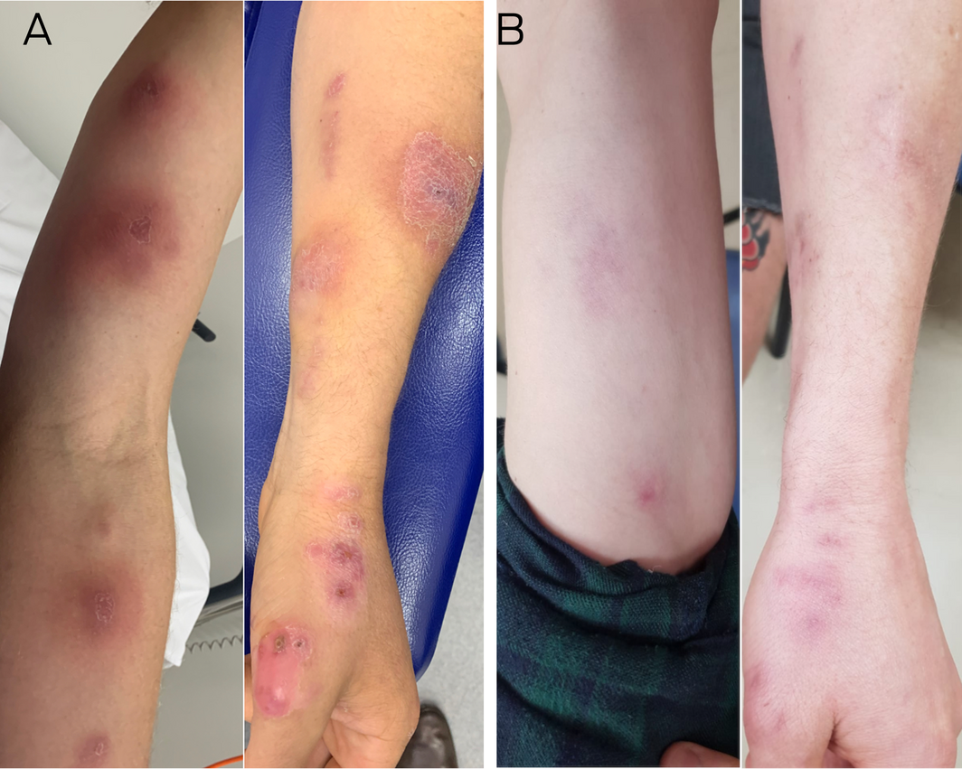
A 34‐year‐old Australian man, with no relevant past medical history, presented to the emergency department with 4 weeks of progressive, nodular skin lesions on his non‐dominant hand and arm (Box 1, A). He was systemically well, without constitutional symptoms, and worked as a plumber in suburban Melbourne. He was an avid gardener and had completed a 4‐week permaculture course in Pisac, Peru, 8 weeks prior. While in Peru, he sustained multiple insect bites and scratches on his hands while working with soil. He had no animal bites or fresh or salt water exposure. He completed a 5‐day course of cephalexin with his local doctor, without improvement, upon the emergence of the first lesion on this thumb.
Examination revealed a well man, with multiple large, tender, violaceous, lesions tracking up his left arm. The lesions were raised, warm and ulcerated. Larger lesions were fluctuant, with serous discharge (Box 1, A). The remainder of his skin and physical examination was normal with no lymphadenopathy and normal vital signs.
Full blood count and renal and liver function tests were normal. Human immunodeficiency virus, Bartonella henselae and rickettsial serology were negative. Punch biopsies of lesions revealed a mixed inflammatory infiltrate, without granulomata or organisms identified on special stains (Box 2). Polymerase chain reaction (PCR) on the direct specimen was negative for Leishmania spp. The biopsy sample was referred for panfungal PCR targeting the internal transcribed spacer (ITS) regions of the ribosomal RNA (Clinical Mycology Reference Laboratory, Centre for Infectious Diseases and Microbiology Laboratory Services, NSW Health Pathology), followed by conventional Sanger sequencing and fungal identification. This detected Sporothrix schenkii complex DNA. Occasional mycotic elements were detected on direct microscopy, and fungal culture revealed a dimorphic fungus, with yeast at 37°C and hyphal form at 25°C (Box 2, A and B). Hyphal morphology was consistent with S. schenkii. The isolate was sent for formal identification, by amplification and sequencing of the ITS1‐5.8S‐ITS2 region of the ribosomal DNA (National Mycology Reference Centre, SA Pathology), which confirmed S. schenkii. Cultures for mycobacteria, Nocardia spp and other bacteria were negative.
The patient was diagnosed with lymphocutaneous sporotrichosis and completed 9 months of itraconazole therapy at 200 mg twice daily, which was well tolerated. He experienced complete resolution of nodular lesions (Box 1, B) over this period and remains well 6 months after the conclusion of therapy.
Discussion
Sporotrichosis is caused by several species of dimorphic fungi in the globally ubiquitous Sporothrix schenkii complex. S. schenkii sensu stricto is the most common causative species and most common subcutaneous mycosis in Latin America, including Peru, where our patient travelled.1 Although uncommon in Australia, more than 200 locally acquired cases have been described across Western Australia, Queensland, New South Wales and the Northern Territory since the 1950s. These include sporadic cases and four larger outbreaks associated with mouldy hay exposure.2
S. schenkii is an environmental saprophyte, surviving on dead or decaying matter, particularly in tropical and subtropical regions. Infection usually occurs by the traumatic inoculation of contaminated soil or animal or plant matter into the skin or mucosa, classically with rose thorns, sphagnum moss, and hay. Sporadic human outbreaks attributed to feline scratches and bites reported in Asia and the Americas demonstrate the potential for zoonotic transmission. In Brazil, there has been an epidemic of sporotrichosis due to S. brasiliensis, with about 5000 human cases, for the past two decades.3 Hence, certain occupational and environmental exposures, including farming, animal handling, and gardening are considered risk factors for acquiring S. schenkii.
Lymphocutaneous sporotrichosis is the most common manifestation, characterised by the emergence of secondary lesions along regional lymphatics in a sporotrichoid pattern. Rarely, disseminated disease involving the lungs, joints, or central nervous system occurs, especially in immunocompromised individuals. The diagnosis of sporotrichosis is frequently delayed due to misdiagnosis or lack of suspicion, which may contribute to additional morbidity, including scarring, secondary spread, or unnecessary antibiotic exposure, as highlighted in this case. However, 3–6 months of daily treatment with 200–400 mg itraconazole is well tolerated, 95% effective, and remains the preferred treatment for this indication.4,5 Hence, clinicians should remain vigilant to this diagnosis in patients who present with typical lesions with sporotrichoid spread, who travel to an endemic region, and who have a typical exposure or injury.
However, the differential diagnosis for sporotrichosis remains broad, particularly in travellers and immunocompromised hosts (Box 3). Therefore, clinicians should biopsy suspicious lesions for histological examination and use special stains for relevant organisms, in addition to bacterial, mycobacterial and fungal culture. Targeted molecular testing, including panfungal, leishmanial or mycobacterial PCRs may be helpful in the appropriate context. Although skin scrapings may be useful for the diagnosis of some superficial mycoses (eg, pityriasis versicolor, tinea, dermatophytosis) and parasitic infections (eg, lice, scabies, cutaneous leishmaniasis), they are not considered as sensitive for the diagnosis of sporotrichosis as tissue biopsy.6 This may be explained by the tendency for Sporothrix spp to infect subcutaneous tissue. Hence, skin scrapings were not performed in this case. A swab of exudate or pus may useful for bacterial or fungal microscopy, with culture and sensitivities, including sporotrichosis, but was negative in this patient.
Confirmatory testing is particularly important due to the evolving global epidemiology of endemic mycoses.7 Several fungal pathogens have emerged outside of previously appreciated areas of endemicity. This is likely due to an expanded pool of travellers, including those with immunocompromising conditions; evolving migration patterns; climate change; and improved recognition and diagnostics. Nevertheless, there are limited data about the incidence of fungal infections in travellers and an absence of formalised surveillance systems. It is important that clinicians are mindful of the risk of endemic mycoses in some travellers, and identify and educate patients about the available mitigation strategies for those at risk.
This case is a timely reminder of sporotrichosis as a subcutaneous mycosis with potential for local and travel‐associated acquisition. Prompt recognition and microbiological diagnosis is important to reduce morbidity. Identification of individuals at risk through travel or environmental exposure may enable education around personal protective equipment.
Lessons from practice
- Sporotrichosis is an endemic mycosis caused by dimorphic fungi in the Sporothrix schenkii complex.
- Sporotrichosis is most common in tropical and subtropical climates, and can be acquired both in Australia and in endemic regions overseas.
- Lymphocutaneous disease is most common, with a broad differential diagnosis, especially in travellers and immunocompromised hosts.
- Diagnosis requires clinical vigilance and appropriate microbiological specimens to confirm sporotrichosis and exclude alternative diagnoses, including other endemic mycoses.
- Pre‐travel counselling to mitigate the risk of travel‐associated endemic mycoses is of growing importance, particularly in immunocompromised travellers.
Box 1 – Lymphocutaneous sporotrichosis on the patient’s left arm and forearm (A) before (January 2019) and (B) after (October 2019) itraconazole therapy
Box 2 – Fungal culture and morphology: dimorphic fungal growth on Sabouraud dextrose agar, with chloramphenicol and gentamicin, illustrating (A) glabrous, white to greyish yellow growth at 37°C and (B) dark, pigmented hyphal form at 25°C. (C) Lacto‐fuchsin stain demonstrating ovoid conidia (spores) arising from conidiophores in hyphal form of fungus*
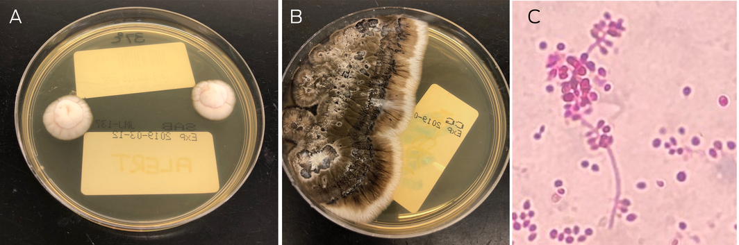
*Additional stains included Gram, Ziehl–Neelsen, Giemsa, periodic acid–Schiff, Grocott, and Wade–Fite stains, which were negative for organisms (not shown). Photos courtesy of Victor Fan.
Box 3 – Differential diagnosis for sporotrichosis and useful investigations
|
|
Diagnoses and investigations |
||||||||||||||
|
|
|||||||||||||||
|
Key infectious differential diagnoses |
|
||||||||||||||
|
Key non‐infectious differential diagnoses |
|
||||||||||||||
|
Preliminary investigations for nodular cutaneous lesions |
|
||||||||||||||
|
|
|||||||||||||||
|
HIV = human immunodeficiency virus; PCR = polymerase chain reaction. * May be locally acquired in Australia. |
|||||||||||||||
Competing interests
No relevant disclosures.
Acknowledgements
We acknowledge Victor Fan, who provided images of the organism and plates in the laboratory; Sarah Kidd and colleagues at the National Mycology Reference Center, SA Pathology; Catriona Halliday and colleagues at the Clinical Mycology Reference Laboratory, ICPMR, NSW Health Pathology, Westmead Hospital; and the mycology scientists and pathologists at Dorevitch Pathology.
References
- Lopes‐Bezerra LM, Mora‐Montes HM, Zhang Y, et al. Sporotrichosis between 1898 and 2017: the evolution of knowledge on a changeable disease and on emerging etiological agents. Med Mycol 2018; 56 (Suppl): 126–143.
- Schauer A, Campbell AJ, Arthur I, Blyth CC, Bowen AC. Spotting sporotrichosis skin infection: The first Australian paediatric case series. J Paediatr Child Health 2020; 56: 408–410.
- Gremião IDF, Miranda LHM, Reis EG, et al. Zoonotic epidemic of sporotrichosis: cat to human transmission. PLoS Pathog 2017; 13: e1006077.
- de Lima Barros MB, Schubach AO, de Vasconcellos Carvalhaes de Oliveira R, et al. Treatment of cutaneous sporotrichosis with itraconazole — study of 645 patients. Clin Infect Dis 2011; 52: e200–e206.
- Kauffman CA, Bustamante B, Chapman SW, Pappas PG; Infectious Diseases Society of America. Clinical practice guidelines for the management of sporotrichosis: 2007 update by the Infectious Diseases Society of America. Clin Infect Dis 2007; 45: 1255–1265.
- Govender NP, Maphanga TG, Zulu TG, et al. An outbreak of lymphocutaneous sporotrichosis among mine‐workers in South Africa. PLoS Negl Trop Dis 2015; 9: e0004096.
- Ashraf N, Kubat RC, Poplin V, et al. Re‐drawing the maps for endemic mycoses. Mycopathologia 2020; 185: 843–865.
Provenance: Not commissioned; externally peer reviewed.
Reorienting Allied Health Into Community-Based Care for People Experiencing Trauma and Social Disadvantage
Simon Rosenbaum, Grace McKeon, Gulsah Kurt, Oscar Lederman, Kemi Wright, Sabuj Kanti Mistry, Jackie E. Curtis, Philip B. Ward, Zachary Steel, Hamish Fibbins, Rachel Morell, Melissa C. Eaton, Andrew Watkins, Ben Harris-Roxas, Brendan Goodger, Eleanor Beck, Megan Teychenne, Joseph Firth, Davy Vancampfort, David Burns, Russell Roberts, Tristan Favaloro, Danielle Weber, Rosanna Barbero, Vasili Maroulis, Melissa Holmes, Stefan Mackenzie, Chiara Mastrogiovanni, Afsana Anwar, Uzma Choudhry, Catherine Sherrington, Jane Currie, Thomas Gadsden, Scott Teasdale
Psychosocial Hazards for Healthcare Workers: Supporting the Second Victim Also Helps the Primary Victims
Sarah Michael
Shifting focus to adolescent wellbeing and inclusive participation in the digital age
Allyson R Todd, Elena Wang, Stephanie R Partridge
Shifting focus to adolescent wellbeing and inclusive participation in the digital age
Jodie Bailie, Helen Dickinson, Briony Lipton, Marissa Shields
The CURE Asthma roadmap
Gary P Anderson, Anthony Flynn, Phil G Bardin, John D Blakey, Shyamali C Dharmage, Paul Foster, Peter G Gibson, Adam Jaffe, Alan James, Christine R Jenkins, Sundram Sivamalai, Peter D Sly, Guy B Marks, Vanessa M McDonald, Judy Wetttenhall