




Volume 215 - Issue 9
A non‐healing ulcer: amelanotic melanoma
Authors: Michelle KY Chen and Deshan Frank Sebaratnam
Med J Aust 2021; 215 (9): 405-405. || doi: 10.5694/mja2.51294
Published online: 1 November 2021
Published online: 1 November 2021
A 73-year-old woman presented with a 7-month history of a non- healing ulcer on her left hallux
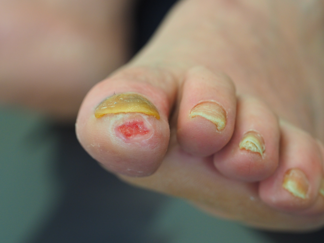
A 73‐year‐old woman presented with a 7‐month history of a non‐healing ulcer on her left hallux (Figure), having seen multiple medical professionals previously. She had no history of diabetes, peripheral vascular disease, or neuropathy. Biopsy demonstrated an amelanotic melanoma with a Breslow thickness of 4 mm and tumour mitotic rate of 1/mm2. No evidence of metastasis was evident on imaging, categorising her disease as stage IIC (T4bN0M0). She proceeded to amputation at the interphalangeal joint and continues lifelong clinical and radiological surveillance. This case demonstrates the importance of skin biopsy for any non‐healing ulcers. Differential diagnoses include neoplastic, infectious, vasculopathic, autoinflammatory and haematological aetiologies.1 Biopsy is recommended for ulcers with an atypical clinical presentation, recalcitrant to standard care and those with dynamic change over a period greater than one month.2,3
Competing interests
No relevant disclosures.
References
- Janowska A, Dini V, Oranges T, et al. atypical ulcers: diagnosis and management. Clin Interv Aging 2019; 14: 2137–2143.
- Alavi A, Sibbald RG, Phillips TJ, et al. What’s new: management of venous leg ulcers: treating venous leg ulcers. J Am Acad Dermatol 2016; 74: 643–664; quiz 665‐666.
- Mar VJ, Chamberlain AJ, Kelly JW, et al. Clinical practice guidelines for the diagnosis and management of melanoma: melanomas that lack classical clinical features. Med J Aust 2017; 207: 348–350. https://www.mja.com.au/journal/2017/207/8/clinical‐practice‐guidelines‐diagnosis‐and‐management‐melanoma‐melanomas‐lack
From Ambition to Action: Closing Policy, Equity and Implementation Gaps in the Australian Cancer Plan's ‘Enhanced Consumer Experience’
Carla Thamm, Elise Button, Oluwaseyifunmi A. Agbejule, Reegan Knowles, Karen van Gorp, Mark B. Pinkham, Meinir Krishnasamy, Alexandra L. McCarthy, Raymond J. Chan
Sovereignty and Survival: Understanding Lung Cancer Outcomes for Aboriginal and Torres Strait Islander People in Australia, a Case–Control Study
Alice R. T. Bergin, Luc te Marvelde, Roger L. Milne, Benjamin J. Solomon, Kris Ivanova, Peter Savas, Stephen J. Luen, Jay Hamann, Andrea Casey, Nicole Watt, Sue M. Evans, Gail Garvey, Sherene Loi
The Changing Indications for Adult Liver Transplantation in Australia: A Narrative Review
Fadak Mohammadi, Alan J. Wigg, Ken Liu, Simone I. Strasser
Uneven Ground: Survival Differences Among Victorian Lung Cancer Patients by Location of Residence (2011–2023): A Retrospective Cohort Study
Evangeline Samuel, Eldho Paul, Mike Lloyd, Sanuki Tissera, Craig Underhill, Sagun Parakh, Phillip Parente, Inger Olesen, Javier Torres, Katharine See, Gavin M. Wright, David Langton, Thomas John, Matthew Conron, James Bartlett, Nicola Atkin, Nikolajs Zeps, Susan V. Harden, Wasek Faisal, John R. Zalcberg, Rob G. Stirling
Early-Onset Colorectal Cancer With Liver-Only Metastases: A Retrospective Cohort Study Integrating Prospectively Collected Real-World Clinical and Molecular Data From an Australian National Database (2009–2024) to Guide Treatment Planning
Savio G. Barreto, Christos S. Karapetis, Shahid Ullah, Matthew Burge, Susan Caird, Angus Campbell, Azim Jalali, Ross Jennens, Muhammad A. Khattak, Belinda Lee, Stephanie H. Lim, Shehara Mendis, Louise Nott, Timothy J. Price, Jeremy D. Shapiro, Jeanne Tie, Javier Torres, Colin Williams, Rachel Wong, Vanessa Wong, Peter Gibbs
Australian Pathways for Specialist Pain Management and Early Palliative Care for People With Pancreatic Cancer: Developed Using a Community Consensus Approach
Jennifer Philip, Melanie R. Lovell, Kylee Bellingham, Gail Garvey, Gregory B. Crawford, Nicole M. Rankin, Kara Burns, Isabel Young, Vivienne Milch, Dorothy Keefe, Katrina Anderson, James Lawson, Meinir Krishnasamy