




Medical education
Lessons from practice
Volume 215 - Issue 8
Congenital syphilis on the rise: the importance of testing and recognition
Authors: Mandy X Wu, Aoife Moore, Mandy Seel, Sumi Britton, Judith Dean, Janet Sharpe, Garry Inglis and Clare B Nourse
Med J Aust 2021; 215 (8): 345-346.e1. || doi: 10.5694/mja2.51270
Published online: 18 October 2021
Published online: 18 October 2021
A 3-month-old boy was admitted with clinical sepsis, hepatosplenomegaly, severe anaemia, transaminitis, high lactate, and coagulopathy
Clinical record
A 3‐month‐old boy was admitted to paediatric intensive care with clinical sepsis, hepatosplenomegaly, severe anaemia, transaminitis, high lactate, and coagulopathy. He was born at 37 weeks’ gestation via emergency caesarean delivery for fetal bradycardia following an uncomplicated pregnancy.
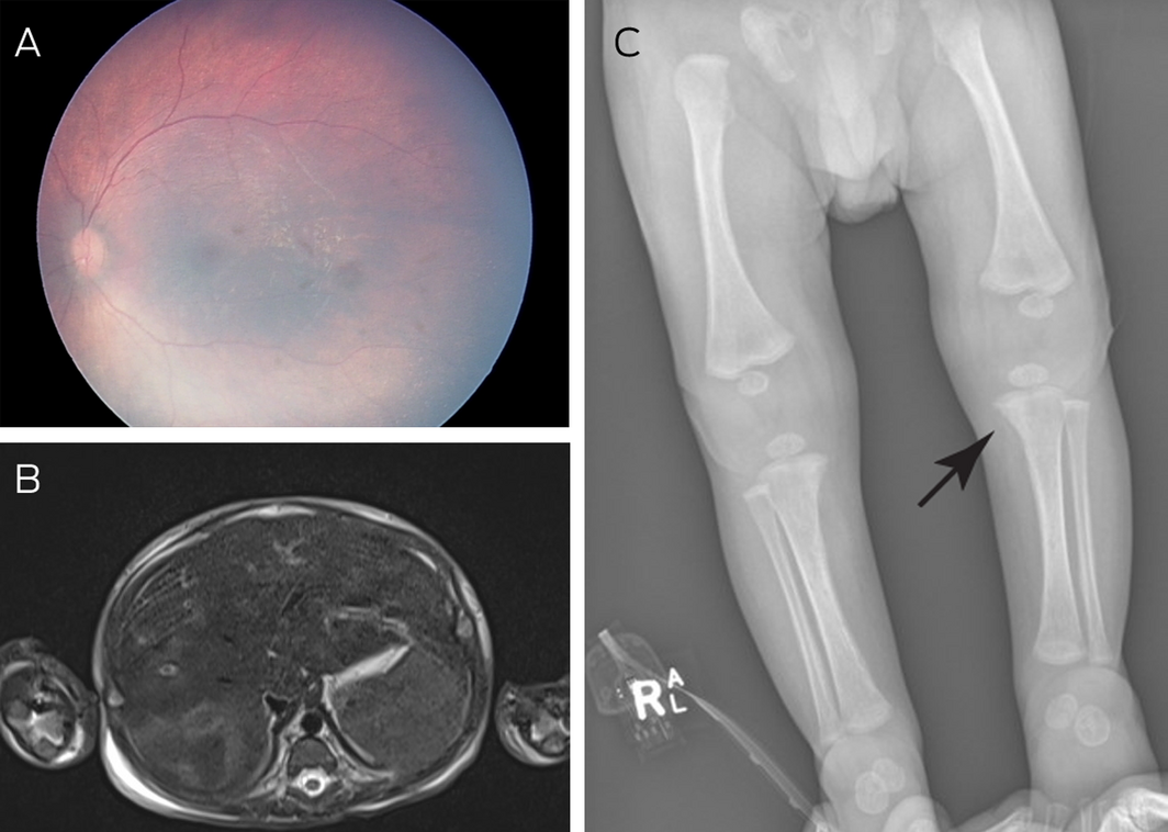
Leukaemia with sepsis was suspected. As part of the extended sepsis screening, syphilis serology was performed on day 5 of admission, which revealed a rapid plasma reagin (RPR) titre of 1:128, reactive Treponema pallidum particle agglutination, positive blood syphilis polymerase chain reaction (PCR) and, subsequently, reactive enzyme immunoassay for IgM antibodies to syphilis. Further assessment revealed mottled retinal pigmentation (Box, A), hepatosplenomegaly with infiltrates (Box, B), and vitreous opacities, osteopenia, periostitis, metaphyseal erosions, and positive Wimberger sign (Box, C) — all typical features of congenital syphilis. Cerebrospinal fluid analysis revealed normal cell count and chemistry and was non‐reactive for venereal disease research laboratory test. T. pallidum DNA was not detected by PCR. Audiology was normal. The infant was treated with intravenous benzylpenicillin 60 mg/kg every 6 hours for 10 days. He responded well and was discharged home after a total of 18 days in hospital. At one year of age, he had normal development and vision despite persistent retinopathy on fundoscopy.
Subsequent parental serology revealed a maternal RPR titre of 1:32 and a paternal RPR titre of 1:64. The mother was a non‐Aboriginal woman who had a negative single antenatal syphilis serology performed at 12 weeks’ gestation. The father was an Aboriginal man. Neither parent described skin lesions consistent with a chancre, but the mother had developed a rash on her abdomen and on the back of her legs at around 30 weeks’ gestation, which persisted until the end of her pregnancy. The father also had a rash on his chest at around the same time.
Discussion
Congenital syphilis is a vertically transmitted bacterial infection caused by T. pallidum and is associated with serious perinatal consequences, such as premature birth, intrauterine growth restriction, miscarriage, stillbirth, and perinatal death.1 The infection is entirely preventable with appropriate antenatal screening and penicillin treatment of infected pregnant women.
Despite the 2007 launch of a World Health Organization global initiative to eliminate congenital syphilis, there remains a persistent increase in the rates of this disease in high income countries which is related to growing rates of infectious syphilis, particularly in pregnancy. Factors contributing to the worldwide re‐emergence of this infection include an increase in higher risk sexual activities, increased travel and migration, and social and economic conditions that limit health care access in some populations.1 National data for Australia reveal that infectious syphilis notifications are increasing significantly in all states except Tasmania. Between 2009 and 2019, there was a fourfold increase in the rate from 5.9 to 23.2 per 100 000 population. This increase was most marked in the Northern Territory, which had an 8.6‐fold increase from 16.8 to 138.6 notifications per 100 000 population.2
Rates of infectious syphilis notifications are about 300 times higher in Aboriginal and Torres Strait Islander peoples, with up to 70% of infections among those aged between 15 and 29 years. Roughly equal numbers of males and females are affected.3 By contrast, infections in non‐Indigenous Australians before 2014 were largely confined to men who have sex with men, and more recent trends show increasing infections in heterosexual persons, with a concomitant increase in incidence among young women of childbearing age. This case highlights the need for clinicians to be aware of the increasing incidence of congenital syphilis and to consider this as a possibility in critically unwell infants presenting with non‐specific symptoms. The prominence of anaemia and thrombocytopenia in this case is reflected in other published literature, with congenital syphilis gaining recognition as a mimicker for haematological and oncological disease.4 Other common features of congenital syphilis include hepatosplenomegaly, fever and rash, but there are a wide range of diverse presentations that vary in nature, age at onset, and severity.
Penicillin remains the only recommended treatment in pregnancy, with adequacy of treatment defined by a fourfold reduction in RPR titre over 6–12 months and completion of treatment course at least 30 days before the delivery of an infant.5 As such, mothers treated for infectious syphilis in pregnancy should have monthly serology testing until delivery to monitor the response to treatment and to exclude reinfection. A further critical aspect of adequate maternal treatment is ensuring that all current sexual partners and their contacts are tested and treated to prevent reinfection. Infants with confirmed congenital syphilis should receive 10 days of intravenous benzylpenicillin.
In addition, infants born to mothers treated for syphilis during pregnancy should have follow‐up serology at 3, 6 and 12 months of age (until RPR remains non‐reactive) even if the initial assessment for congenital syphilis at birth is negative.5
This case demonstrates that congenital syphilis can occur following syphilis infection in women who have negative antenatal testing and who have no specific risk factors for syphilis infection identified during pregnancy. The case described also demonstrates insufficient retesting of women with specific risk factors — in this case, an Aboriginal partner. These specific risk factors are comprehensively listed in Queensland’s recently published Syphilis in pregnancy guidelines5 and include Aboriginal and/or Torres Strait Islander woman or partner, migrants or refugees from high prevalence countries, adolescent pregnancy, woman or partner with sexual practices resulting in increased risk, women with late or no antenatal care, unwanted pregnancy, and substance use. All women should be tested at the first antenatal visit, preferably before 10 weeks’ gestation, and again if the risk of exposure occurs or if symptomatic. However, women at higher risk should have up to five tests during pregnancy: in the early first trimester and at 16–26, 26–28 and 34–36 weeks’ gestation, and again at birth in particular situations.5 One of the challenges is to identify risk factors during pregnancy, which requires a clinical context and specific skills in history taking, including taking sexual histories. Screening intervals could also be considered for the general population in areas of increasing transmission, accepting that multiple factors would need to be balanced. Further discussion of the merits and risks of a second test later in pregnancy for all women is warranted.
Lessons from practice
- The incidence of congenital syphilis is on the rise in Australia and it can occur in the setting of negative maternal antenatal screening.
- Congenital syphilis is an important differential to consider in any critically unwell infant, and symptoms may include non‐specific signs such as anaemia, thrombocytopenia, hepatosplenomegaly, fever and rash.
- A diagnosis of congenital syphilis can be made through clinical findings and parallel testing of infant and maternal rapid plasma reagin (RPR), infant antibodies to syphilis via enzyme immunoassay for IgM, and placental syphilis polymerase chain reaction.
- Treatment of congenital syphilis involves 10 days of intravenous benzylpenicillin and follow‐up serology at 3, 6 and 12 months of age or until RPR is non‐reactive to ensure that the treatment is effective.
Competing interests
No relevant disclosures.
References
- Stamm LV. Syphilis: Re‐emergence of an old foe. Microb Cell 2016; 3: 363–370.
- Australian Government, Department of Health. National Notifiable Diseases Surveillance System, 2020. http://www9.health.gov.au/cda/source/rpt_4_sel.cfm (viewed Feb 2021).
- Australian Government, Department of Health. Infectious syphilis outbreak, 2020. https://www1.health.gov.au/internet/main/publishing.nsf/Content/ohp‐infectious‐syphilis‐outbreak.htm (viewed Feb 2021).
- Keuning MW, Kamp GA, Schonenberg‐Meinema D, et al. Congenital syphilis, the great imitator — case report and review. Lancet Infect Dis 2020; 20: e173–e179.
- Queensland Health. Queensland Clinical Guidelines: syphilis in pregnancy, 2018. https://www.health.qld.gov.au/__data/assets/pdf_file/0035/736883/g‐sip.pdf (viewed Feb 2021).
Linked content
-
MJA Letter: Congenital syphilis on the rise: the importance of testing and recognition
Provenance: Not commissioned; externally peer reviewed.
Forty Years in the Making: Reporting on the Co-Creation of a National Roadmap for Birthing on Country Services for the Best Start to Life
Res McCalman, Anneka J. Bowman, Roianne West, Kristie Watego, Jyai Allen, Claire Clack, Sue Kildea, Sue Kruske, Melanie Briggs, Cleone Wellington, Rebecca Coddington, Yu Gao, Isabella Garti, Sascha Kowalenko, Sarah Ireland, Catherine Austin, John D. Boffa, Marah Prior, Kelsie Kahl, Bettina Chaseling, Sarah Khaw, Emily Armstrong, Donna Hartz, Yvette Roe
Temporal Trends in Preterm Birth Associated With Hypertensive Disorders of Pregnancy in Victoria, Australia: A Population-Based Interrupted Time-Series Study
Melvin Marzan, Heng Jiang, Daniel Lorber Rolnik, Joanne M. Said, Lisa Hui
Linguistic Manoeuvres: Obstetric Violence Camouflages Harm and Loss of Consent From Birth
Liz Sutton, Harsha Ananthram, Rebecca Matthews
Linguistic Manoeuvres: Obstetric Violence Camouflages Harm and Loss of Consent From Birth
Bashi Kumar-Hazard, Andrew M. Bisits, Sharon L. Settecasse, Jenny Gamble, Hazel Keedle, Hannah G. Dahlen
Linguistic Manoeuvres: Obstetric Violence Camouflages Harm and Loss of Consent From Birth
Wendy Pollock, Alissa Fleming, Ensieh Fooladi, Joy Kloester, Anne Tremayne, Yasmin Zisin, Bethany Carr, Kym Davey, Suzanne Willey, Jenny Gamble
Getting on the Same Page: Why Australia Needs a National Maternity Early Warning System (MEWS) Chart
Briony A. Cutts, Lucy Bowyer, Nisha Khot, Sandra Lowe, Stefan C. Kane