




Medical education
Lessons from practice
Volume 210 - Issue 8
Mycobacterial mimicry in a man from Myanmar
Authors: David WJ Griffin, G Khai Lin Huang, Philippe Lachapelle, Steven YC Tong and Siddhartha Mahanty
Med J Aust 2019; 210 (8): 349-351.e1. || doi: 10.5694/mja2.50133
Published online: 6 May 2019
Published online: 6 May 2019
A 26-year-old refugee from Myanmar was referred to the infectious diseases unit of an Australian teaching hospital
Clinical record
A 26‐year‐old refugee from Myanmar was referred to the infectious diseases unit of an Australian teaching hospital for assessment of suspected recurrent pulmonary tuberculosis (TB). He had arrived in Australia 3 months earlier, after spending the preceding 5 years in Malaysia. He was diagnosed with presumed pulmonary TB in Malaysia in 2013, in the context of a productive cough and suspicious chest x‐ray findings, without microbiological confirmation. He completed treatment with 6 months of first line anti‐TB therapy (2 months of rifampicin, isoniazid, pyrazinamide and ethambutol, followed by 4 months of rifampicin and isoniazid).
He described a cough, present over the past few years, with expectoration of dark, black‐flecked sputum, without frank haemoptysis, wheeze or dyspnoea. He was a non‐smoker, denied constitutional symptoms and took no regular medications. Examination was unremarkable.
Outpatient computed tomography (CT) imaging of the chest demonstrated left lower lobe consolidation, with cavitation (Box 1, A), raising the concern of TB recurrence. Cavitation was not evident on chest x‐ray (Box 2). The baseline chest x‐ray from Malaysia could not be traced for comparison.
Three morning sputum samples and subsequent bronchial washings by bronchoscopy were smear negative for acid‐fast bacilli and Xpert MTB/RIF (Cepheid) assay was negative for Mycobacterium tuberculosis. Full blood count was normal.
Mycobacterial and fungal cultures remained negative at review 6 weeks later, and the patient's symptomatology was unchanged. Additional testing for human immunodeficiency virus, vasculitides and malignancy was negative (Box 3). Repeat chest CT was similar to previous imaging, but with apparent medial migration of and track formation adjacent to the original cavitating lesion (Box 1, B). The possibility of paragonimiasis was raised, and sputum was sent for microscopy, seeking ova, cysts and parasites.
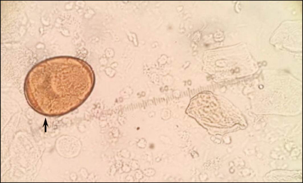
Sputum microscopy revealed the presence of Paragonimus sp. eggs (Box 4), confirming a diagnosis of pulmonary paragonimiasis. The patient was treated with oral praziquantel 1500 mg 75mg/kg/day for three doses over 3 days, with concurrent tapering doses of oral dexamethasone (6 mg, 4 mg, 2 mg, 1 mg, 1 mg over 5 days to reduce the risk of post‐treatment inflammation. At clinical review, one week later, he reported a rapid improvement in symptomatology and had tolerated treatment well.
Three months after treatment, a CT scan revealed near‐complete radiological resolution of pulmonary parenchymal changes (Box 1, C). The patient experienced a complete recovery from symptomatology, and remained well 3 months after treatment.
Discussion
Paragonimiasis is a zoonotic infection caused by several species of lung fluke in the genus Paragonimus (most commonly P. westermanii), which are endemic to South‐East Asia, Africa and the Americas. Paragonimus westermanii has a wide geographical distribution, including India, China, Japan, Korea and South‐East Asia. While the exact prevalence of the infection is unknown due to lack of surveillance, an estimated 20 million people are infected worldwide;1 however, it is uncommon in Australia. Infection typically follows the ingestion of metacercariae (encysted larvae; the parasite stage capable of infecting mammalian hosts) from inadequately cooked or pickled crustaceans, or their juices.2 Indeed, our patient reported frequent ingestion of raw crabs in childhood. After ingestion, metacercariae hatch and larvae typically migrate to the lung, establishing pulmonary infection with adult worms.
The diagnosis of paragonimiasis is frequently delayed. However, it is an important differential diagnosis for TB in the appropriate context, with similar clinical and radiological features, and overlapping epidemiological risk factors, including travel to or emigration from an endemic country. Like TB, paragonimiasis may only become evident many years after exposure, manifesting with productive cough, haemoptysis, cavitating lesions or pleural effusion on radiography.3 While TB most commonly affects the upper lobe(s) in immunocompetent hosts, paragonimiasis lesions are typically peripheral and affect the middle and lower lobes.4,5 The presence of a worm migration tract, as seen in our patient, is a characteristic radiological finding of pulmonary paragonimiasis.4 Although the location of cavitation is not diagnostic, it may increase suspicion for a particular diagnosis.
When paragonimiasis is suspected, clinicians should request examination of sputum and stool specimens for ova, cysts and parasites, with relevant clinical history to guide laboratory staff (Box 3). Ova have a characteristic size and morphology (Box 4), but can be difficult to detect in sputum or faeces. Hence, serodiagnosis can help to confirm infection and monitor treatment, but is not easily accessible in Australia. The United States Centers for Disease Control and Prevention offers a Western blot assay with a sensitivity of 96% and specificity of > 99%.6 Short course treatment with 3 days of praziquantel 75 mg/kg/day has an efficacy close to 100%, with few side effects.7
This case highlights the importance of considering paragonimiasis as a differential diagnosis for TB in patients with chronic pulmonary symptoms and epidemiological risk factors. Timely diagnosis enables appropriate therapy, thus minimising potential morbidity and mortality associated with both the disease and empirically prescribed anti‐TB drugs.
Lessons from practice
- Paragonimiasis is rare in Australia but endemic in many parts of the world, including South‐East Asia. It should be considered in the differential diagnosis of patients with a chronic productive cough and cavitating lung lesions, who are recent migrants, travellers or refugees from endemic areas, especially where investigations for tuberculosis are negative.
- Evaluation for paragonimiasis should include examination of the sputum and stool for Paragonimus eggs.
- The presence of migrating cavitating lesions on sequential radiology, affecting the lower or middle lobes, with a worm migration tract, should raise suspicion for pulmonary paragonimiasis.
- Treatment with 3 days of oral praziquantel is well tolerated and effective for pulmonary paragonimiasis.
Box 1 – Computed tomography chest scans
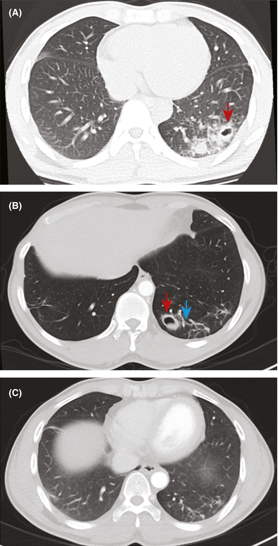
Scans taken in July 2017 (A), January 2018 (B) and May 2018 (C) show radiological evolution (A and B) and resolution (C) of pulmonary parenchymal lesions adjacent to the pleura. Note the apparent midline migration of the cystic pleural‐based lesion in the left lower lobe, with adjacent scarring, and with adjacent track formation in repeat imaging (B) compared with original imaging (A). Arrows indicate cavitating lesions (red) with evidence of migration tract (blue) typical of paragonimiasis.♦
Box 2 – Chest x‐rays
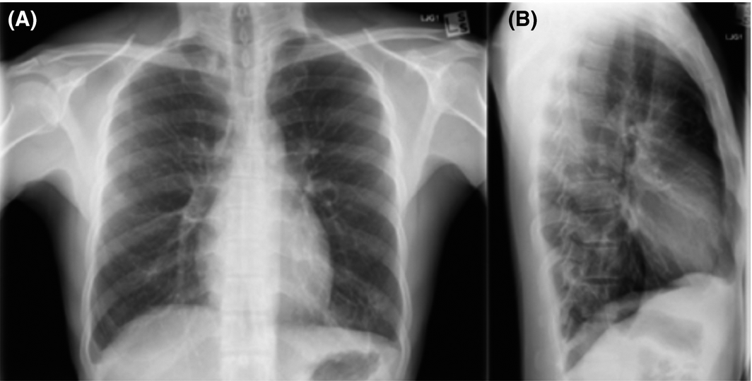
Posterioanterior (A) and left lateral (B) x‐rays (August 2017) show patchy opacification of the left lower lobe, without the evidence of cavitation shown on computed tomography imaging.♦
Box 3 – Possible diagnostic investigations in travellers, immigrants and refugees with chronic productive cough and cavitating lung lesions
To confirm or exclude key differential diagnoses- Computed tomography of the chest
- Sputum microscopy culture and sensitivity for bacterial infection
- Sputum Mycobacterium tuberculosis‐specific polymerase chain reaction (Xpert MTB/RIF), acid‐fast stain and mycobacterial cultures
- Sputum fungal culture and serological testing for histoplasmosis, aspergillosis or coccidioidomycosis
- HIV serology
- Serum cryptococcal antigen
- Sputum cytology for malignancy (consider bronchoscopy)
- Antineutrophil cytoplasmic antibodies for vasculitides (eg, granulomatosis with polyangiitis, or eosinophilic granulomatosis with polyangiitis)
Competing interests
No relevant disclosures.
Acknowledgements
David Griffin and Khai Huang contributed equally to the authorship of this manuscript. We would like to thank the staff in the Department of Microbiology at Melbourne Health.
References
- Foodborne Diseases Burden Epidemiology Reference Group 2007‐2015. WHO estimates of the global burden of foodborne diseases. Geneva: World Health Organization, 2015. http://www.who.int/foodsafety/publications/foodborne_disease/fergreport/en/ (viewed Oct 2018).
- Procop GW. North American paragonimiasis (caused by Paragonimus kellicotti) in the context of global paragonimiasis. Clin Microbiol Rev 2009; 22: 415–446.
- Barennes H, Slesak G, Buisson Y, Odermatt P. Paragonimiasis as an important alternative misdiagnosed disease for suspected acid‐fast bacilli sputum smear‐negative tuberculosis. Am J Trop Med Hyg 2014; 90: 384–385.
- Seon HJ, Kim YI, Lee JH, et al. Differential chest computed tomography findings of pulmonary parasite infestation between the paragonimiasis and nonparagonimiatic parasite infestation. J Comput Assist Tomog 2015; 39: 956–961.
- Rozenshtein A, Hao F, Starc MT, Pearson GD. Radiographic appearance of pulmonary tuberculosis: dogma disproved. Am J Roentgenol 2015; 204: 974–978.
- Centers for Disease Control and Prevention. DPDx ‐ Laboratory Identification of Parasites of Public Health Concern. Paragonimiasis. https://www.cdc.gov/dpdx/paragonimiasis/index.html (viewed Oct 2018).
- Udonsi JK. Clinical field trials of praziquantel in pulmonary paragonimiasis due to Paragonimus uterobilateralis in endemic populations of the Igwun Basin, Nigeria. Trop Med Parasitol 1989; 40: 65–68.
Provenance: Not commissioned; externally peer reviewed.
Costs of Delivering the Hep B PAST Model of Care in Remote Australia: A Retrospective Pre–Post Study
Hoa Nguyen, Anh Le Tuan Nguyen, Paula Binks, Melita McKinnon, Emily Vintour-Cesar, Karen Wills, Nicola Stephens, Benjamin Cowie, Joshua S. Davis, Yuejen Zhao, Peter Nihill, Julie A. Campbell, Andrew J. Palmer, Jane Davies, Barbara de Graaff
Buruli Ulcer Acquired From Ringtail Possum Rescue
Daniel P. O'Brien, Naomi Clarke, Sara Bonnici, Mohammad A. Hussain
Q Fever Visceral Abscesses After Contact With Native Australian Marsupials
Samantha J. Laws, Enzo Binotto, James Stewart, Jennifer Ho, Trent Yarwood, Josh Hanson, Simon Smith
Hospital-Admitted Injection-Related Infections Among Incarcerated People Who Inject Drugs in Australia: A Retrospective Cohort Study
Andrew Palmer, Matthew Carter, Jeremy Yeo, Cecilia Shim, Jason Connor, Jeremy Hayllar, Gerald Holtmann, Naomi Moy, Elliott G. Playford, Naomi Runnegar, Paul J. Clark
The Live Attenuated Influenza Vaccine in Australia: An Additional Tool for Influenza Prevention
Cyra Patel, Alexis Pillsbury, Tran Nguyen, Xia Wang, Helen E. Quinn, Clayton K. Chiu, Allen C. Cheng, Katie L. Flanagan, Zhicheng Wang
Reflex Testing for Hepatitis D Infection: A Unique Opportunity to Reduce Hepatitis D-Related Chronic Liver Disease Deaths in Australia
Jessica Howell, Lauren Andersson, Miriam T. Levy, James O'Beirne, Leon Adams, Katharine Irvine, Avik Majumdar, Golo Ahlenstiel, Kathy Jackson, Krispin Hajkowicz, Joseph Doyle, Jane Davies, Sarah Cherian, Wayne Dimech, Alexander J. Thompson