




Volume 210 - Issue 8
Advances in stroke medicine
Author: Bruce CV Campbell
Med J Aust 2019; 210 (8): 367-374. || doi: 10.5694/mja2.50137
Published online: 6 May 2019
Published online: 6 May 2019
Reperfusion therapies for ischaemic stroke have transformed the prognosis for long term disability, but maximising patient benefit requires increased community recognition and faster treatment
Summary
- In recent years, reperfusion therapies such as intravenous thrombolysis and endovascular thrombectomy for ischaemic stroke have dramatically reduced disability and revolutionised stroke management.
- Thrombolysis with alteplase is effective when administered to patients with potentially disabling stroke, who are not at high risk of bleeding, within 4.5 hours of the time the patient was last known to be well. Emerging evidence suggests that other thrombolytics such as tenecteplase may be even more effective. Treatment may be possible beyond 4.5 hours in patients selected using brain imaging.
- Endovascular thrombectomy (via angiography) effectively reduces risk of death or dependency in patients with large vessel occlusion (internal carotid, proximal middle cerebral and basilar arteries) if applied within 6 hours of the time they were last known to be well.
- Endovascular thrombectomy is also beneficial 6–24 hours from the last known well time in selected patients with favourable brain imaging. Thus, some patients with wake‐up stroke are now treatable, and protocols for stroke need to include computed tomography (CT) perfusion scan and CT angiography as routine, in addition to the non‐contrast CT brain scan.
- Optimised pre‐hospital and emergency department systems (eg, code stroke response teams, pre‐notification by ambulance, direct transport from triage to CT scanner) are essential to maximise the benefit of these strongly time‐dependent therapies. Telemedicine is increasingly providing specialist guidance for these more complex treatment decisions in rural areas.
- Important developments in secondary stroke prevention include the use of direct oral anticoagulants or left atrial appendage occlusion for atrial fibrillation, and endovascular closure of patent foramen ovale.
Acute ischaemic stroke caused by occlusion of a cerebral artery is a leading cause of disability globally. In Australia, there are over 56 000 stroke cases annually.1 Stroke care has been revolutionised in recent years with the advent of effective reperfusion therapies. It is now possible for some patients with severe stroke to recover and be discharged home in a matter of days. However, major challenges remain in the prevention and treatment of stroke. This review outlines the current state of evidence and practice in Australia.
The literature review was performed searching the PubMed online database, including articles published between 2010 and 2018, and the Australian stroke guidelines (www.informme.org.au/guidelines). Randomised controlled trials in the areas of acute stroke treatment and secondary prevention were selected based on personal assessment of importance and relevance.
Evidence‐based therapies for stroke
The basis of all stroke care is management in a geographically defined, specialist‐led stroke unit (Box 1). This “black box” of skilled personnel reduces disability and death for all stroke subtypes and severities through a combination of acute treatment, prevention of complications and early rehabilitation.2 Aspirin remains an important, generalisable and inexpensive therapy to reduce early ischaemic stroke recurrence, with a modest number needed to treat.3,4 In contrast, hemicraniectomy is a treatment for patients selected on a case‐by‐case basis who have large hemispheric infarction to prevent mass effect causing tonsillar herniation. The number needed to treat to reduce disability is 4.6, and 2 to save a life, but relatively few patients require the procedure.5 Thrombolysis for ischaemic stroke was first shown to be effective in 19956 but remains underutilised globally. Recent randomised trials suggest that selection by tissue status using advanced brain imaging rather than time may allow additional patients to receive treatment.7,8 Endovascular thrombectomy came of age in 2015 with the publication of five randomised controlled trials demonstrating treatment efficacy within 6 hours of stroke onset.9,10,11,12,13 In 2018, that time window was extended to 24 hours in patients selected based on advanced brain imaging to identify salvageable ischaemic penumbra. 14,15Box 2 summarises the indications and evidence of benefit for each therapy.
Reperfusion therapies for ischaemic stroke
Intravenous thrombolysis
Intravenous alteplase was initially established as effective within 3 hours of symptom onset6 and subsequently extended to 4.5 hours.20 An individual patient data meta‐analysis of all randomised trials of alteplase clarified the relationship between longer time to treatment and poorer outcome.16
Thrombolysis with 0.9 mg/kg alteplase within 4.5 hours of stroke onset is now recommended in stroke guidelines globally.21,22,23,24 The benefits are generalisable across all ages and clinical severities for patients with a potentially disabling stroke.16 Thrombolysis beyond 4.5 hours is not recommended in current guidelines. However, the recent WAKE‐UP randomised trial used magnetic resonance imaging (MRI) to identify patients with a diffusion lesion that was not yet visible on fluid‐attenuated inversion recovery imaging as a marker of recent onset in patients with stroke of unknown onset and it demonstrated improved outcomes with alteplase versus placebo.7 Urgent MRI is not practical in most Australian centres. Fortunately, the Australian‐led EXTEND randomised trial, which selected patients based on the presence of salvageable brain tissue predominantly using computed tomography (CT) perfusion also showed improved functional outcomes with alteplase in patients within 9 hours of the last known well time or from the midpoint of going to bed and waking with stroke symptoms.8 These results will likely be incorporated into treatment guidelines and allow the treatment of additional patients. However, the imperative to minimise treatment delays is not reduced by this expansion in treatment time window as neuronal death continues rapidly and the prevalence of favourable brain imaging decreases as time passes.
About 20% of all patients with ischaemic stroke are eligible for thrombolysis based on the 4.5‐hour time window in data from the most active centres, but the current Australia‐wide rate is 13%.25 Thrombolysis reduces disability, with a number needed to treat to achieve an additional excellent patient outcome (ie, return to all their usual activities, modified Rankin Scale score 0–1) of 4.5 when treated within 90 minutes, 9 when treated within 90–180 minutes and 14 when treated within 180–270 minutes.26 Clearly, the earlier the treatment, the greater the benefit. Unfortunately, only 30% of Australian patients with stroke treated with thrombolysis receive it within 60 minutes of hospital arrival.25 This rate compares with approximately 60% of thrombolysis patients treated within 60 minutes in the United States and the United Kingdom.25
Risks
The main risk of alteplase is symptomatic intracerebral haemorrhage. Early trial definitions of symptomatic haemorrhage included patients with any bleeding within a large infarct, in whom the bleeding was clearly not the cause of neurological deterioration. Indeed, haemorrhagic transformation is part of the natural history of large infarcts. Current definitions of intracerebral haemorrhage require that the bleeding occupy at least 30% of the infarct volume and cause mass effect.27 Using that definition, current real‐world registry data indicate a 1.7% risk of symptomatic intracerebral haemorrhage.27
Serious systemic bleeding is rare in appropriately screened patients, and endovascular thrombectomy is an option for patients with large vessel occlusion (internal carotid, proximal middle cerebral and basilar arteries) and systemic bleeding risk. In some patients with post‐operative stroke, thrombolysis can be considered, in discussion with the surgeon, based on careful consideration of individual risk and benefit. Orolingual angioedema (usually affecting unilateral tongue and palate on the paretic side) occurs in about 1% of patients (5% if taking angiotensin‐converting enzyme inhibitors) and is mediated by bradykinin.28 Icatibant (a bradykinin receptor antagonist) may be used for severe cases to prevent the need for intubation.29
Mild stroke with non‐disabling symptoms
In patients with mild stroke with non‐disabling symptoms, there is a particularly difficult balance of risk and benefit, and 30% of patients deemed “too mild to treat” subsequently deteriorate (beyond the time frame for treatment) and become disabled.30 The risk of symptomatic haemorrhage is lower in patients with smaller strokes but the natural history is also better. The recent PRISMS (Potential of rtPA for Ischemic Strokes With Mild Symptoms) randomised controlled trial in patients with minor, non‐disabling stroke (eg, isolated sensory loss, facial droop or dysarthria) showed that thrombolysis with alteplase was not beneficial.31 However, PRISMS did not use brain imaging to demonstrate a target for thrombolysis. As patients with arterial occlusion are most at risk of deterioration despite initially mild symptoms,32 trials of thrombolysis in mild stroke that use imaging to select patients with arterial occlusion are in progress. Importantly, this uncertainty does not apply to patients with isolated motor weakness, aphasia or hemianopia that would be regarded as potentially disabling. These patients should be considered candidates for thrombolysis.
Ongoing research is investigating whether intravenous thrombolysis efficacy can be improved. Tenecteplase has shown superior outcomes compared with alteplase in two trials that enrolled patients with vessel occlusion.33,34 Other trials that did not select patients with vessel occlusion showed similar results to alteplase.35,36 This is consistent with the minimal benefit of thrombolysis in patients without demonstrable vessel occlusion seen in multiple studies.37
Endovascular thrombectomy
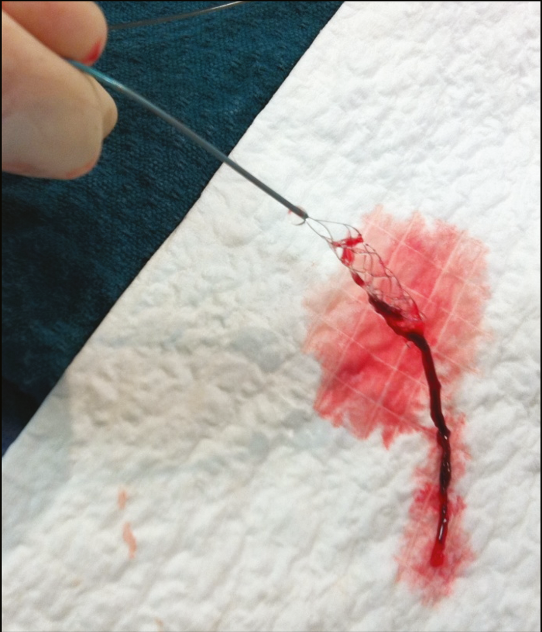
The concept of endovascular treatment of stroke via angiography preceded intravenous thrombolysis but, despite a positive trial in 1999,38 it did not enter mainstream practice. Initially, treatment was provided with intra‐arterial thrombolytics, but mechanical clot retrieval devices appeared in the first decade of the 21st century. Three neutral randomised trials published in 201339,40,41 dampened the enthusiasm but also reset clinician equipoise and facilitated recruitment into the next generation of trials. A new generation of devices was developed: retrievable stents that are deployed within the clot and then retrieved under negative pressure aspiration (Box 3). In 2015, five positive trials of endovascular thrombectomy using predominantly stent‐retriever devices essentially doubled the rate of successful reperfusion compared with previous generation devices, and also incorporated a variety of important imaging selection and streamlined workflow approaches to reduce treatment delays.9,10,11,12,13
A subsequent individual patient data meta‐analysis has demonstrated remarkable consistency of treatment benefit across the spectrum of age, clinical severity, and imaging characteristics of participants.17 As in most conditions, increasing age and more severe clinical presentations were associated with worse outcomes. However, older and severely affected patients benefitted from endovascular thrombectomy compared with medical therapy, with effect sizes similar to other subgroups. There were trends to greater mortality reduction in older patients. Using general anaesthesia was associated with worse outcomes than performing the procedure with the patient awake,42 in contrast to single centre randomised trials that showed little difference.43,44,45 However, the randomised trials were performed by specialist neuroanaesthetic teams with strict protocols to maintain physiological parameters and minimal delay (approximately 9 minutes) to achieve general anaesthesia. These practices are not routine in most centres.
Brain imaging is key to stroke diagnosis but also provides important prognostic information regarding the extent of irreversible injury and tissue at risk. Individual patient data meta‐analyses of centrally reviewed imaging data from all the endovascular thrombectomy trials have examined the effect of non‐contrast CT hypodensity, collateral blood flow quality,46 CT perfusion and MRI47 parameters on prognosis and benefit from endovascular thrombectomy versus medical therapy. Even patients with extensive injury on pre‐treatment imaging can still benefit from thrombectomy. Further randomised trials are specifically addressing the treatment benefit of endovascular thrombectomy in patients with a large irreversibly injured ischaemic core, but at present there is no reason to exclude a patient with large vessel occlusion and onset within 6 hours from endovascular therapy based purely on imaging appearances.
Time to reperfusion remains a crucial determinant of outcome, with every 4‐minute delay after reaching the emergency department associated with one in 100 patients having a worse outcome.48 Despite this time‐sensitivity, individual patients can have prolonged survival of salvageable brain due to good collateral blood flow. These patients can be identified using CT perfusion or perfusion‐diffusion MRI (Box 4). CT perfusion dynamically tracks the passage of an intravenous bolus of contrast, identifying regions of the brain with delayed contrast arrival due to arterial occlusion with retrograde filling via collateral pathways. Significant delay indicates tissue at risk of infarction, and regions with severely reduced flow are likely to already be irreversibly injured. The use of automated processing software to apply delay and blood flow thresholds to CT perfusion allows reasonably accurate determination of tissue viability in individual patients.
The 2018 DAWN14 and DEFUSE 315 randomised controlled trials showed large absolute benefits of thrombectomy in patients up to 24 hours after the time they were last seen well if they had favourable advanced brain imaging. Recovery to independence occurred in 47% of patients with thrombectomy compared with 15% of controls, and there was significant improvement across the spectrum of disability14,15 Having broader, simpler eligibility criteria, essentially requiring < 70 mL irreversible injured brain, DEFUSE 3 allowed inclusion of additional patients, who experienced comparable treatment benefit.15 This has led to a major expansion in access to endovascular thrombectomy for patients with wake‐up onset or delayed presentation, including rural patients with stroke. However, the proportion of patients with favourable imaging declines rapidly, so the imperative to treat as fast as possible remains.
Risks
The risks of endovascular thrombectomy are relatively low when performed by expert neurointerventionalists. Arterial injury or perforation occurs in less than 2% of patients.42 Symptomatic intracerebral haemorrhage occurred in 4.3% of patients in the HERMES meta‐analysis, similar to the control group.17 Intravenous thrombolysis was administered to all eligible patients in the trials. Ongoing studies are testing whether intravenous thrombolysis is beneficial in patients who have immediate access to on‐site thrombectomy. However, available observational data suggest improved recanalisation and outcomes in patients pre‐treated with thrombolysis, without increased risk of symptomatic intracerebral haemorrhage.49 Currently, most Australian patients with stroke do not present directly to an endovascular‐capable hospital; therefore, “bridging” thrombolysis preceding thrombectomy remains standard — analogous to the pharmaco‐invasive strategy for ST‐elevation myocardial infarction when a delay of more than 90 minutes from first medical contact to reperfusion is expected.50 Stroke thrombectomy has also not yet achieved the over 95% success rate of percutaneous coronary intervention for ST‐elevation myocardial infarction, with about 75% of patients achieving more than 50% reperfusion, but only 32% of patients achieving more than 90% reperfusion in the positive trials.17 Therefore, intravenous thrombolysis may provide some benefit for patients in whom thrombectomy fails to fully reperfuse the brain.
Systems of care
The evidence from randomised trials has indicated a strong time dependence of reperfusion therapies. It therefore follows that the greatest value for money in maximising benefits for patients comes from delivering these proven therapies faster and to a larger proportion of appropriate patients.
Ambulance assessment and pre‐notification
Ambulance paramedics should use validated stroke screening tools to identify suspected stroke.21 Patients within potential treatment time frames (now 24 hours) should be transported to the nearest stroke‐capable hospital and pre‐notified to the receiving emergency department so that the stroke team can prepare in advance of the patient's arrival and clear the CT scanner. Pre‐notification should include clinical details, name and date of birth to allow record searching and pre‐ordering of the CT scan. On arrival, after a brief check to ensure the patient is stable from a cardiorespiratory viewpoint, the patient should be transported directly to the CT scanner for imaging, as this is the rate‐limiting step in determining treatment. Clinical examination can occur en route to the scanner. These simple workflow changes, rather than taking the patient to an emergency cubicle, offloading from the ambulance stretcher and assessing the patient extensively before the CT scan, save significant time and translate to improved patient outcomes.51,52
With the advent of endovascular thrombectomy and the potential for treatment beyond 6 hours, routine acquisition of CT perfusion and CT angiography of the aortic arch to the cerebral vertex to identify a therapeutic target is advisable, in addition to non‐contrast CT brain scan (Box 4).53
Pre‐hospital triage
As in‐hospital workflow improves, the bulk of the onset to treatment time for patients with stroke now elapses pre‐hospital. Community awareness is a prerequisite and, ideally, everyone would know the “facial droop, arm weakness, speech disturbance, time to call 000” (FAST) stroke recognition message and dial “000” immediately. The standard pre‐hospital approach to patients with suspected stroke is to transport them directly to the nearest thrombolysis‐equipped stroke unit if that is possible within a reasonable time frame (often < 60‐minute drive time). States such as Victoria are well served with a network of such hospitals linked by telemedicine, and 99% of the population are within a 60‐minute drive time of a stroke centre. However, other regions face greater geographical challenges. Even in metropolitan areas, endovascular thrombectomy is only available at selected centres. If a patient with large vessel occlusion first presents to a primary (thrombolysis‐only) stroke centre, the process of assessment and transfer to a comprehensive (endovascular‐capable) stroke centre introduces a delay of about 2 hours, which has major consequences for patient outcomes.54 For this reason, various clinical triage tools have been introduced to assist paramedics in identifying likely large vessel occlusion stroke so that they can bypass directly to an endovascular‐capable hospital. In Western Australia, the Rapid Arterial Occlusion Evaluation (RACE) scale (developed in Catalonia, Spain)55 has been successfully implemented. In Victoria, a simpler algorithmic approach called Ambulance Clinical Triage for Acute Stroke Treatment (ACT‐FAST) is currently being trialled (Box 5).56
Mobile stroke units
The high technology solution to reducing treatment times is to bring the traditionally hospital‐based elements of care to the pre‐hospital environment. Mobile stroke units are ambulances equipped with CT scanners and clinicians specialised in stroke.57 The CT brain scan allows definitive diagnosis of intracerebral haemorrhage versus ischaemic stroke, permitting pre‐hospital thrombolysis. CT angiography allows definitive diagnosis of large vessel occlusion requiring thrombectomy, permitting bypass directly to an endovascular‐capable hospital. Blood pressure management, reversal of anticoagulants and treatment of seizures are also possible. Pre‐hospital thrombolysis and triage to a thrombectomy‐capable hospital cuts treatment delays substantially, and trials to evaluate the clinical effectiveness of such interventions are ongoing.
Intracerebral haemorrhage
About 15% of strokes in Australia are due to intracerebral haemorrhage but these contribute to 50% of the mortality from stroke.58 In other regions (eg, western China) intracerebral haemorrhage causes up to 40% of all stroke cases.59 Stroke unit care benefits all stroke subtypes, including intracerebral haemorrhage. The Australian‐led INTERACT‐2 randomised controlled trial showed a modest 3% absolute reduction in death and disability, with no safety concerns when the systolic blood pressure was lowered to 140 mmHg.19 The guidelines recommend lowering blood pressure to this level but not substantially below,21 as results of the more intensive ATACH‐2 randomised controlled trial did not show benefit of targeting a systolic blood pressure of 120 mmHg, which was associated with increased renal adverse events.60 Ongoing trials of haemostatic agents (eg, tranexamic acid, recombinant activated factor VII) and minimally invasive surgery provide hope for future effective treatments of this major cause of disability.
Cytoprotection and neurorecovery
As yet, there are no proven strategies to protect the brain from the effects of stroke other than reperfusion. Multiple trials of neuroprotective agents have been unsuccessful. However, with the advent of effective reperfusion with endovascular thrombectomy, human stroke treatment more closely resembles the temporary middle cerebral artery occlusion model used in animal studies, and a new wave of trials are underway to re‐examine whether cytoprotection is feasible in humans.
Secondary prevention
There is a strong evidence base for secondary stroke prevention, which relies on accurate determination of stroke mechanism. The guidelines now recommend the imaging of the vessels from aortic arch to cerebral vertex with CT angiography as the preferred vascular imaging approach to identify atherosclerosis, dissection and other arterial lesions.21 Cardiac investigations search for a cardiac source of embolism. In particular, prolonged monitoring for paroxysmal atrial fibrillation is increasingly used, as the traditional 24‐hour Holter monitoring has very limited sensitivity for this high risk cause of stroke that warrants anticoagulation. Implantable loop recorders can provide 3 years of continuous cardiac monitoring with an atrial fibrillation yield of 30% in patients with stroke without clear cause for their stroke, compared with 3% using standard clinical surveillance.61 Consumer electronics have increasingly sophisticated electrocardiogram monitoring capabilities and may play an important role in atrial fibrillation detection.
High blood pressure is a risk factor for cardiovascular disease in general, but particularly for intracerebral haemorrhage and ischaemic stroke. Most patients who have had a stroke should take blood pressure‐lowering medication unless contraindicated by symptomatic hypotension.21 The SPRINT trial, which showed that a systolic blood pressure target of 120 mmHg improved outcome versus a 140 mmHg target, specifically excluded patients with stroke, but did reduce the incidence of stroke.62 Post hoc analysis of the PROGRESS trial of perindopril–indapamide for secondary stroke prevention also suggested the benefit of lowering the blood pressure, regardless of the baseline blood pressure.63
Cholesterol‐lowering also reduces the risk of ischaemic stroke. Atorvastatin 80 mg showed significant benefits64 and current guidelines recommend the use of a high dose, high potency statin.21 The role of proprotein convertase subtilisin/kexin type 9 (PCSK9) inhibitors is evolving but may certainly benefit patients with stroke, particularly those who are intolerant of statins.
Antithrombotic therapy is essential after ischaemic stroke. Recent publications have re‐emphasised the value of early aspirin commencement.65 Clopidogrel or aspirin–dipyridamole provide slightly greater efficacy than aspirin alone.21 Combined aspirin and clopidogrel for about 3 weeks reduces the risk of recurrent stroke,66,67 but longer term dual antiplatelet therapy is not used in stroke prevention due to increased bleeding and minimal benefit.21
For patients with atrial fibrillation, aspirin has no role in stroke prevention.21 Anticoagulation for atrial fibrillation remains underutilised in Australia and globally. The guidelines recommend first‐line direct oral anticoagulants for non‐valvular atrial fibrillation (ie, no mechanical prosthetic heart valve or moderate to severe mitral stenosis), provided the creatinine clearance rate is adequate (> 30 mL/min for dabigatran and rivaroxaban; > 25 mL/min for apixaban using the Cockcroft–Gault formula).21 Warfarin (target international normalised ratio, 2.0–3.0) is still initiated in patients who are unable to take direct oral anticoagulants. Common problems with anticoagulation management leading to preventable strokes are the overestimation of the risk of bleeding (eg, falls) relative to the risk of stroke, leading to underutilisation of anticoagulation, and the use of lower doses than recommended in the applicable product information. For patients with genuine contraindications to anticoagulation, left atrial appendage occlusion is emerging as a relevant treatment option.68 Anticoagulation in suspected embolic stroke without proving the diagnosis of atrial fibrillation has not been supported by recent randomised trials.69
Patent foramen ovale has been a controversial potential cause of stroke. However, new trials published in 2017 using improved patient selection to thoroughly exclude alternative stroke mechanisms have demonstrated the unequivocal benefit of percutaneous device closure of patent foramen ovale in highly selected patients.70,71,72 The absolute benefit is about 1% per annum. However, unlike front‐loaded atherosclerotic risk that decreases over time, risk of stroke related to patent foramen ovale appears to accumulate at a constant rate, implying a major lifetime risk reduction for young patients.
Conclusion
Stroke medicine is an exciting field with major advances in recent years. In particular, reperfusion therapies for ischaemic stroke have transformed the prognosis for long term disability. However, optimal implementation to maximise patient benefit requires increased community recognition, immediate “000” activation, faster emergency treatment systems and innovative therapeutic developments to further improve outcomes.
Box 1 – Ischaemic stroke pathophysiology*
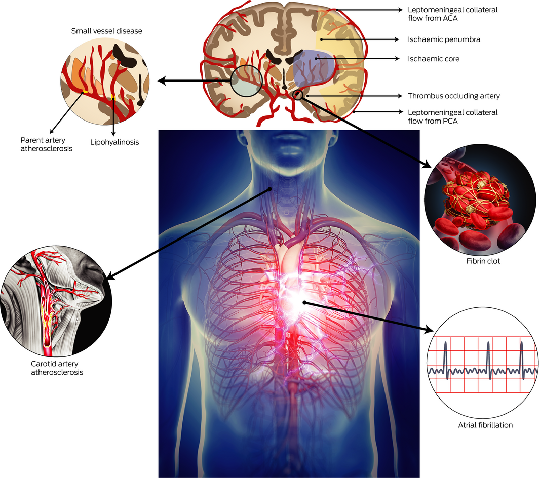
ACA = anterior cerebral artery. PCA = posterior cerebral artery. * Most ischaemic stroke is thromboembolic: a thrombus forms in the heart or large arteries and then migrates to the intracranial circulation. When this occurs, symptoms develop immediately as neurons lose the oxygen and glucose required for electrical function. However, collateral vessels are often able to sustain metabolic viability in the affected brain for some time. This hibernating but salvageable brain is termed the ischaemic penumbra and is the reason that rapid reperfusion can dramatically alter the natural history of an otherwise disabling stroke. Collateral circulation is highly variable between individuals and the genetic and environmental determinants are poorly understood. Over time, collateral circulation tends to fail leading to growth in the irreversibly injured ischaemic core. The patient's clinical deficit generally remains stable but it has now become irreversible. One of the important insights from recent endovascular reperfusion trials in patients 6–24 hours after stroke onset is that good collaterals and ischaemic penumbra can persist for many hours in a subgroup of patients. However, despite this initial resilience, without treatment the outcome is very poor with recovery to independence in about 15% of patients. ◆
Box 2 – Evidence‐based acute therapies for stroke
|
Intervention |
Indication |
Outcome* |
Relative risk |
NNT |
|||||||||||
|
|
|||||||||||||||
|
All stroke types and severities |
|||||||||||||||
|
Stroke unit care2 |
All admitted patients with stroke |
Death/dependency |
|
0.94 (0.88–0.99) |
28 |
||||||||||
|
Ischaemic stroke |
|||||||||||||||
|
IV alteplase 0‐4.5 h16 (thrombolysis) |
Potentially disabling stroke without excessive bleeding risk |
Return to all usual activities (mRS, 0–1) |
Treatment within 0–3 h |
1.42 (1.21–1.68) |
10 |
||||||||||
|
|
|
|
Treatment within 3–4.5 h |
1.17 (1.05–1.31) |
19 |
||||||||||
|
Endovascular thrombectomy 0‐6 h17 |
Large vessel occlusion (internal carotid or proximal middle cerebral artery)† |
Death/dependency |
|
0.74 (0.67–0.80) |
5 |
||||||||||
|
Large vessel occlusion and favourable CT perfusion/MRI (core < 70 mL) |
Death/dependency |
|
0.62 (0.53–0.71) |
3 |
|||||||||||
|
All ischaemic stroke (unless atrial fibrillation or allergy) |
Recurrent stroke/death |
|
0.90 (0.84–0.96) |
109 |
|||||||||||
|
Hemicraniectomy 0‐48 h5 |
Malignant MCA infarction (mass effect, reduced conscious state), age < 60 years |
Death/severe disability‡ |
|
0.72 (0.54–0.96) |
4.6 |
||||||||||
|
Intracerebral haemorrhage |
|||||||||||||||
|
BP‐lowering in ICH (target 140 mmHg)19 |
BP > 150 mmHg (maintain at 140 mmHg for the first 7 days) |
Death/dependency |
|
0.87 (0.75–1.01) |
28 |
||||||||||
|
|
|
Disability severity§ |
|
0.87 (0.77–1.00) |
na |
||||||||||
|
|
|||||||||||||||
|
BP = blood pressure. CT = computed tomography. ICH = intracerebral haemorrhage. IV = intravenous. MCA = middle cerebral artery. MRI = magnetic resonance imaging. mRS = modified Rankin Scale. na = not applicable. NNT = number needed to treat. * Outcomes and NNT assessed at 3 months, except hemicraniectomy (12 months). † Basilar artery occlusion excluded from these trials. ‡ mRS 4–6: mRS 6 = death, mRS 5 = nursing home, mRS 4 = need for assistance with personal activities of daily living. § Change by at least one level on the mRS. ◆ |
|||||||||||||||
Box 4 – Imaging in a patient* who underwent endovascular thrombectomy†
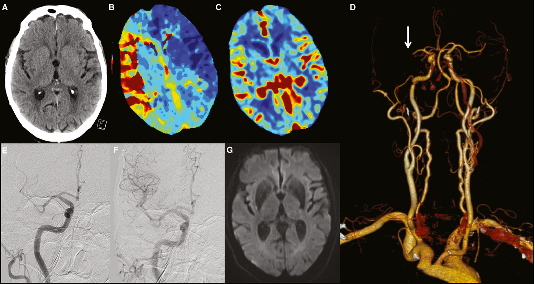
* An older patient living independently at home presented one hour after onset of left hemiparesis, dysarthria, hemianopia and inattention. † Multimodal computed tomography (CT) including (A) non‐contrast scan to exclude haemorrhage and established infarction, CT perfusion confirming diagnosis of ischaemic stroke with (B) delayed blood flow arrival in right middle cerebral artery territory (ie, territory at risk), and (C) preserved cerebral blood volume suggesting salvageable tissue. (D) CT angiography for confirmation of right middle cerebral artery occlusion (arrow) and thrombectomy planning. (E) Pre‐thrombectomy and (F) post‐thrombectomy angiography showing complete reperfusion 2 hours after onset. (G) Follow‐up diffusion magnetic resonance imaging scan showing successful tissue salvage. The patient was discharged directly home on Day 3. ◆
Box 5 – Pre‐hospital identification of suspected large vessel occlusion
|
RACE* score |
Points |
ACT‐FAST* algorithm |
|||||||||||||
|
|
|||||||||||||||
|
Facial droop |
|
Step 1: arm drift to stretcher within 10 seconds
|
|||||||||||||
|
Absent |
0 |
||||||||||||||
|
Mild |
1 |
||||||||||||||
|
Severe |
2 |
||||||||||||||
|
Arm drift |
|
Step 2:
|
|||||||||||||
|
Absent/mild |
0 |
||||||||||||||
|
Drifts to stretcher |
1 |
||||||||||||||
|
Cannot get antigravity |
2 |
||||||||||||||
|
Head and gaze deviation |
|
||||||||||||||
|
Absent |
0 |
||||||||||||||
|
Present |
1 |
||||||||||||||
|
Aphasia (if right arm weak)† |
|
||||||||||||||
|
Both tasks performed correctly |
0 |
||||||||||||||
|
One error |
1 |
||||||||||||||
|
Two errors |
2 |
||||||||||||||
|
Agnosia (if left arm weak)‡ |
|
Step 3: eligibility and mimic exclusion (all criteria needs to be fulfilled)
|
|||||||||||||
|
Both tasks performed correctly |
0 |
||||||||||||||
|
One error |
1 |
||||||||||||||
|
Two errors |
2 |
||||||||||||||
|
RACE score ≥ 5 indicates high probability of large vessel occlusion |
|
All 3 ACT‐FAST steps positive indicates high probability of large vessel occlusion |
|||||||||||||
|
|
|||||||||||||||
|
ACT‐FAST = Ambulance Clinical Triage for Acute Stroke Treatment. RACE = Rapid Arterial Occlusion Evaluation. * Both scales test arm drift by positioning both arms at 90 degrees and observing for unilateral drift over 10 seconds. † RACE tests aphasia by asking the patient “close your eyes” and “make a fist” and evaluating if the patient obeys. ‡ RACE tests agnosia by asking the patient, while showing them the paretic arm, “whose arm is this” and evaluating if the patient recognises their own arm; and by asking the patient “can you lift both arms and clap” and evaluating if the patient recognises their functional impairment. ◆ |
|||||||||||||||
Competing interests
Bruce Campbell has received research support from the National Health and Medical Research Council (GNT1043242, GNT1035688), the Royal Australasian College of Physicians, the Royal Melbourne Hospital Foundation, the National Heart Foundation and the Stroke Foundation. He has received unrestricted grant funding for the EXTEND‐IA trial to the Florey Institute of Neuroscience and Mental Health from Medtronic. He co‐chaired the 2017 Australian Stroke Guidelines content working party.
References
- Deloittte Access Economics. The economic impact of stroke in Australia. Deloittte Access Economics, 2013. https://strokefoundation.org.au/What-we-do/Research/Economic-impact-of-stroke-in-Australia (viewed Aug 2018).
- Stroke Unit Trialists’ Collaboration. Collaborative systematic review of the randomised trials of organised inpatient (stroke unit) care after stroke. BMJ 1997; 314: 1151–1159.
- CAST (Chinese Acute Stroke Trial) Collaborative Group. CAST: randomised placebo‐controlled trial of early aspirin use in 20,000 patients with acute ischaemic stroke. Lancet 1997; 349: 1641–1649.
- International Stroke Trial Collaborative Group. The International Stroke Trial (IST): a randomised trial of aspirin, subcutaneous heparin, both, or neither among 19435 patients with acute ischaemic stroke. Lancet 1997; 349: 1569–1581.
- Vahedi K, Hofmeijer J, Juettler E, et al. Early decompressive surgery in malignant infarction of the middle cerebral artery: a pooled analysis of three randomised controlled trials. Lancet Neurol 2007; 6: 215–222.
- The National Institute of Neurological Disorders and Stroke rt‐PA Stroke Study Group. Tissue plasminogen activator for acute ischemic stroke. N Engl J Med 1995; 333: 1581–1587.
- Thomalla G, Simonsen CZ, Boutitie F, et al. MRI‐guided thrombolysis for stroke with unknown time of onset. N Engl J Med 2018; 379: 611–622.
- Ma H, Campbell BCV, Parsons MW, et al. Thrombolysis up to 9 hours after stroke onset guided by perfusion imaging. N Engl J Med 2019. In press.
- Berkhemer OA, Fransen PS, Beumer D, et al. A randomized trial of intraarterial treatment for acute ischemic stroke. N Engl J Med 2015; 372: 11–20.
- Campbell BC, Mitchell PJ, Kleinig TJ, et al. Endovascular therapy for ischemic stroke with perfusion‐imaging selection. N Engl J Med 2015; 372: 1009–1018.
- Goyal M, Demchuk AM, Menon BK, et al. Randomized assessment of rapid endovascular treatment of ischemic stroke. N Engl J Med 2015; 372: 1019–1030.
- Saver JL, Goyal M, Bonafe A, et al. Stent‐retriever thrombectomy after intravenous t‐PA vs. t‐PA alone in stroke. N Engl J Med 2015; 372: 2285–2295.
- Jovin TG, Chamorro A, Cobo E, et al. Thrombectomy within 8 hours after symptom onset in ischemic stroke. N Engl J Med 2015; 372: 2296–2306.
- Nogueira RG, Jadhav AP, Haussen DC, et al. Thrombectomy 6 to 24 hours after stroke with a mismatch between deficit and infarct. N Engl J Med 2018; 378: 11–21.
- Albers GW, Marks MP, Kemp S, et al. Thrombectomy for stroke at 6 to 16 hours with selection by perfusion imaging. N Engl J Med 2018; 378: 708–718.
- Emberson J, Lees KR, Lyden P, et al. Effect of treatment delay, age, and stroke severity on the effects of intravenous thrombolysis with alteplase for acute ischaemic stroke: a meta‐analysis of individual patient data from randomised trials. Lancet 2014; 384: 1929–1935.
- Goyal M, Menon BK, van Zwam WH, et al. Endovascular thrombectomy after large‐vessel ischaemic stroke: a meta‐analysis of individual patient data from five randomised trials. Lancet 2016; 387: 1723–1731.
- Chen ZM, Sandercock P, Pan HC, et al. Indications for early aspirin use in acute ischemic stroke: a combined analysis of 40 000 randomized patients from the Chinese acute stroke trial and the international stroke trial. On behalf of the CAST and IST collaborative groups. Stroke 2000; 31: 1240–1249.
- Anderson CS, Heeley E, Huang Y, et al. Rapid blood‐pressure lowering in patients with acute intracerebral hemorrhage. N Engl J Med 2013; 368: 2355–2365.
- Hacke W, Kaste M, Bluhmki E, et al. Thrombolysis with alteplase 3 to 4.5 hours after acute ischemic stroke. N Engl J Med 2008; 359: 1317–1329.
- Stroke Foundation. Clinical guidelines for stroke management 2017 [website]. https://informme.org.au/Guidelines (viewed Aug 2018).
- Powers WJ, Rabinstein AA, Ackerson T, et al. 2018 Guidelines for the early management of patients with acute ischemic stroke: a guideline for healthcare professionals from the American Heart Association/American Stroke Association. Stroke 2018; 49: e46–e110.
- Casaubon LK, Boulanger JM, Blacquiere D, et al. Canadian stroke best practice recommendations: hyperacute stroke care guidelines, update 2015. Int J Stroke 2015; 10: 924–940.
- Ringleb P, Schellinger PD, Hacke W, Europaischen S. European Stroke Organisation 2008 guidelines for managing acute cerebral infarction or transient ischemic attack. Part 1 [German]. Nervenarzt 2008; 79: 936–957.
- Stroke Foundation. National stroke audit acute services 2017 [website]. https://informme.org.au/stroke-data/Acute-audits (viewed Aug 2018).
- Lees KR, Bluhmki E, von Kummer R, et al. Time to treatment with intravenous alteplase and outcome in stroke: an updated pooled analysis of ECASS, ATLANTIS, NINDS, and EPITHET trials. Lancet 2010; 375: 1695–1703.
- Wahlgren N, Ahmed N, Davalos A, et al. Thrombolysis with alteplase for acute ischaemic stroke in the Safe Implementation of Thrombolysis in Stroke‐Monitoring Study (SITS‐MOST): an observational study. Lancet 2007; 369: 275–282.
- Myslimi F, Caparros F, Dequatre‐Ponchelle N, et al. Orolingual angioedema during or after thrombolysis for cerebral ischemia. Stroke 2016; 47: 1825–1830.
- Cheong E, Dodd L, Smith W, Kleinig T. Icatibant as a potential treatment of life‐threatening alteplase‐induced angioedema. J Stroke Cerebrovasc Dis 2018; 27: e36–e37.
- Smith EE, Abdullah AR, Petkovska I, et al. Poor outcomes in patients who do not receive intravenous tissue plasminogen activator because of mild or improving ischemic stroke. Stroke 2005; 36: 2497–2499.
- Khatri P, Kleindorfer DO, Devlin T, et al. Effect of alteplase vs aspirin on functional outcome for patients with acute ischemic stroke and minor nondisabling neurologic deficits: the PRISMS randomized clinical trial. JAMA 2018; 320: 156–166.
- Coutts SB, Modi J, Patel SK, et al. CT/CT angiography and MRI findings predict recurrent stroke after transient ischemic attack and minor stroke: results of the prospective CATCH study. Stroke 2012; 43: 1013–1017.
- Parsons M, Spratt N, Bivard A, et al. A randomized trial of tenecteplase versus alteplase for acute ischemic stroke. N Engl J Med 2012; 366: 1099–1107.
- Campbell BCV, Mitchell PJ, Churilov L, et al. Tenecteplase versus alteplase before thrombectomy for ischemic stroke. N Engl J Med 2018; 378: 1573–1582.
- Huang X, Cheripelli BK, Lloyd SM, et al. Alteplase versus tenecteplase for thrombolysis after ischaemic stroke (ATTEST): a phase 2, randomised, open‐label, blinded endpoint study. Lancet Neurol 2015; 14: 368–376.
- Logallo N, Novotny V, Assmus J, et al. Tenecteplase versus alteplase for management of acute ischaemic stroke (NOR‐TEST): a phase 3, randomised, open‐label, blinded endpoint trial. Lancet Neurol 2017; 16: 781–788.
- Mair G, von Kummer R, Adami A, et al. Arterial obstruction on computed tomographic or magnetic resonance angiography and response to intravenous thrombolytics in ischemic stroke. Stroke 2017; 48: 353–360.
- Furlan A, Higashida R, Wechsler L, et al. Intra‐arterial prourokinase for acute ischemic stroke. The PROACT II study: a randomized controlled trial. Prolyse in Acute Cerebral Thromboembolism. JAMA 1999; 282: 2003–2011.
- Broderick JP, Palesch YY, Demchuk AM, et al. Endovascular therapy after intravenous t‐PA versus t‐PA alone for stroke. N Engl J Med 2013; 368: 893–903.
- Ciccone A, Valvassori L, Nichelatti M, et al. Endovascular treatment for acute ischemic stroke. N Engl J Med 2013; 368: 904–913.
- Kidwell CS, Jahan R, Gornbein J, et al. A trial of imaging selection and endovascular treatment for ischemic stroke. N Engl J Med 2013; 368: 914–923.
- Campbell BCV, van Zwam WH, Goyal M, et al. Effect of general anaesthesia on functional outcome in patients with anterior circulation ischaemic stroke having endovascular thrombectomy versus standard care: a meta‐analysis of individual patient data. Lancet Neurol 2018; 17: 47–53.
- Lowhagen Henden P, Rentzos A, Karlsson JE, et al. General anesthesia versus conscious sedation for endovascular treatment of acute ischemic stroke: the AnStroke trial (Anesthesia During Stroke). Stroke 2017; 48: 1601–1607.
- Schonenberger S, Uhlmann L, Hacke W, et al. Effect of conscious sedation vs general anesthesia on early neurological improvement among patients with ischemic stroke undergoing endovascular thrombectomy: a randomized clinical trial. JAMA 2016; 316: 1986–1996.
- Simonsen CZ, Yoo AJ, Sorensen LH, et al. Effect of general anesthesia and conscious sedation during endovascular therapy on infarct growth and clinical outcomes in acute ischemic stroke: a randomized clinical trial. JAMA Neurol 2018; 75: 470–477.
- Roman LS, Menon BK, Blasco J, et al. Imaging features and safety and efficacy of endovascular stroke treatment: a meta‐analysis of individual patient‐level data. Lancet Neurol 2018; 17: 895–904.
- Campbell BCV, Majoie C, Albers GW, et al. Penumbral imaging and functional outcome in patients with anterior circulation ischaemic stroke treated with endovascular thrombectomy versus medical therapy: a meta‐analysis of individual patient‐level data. Lancet Neurol 2018; 18: 46–55.
- Saver JL, Goyal M, van der Lugt A, et al. Time to treatment with endovascular thrombectomy and outcomes from ischemic stroke: a meta‐analysis. JAMA 2016; 316: 1279–1288.
- Mistry EA, Mistry AM, Nakawah MO, et al. Mechanical thrombectomy outcomes with and without intravenous thrombolysis in stroke patients: a meta‐analysis. Stroke 2017; 48: 2450–2456.
- O'Gara PT, Kushner FG, Ascheim DD, et al. 2013 ACCF/AHA guideline for the management of ST‐elevation myocardial infarction: a report of the American College of Cardiology Foundation/American Heart Association Task Force on Practice Guidelines. Circulation 2013; 127: e362–e425.
- Meretoja A, Strbian D, Mustanoja S, et al. Reducing in‐hospital delay to 20 minutes in stroke thrombolysis. Neurology 2012; 79: 306–313.
- Meretoja A, Weir L, Ugalde M, et al. Helsinki model cut stroke thrombolysis delays to 25 minutes in Melbourne in only 4 months. Neurology 2013; 81: 1071–1076.
- Campbell BCV. Stroke imaging: do it right the first time. JAMA Neurol 2017; 74: 1298–1300.
- Ng FC, Low E, Andrew E, et al. Deconstruction of interhospital transfer workflow in large vessel occlusion: real‐world data in the thrombectomy era. Stroke 2017; 48: 1976–1979.
- Perez de la Ossa N, Carrera D, Gorchs M, et al. Design and validation of a prehospital stroke scale to predict large arterial occlusion: the rapid arterial occlusion evaluation scale. Stroke 2014; 45: 87–91.
- Zhao H, Pesavento L, Coote S, et al. Ambulance clinical triage for acute stroke treatment: paramedic triage algorithm for large vessel occlusion. Stroke 2018; 49: 945–951.
- Fassbender K, Grotta JC, Walter S, et al. Mobile stroke units for prehospital thrombolysis, triage, and beyond: benefits and challenges. Lancet Neurol 2017; 16: 227–237.
- Murray CJ, Vos T, Lozano R, et al. Disability‐adjusted life years (DALYs) for 291 diseases and injuries in 21 regions, 1990‐2010: a systematic analysis for the Global Burden of Disease Study 2010. Lancet 2012; 380: 2197–2223.
- Zhang LF, Yang J, Hong Z, Yuan GG, Zhou BF, Zhao LC, Huang YN, Chen J, Wu YF; Collaborative Group of China Multicenter Study of Cardiovascular Epidemiology. Proportion of different subtypes of stroke in China. Stroke 2003; 34: 2091–2096.
- Qureshi AI, Palesch YY, Barsan WG, et al. Intensive blood‐pressure lowering in patients with acute cerebral hemorrhage. N Engl J Med 2016; 375: 1033–1043.
- Sanna T, Diener HC, Passman RS, et al. Cryptogenic stroke and underlying atrial fibrillation. N Engl J Med 2014; 370: 2478–2486.
- Group SR, Wright JT, Jr., Williamson JD, et al. A randomized trial of intensive versus standard blood‐pressure control. N Engl J Med 2015; 373: 2103–2116.
- Arima H, Chalmers J, Woodward M, et al. Lower target blood pressures are safe and effective for the prevention of recurrent stroke: the PROGRESS trial. J Hypertens 2006; 24: 1201–1208.
- Amarenco P, Bogousslavsky J, Callahan A, et al. High‐dose atorvastatin after stroke or transient ischemic attack. N Engl J Med 2006; 355: 549–559.
- Rothwell PM, Algra A, Chen Z, et al. Effects of aspirin on risk and severity of early recurrent stroke after transient ischaemic attack and ischaemic stroke: time‐course analysis of randomised trials. Lancet 2016; 388: 365–375.
- Wang Y, Zhao X, Liu L, et al. Clopidogrel with aspirin in acute minor stroke or transient ischemic attack. N Engl J Med 2013; 369: 11–19.
- Johnston SC, Easton JD, Farrant M, et al. Clopidogrel and aspirin in acute ischemic stroke and high‐risk TIA. N Engl J Med 2018; 379: 215–225.
- Nishimura M, Sab S, Reeves RR, Hsu JC. Percutaneous left atrial appendage occlusion in atrial fibrillation patients with a contraindication to oral anticoagulation: a focused review. Europace 2017; 20: 1412–1419.
- Hart RG, Sharma M, Mundl H, et al. Rivaroxaban for stroke prevention after embolic stroke of undetermined source. N Engl J Med 2018; 378: 2191–2201.
- Mas JL, Derumeaux G, Guillon B, et al. Patent foramen ovale closure or anticoagulation vs. antiplatelets after stroke. N Engl J Med 2017; 377: 1011–1021.
- Sondergaard L, Kasner SE, Rhodes JF, et al. Patent foramen ovale closure or antiplatelet therapy for cryptogenic stroke. N Engl J Med 2017; 377: 1033–1042.
- Saver JL, Carroll JD, Thaler DE, et al. Long‐term outcomes of patent foramen ovale closure or medical therapy after stroke. N Engl J Med 2017; 377: 1022–1032.
Linked content
-
MJA Editorial: The power of networks
-
MJA Podcast: Professor Bruce Campbell
-
InSight+: Stroke care: “tissue clock” may replace “time clock”
Provenance: Commissioned; externally peer reviewed.
Motor Neuron Disease Mortality Trends in Australia From 1986 to 2023: A Population-Based Study
Carol M. Y. Lee, Rupendra N. Shrestha, Julian Gold, Mark Stevenson, Kelly L. Williams, Lyndal Henden, Sandrine K. K. Chan Moi Fat, Zoe Zussa, Amanda Wright, Martin Jude, Mark A. S. Laidlaw, Willem J. R. Fokkink, Dominic B. Rowe
Mild traumatic brain injury and concussion and persisting post‐concussion symptoms: new guidelines to support evidence‐based assessment and management in Australia and Aotearoa New Zealand
Karen M Barlow, Jennie L Ponsford, Alice Theodom, Gill Cowen, Gavin A Davis, Vicki Anderson, Franz E Babl, David Cole, Jennifer Cullen, Stuart R Dalziel, Melinda Fitzgerald, Howard Flavell, Caroline Yates, Rebecca Kimble, John H Olver, Rhonda Orr, Mark Ralfe, Michael Rose, Nick Rushworth, Julia Treleaven, Gary Browne, Nathan Delang, Sarah Harris, Gary Mitchell, Sean Tweedy
The prevalence of and variation in indicators of the quality and safety of long term aged care in Australia, 2019: a cross‐sectional population‐based study
Tesfahun C Eshetie, Gillian E Caughey, Catherine Lang, Olivia Ryan, Renuka Visvanathan, Craig Whitehead, Keith Evans, Janet K Sluggett, Jyoti Khadka, Carolyn Dawkins, Helena Williams, Miranda Starke, Sara Blunt, Anne Liddell, Megan Corlis, Anna Sheppeard, Penelope Lello, Marilyn Thien, Steven L Wesselingh, Maria C Inacio
Health and economic benefits of improving pre‐hospital identification of stroke in Australian women: a modelling study
Thomas Gadsden, Lei Si, Emily R Atkins, Cheryl Carcel, Xia Wang, Stephen Jan, Mark Woodward, Laura E Downey
Symptom burden, quality of life, and diagnostic journey of people with postural orthostatic tachycardia syndrome, Australia, 2021–24: a descriptive patient registry data study
Marie‐Claire Seeley, Gemma Wilson, Eric Ong, Amy Langdon, Jonathan Chieng, Danielle Bailey, Kristina Comacchio, Amanda Page, Dennis H Lau, Celine Gallagher
The intersection of rurality and dementia prevalence in Australia for Aboriginal and Torres Strait Islander and non‐Indigenous peoples
Antonia J Clarke, Marwan Ahmed, Judith M Katzenellenbogen, John Towney (Wiradjuri), Anna H Balabanski, Adrienne Withall (Dharawal Yuin), Kylie Radford, Amy Brodtmann