




Volume 210 - Issue 8
Biology and therapy of multiple myeloma
Authors: Douglas E Joshua, Christian Bryant, Caroline Dix, John Gibson and Joy Ho
Med J Aust 2019; 210 (8): 375-380. || doi: 10.5694/mja2.50129
Published online: 6 May 2019
Published online: 6 May 2019
Due to the remarkable improvements in the diagnosis and management of myeloma over the past decade, we can look forward to regimens that will allow the eradication of residual disease and result in the cure of myeloma
Summary
- Genetic sequencing of the myeloma genome has not revealed a specific disease‐determining genetic alteration.
- Multiple disease subclones exist at diagnosis and vary in clinical importance with time and drug sensitivity.
- New diagnostic criteria have identified indications for early introduction of therapy.
- Autologous stem cell transplantation remains an essential component of therapy in young and fit patients.
- The use of continual suppressive (maintenance) therapy has been established as an important component in therapy.
- Immune therapies and the harnessing of the innate immune system offer great promise for future treatments.
- Since 2005, quality of life, supportive therapies, and survival have dramatically improved over a decade of remarkable progress.
- The common manifestations of multiple myeloma, such as bone pain, fatigue and weight loss, may be non‐specific and are often initially ignored or missed by patients and medical practitioners.
Multiple myeloma is a malignancy of plasma cells originating from the bone marrow; it is a clonal plasma cell disorder that produces excess monoclonal immunoglobulin. The disease most commonly presents with hypercalcaemia, renal failure, anaemia and bone lesions (CRAB features) (Box 1).1 Myeloma accounts for about 10% of all haematological malignancies. The annual incidence of myeloma in Australia is about five cases per 100 000 population, and there are about 1200 new patients diagnosed each year; the median age of diagnosis is the mid‐60s.4 There is variation in incidence among the different ethnic groups, with myeloma being twice as common in African Americans than in white people and less common in Asians.1,5 Myeloma is preceded by an indolent phase termed monoclonal gammopathy of undetermined significance (MGUS), which is defined by the presence of a monoclonal protein (< 3 g/dL) without any end organ damage or features of myeloma. The cause of MGUS is currently unknown, but this disorder can evolve into symptomatic myeloma. The risk of progression to myeloma is about 1% per year, with risk factors being a high monoclonal protein level, high percentage of plasma cells in the bone marrow, presence of IgA monoclonal protein, and an abnormal free light chain ratio.6 The prevalence of MGUS increases with age, with 3.2% of cases presenting in persons aged over 50 years, and 5.3% of cases in persons aged over 70 years.7 From the early 1960s until the early 2000s, melphalan chemotherapy with addition of steroids (prednisone or dexamethasone) formed the basis for treating multiple myeloma. Melphalan was also used both in conditioning chemotherapy, which ablates the bone marrow before autologous stem cell transplantation, and for the treatment of patients deemed unsuitable for transplantation. There has recently been progress in the treatment of myeloma with the development of new targeted therapies, which include thalidomide, lenalidomide and bortezomib (Box 2). These newer agents have significantly changed the treatment strategies (Box 3).
In the decade following 2005,1 there have been remarkable advances in therapy, which have resulted in improved survival rates in all age groups, but especially in younger patients (aged < 65 years), with median survival rates in a real‐world situation of 7–8 years.8,9 This evolution in understanding of myeloma has led to new diagnostic criteria for myeloma and high risk smouldering multiple myeloma and to the development of concepts of continual suppressive therapy akin to protocols used in acute lymphoblastic leukaemia, with induction, consolidation and maintenance phases of treatment.
This progress has been made possible by high dose therapy and stem cell transplantation, the development of novel drugs, targeted therapies, and the ability to harness the patient's immune system. There has been a progressive evolution of novel immunomodulatory drugs from thalidomide to lenalidomide and pomalidomide, proteasome inhibitors from bortezomib to carfilzomib and ixazomib, and targeted monoclonal antibodies (elotuzumab and daratumumab) as well as the bone‐protective agents zolendronic acid and denosumab.
In addition, new immunomodulatory therapeutic endeavours using chimeric antigen receptor T cells (CAR T cells), bispecific T cell engagers and immune checkpoint inhibitors are under active investigation overseas and in Australia. In 2017, the United States granted approval to CAR T cell‐based therapies for acute lymphoblastic leukaemia and lymphoma, together with checkpoint inhibitors, which may herald their future possible use in myeloma.
In this Narrative Review, we discuss new concepts of the biology of myeloma, including the process of clonal evolution and the new criteria for introducing therapy, as proposed by the International Myeloma Working Group. We performed a search of online databases including MEDLINE, PubMed and BMJ Clinical Evidence, using the term “myeloma”, and searched recent conference proceedings from 2005 until the present.
Myeloma genomics
The sequencing of the myeloma genome in 201110 failed to identify a specific defect such as is seen in Waldenstrom macroglobulinaemia.11 Instead, a wide range of molecular abnormalities was found. In addition, all patients at diagnosis demonstrated multiple different subclones, including mutations in the driver genes KRAS, NRAS and BRAF,12,13 and whole exome sequencing revealed similar findings.14 Clones undergo varying prominence with disease progression and response to therapy. This finding has changed our concept of myeloma as a linearly progressive disease with increasing resistance to treatment to one in which a “Darwinian” or “branching” process occurs, such that some clones may be suppressed by chemotherapy but, eventually, new clones resistant to chemotherapy dominate.15,16,17
Mutations in myeloma are complex and the median missense mutational load is about 60 per patient.17 The myeloma genome has fewer mutations compared with those observed in carcinogen‐induced tumours such as melanoma and lung cancer.18 The clinical relevance of this is that it may explain the relatively poor response of myeloma to the newer immunomodulatory checkpoint inhibitors, which rely on immune recognition of cancer neoantigens by cytotoxic T cells compared with melanoma and lung cancer, in which such drugs play an important therapeutic role.19
Classification
Patients are currently classified into high risk and low risk genetic groups based on simple cytogenetic and fluorescent in situ hybridisation analysis, with implications for expected length of survival (Box 4). The Revised International Staging System (R‐ISS) risk stratification model forms the basis for this classification.22 High risk cytogenetic features include the presence of t(14;16), t(14;20) and del(17p), while patients with t(4;14) and amp(1q) have intermediate risk. Other anomalies, such as hyperdiploidy (which occurs in about 50% of patients), are considered standard risk. The term “good risk” in myeloma is still an oxymoron.20 Gene expression profiling can also identify significant prognostic groupings. While gene expression profiling may provide more detailed insight into an individual patient than standard fluorescent in situ hybridisation analysis, these analyses are complex and infrequently used in standard practice in Australia.23
Diagnosis: smouldering multiple myeloma and active myeloma
Traditional diagnostic criteria for the diagnosis and indications for the introduction of therapy in myeloma have defined active myeloma by the presence of end organ effects (Box 1). These have now been expanded by the International Myeloma Working Group from the analysis of the risk of progression in large cohorts of patients with smouldering multiple myeloma2 (Box 1). These new criteria have extended the diagnostic criteria to include the presence of free light chain abnormality (ratio of involved free light chain to non‐involved free light chain > 100 mg/L; reference interval, 0.26–1.65 mg/L), bone marrow involvement demonstrating more than 60% plasma cells and the presence of more than one lytic lesions on magnetic resonance imaging (MRI) scan, in addition to the previous standard criteria.
The new criteria have facilitated the introduction of therapeutic concepts relating to smouldering multiple myeloma, which can now be subdivided into a group with high risk smouldering multiple myeloma, with an expected transformation rate to active myeloma of over 80% in the next 2 years. This is based on a marrow infiltration of more than 10% malignant plasma cells and one of the following: paraprotein level greater than 30 g/dL or increasing paraprotein, free light chain ratio greater than 8 mg/L but less than 100 mg/L, immunoparesis of non‐involved immunoglobulins, 50–60% bone marrow plasma cells or circulating plasma cells, abnormal plasma cell phenotype, high risk genetics (Box 4), and positron emission tomography (PET) and MRI scans abnormalities.24,25
With the availability of new novel agents, the question of whether patients with high risk smouldering multiple myeloma should be treated is now legitimately being tested in controlled clinical trials. For example, the Spanish Myeloma Group found that early treatment with a combination of lenalidomide and dexamethasone, compared with standard therapy of close monitoring, resulted in a significant benefit in delaying progression to myeloma and improved overall survival without unexpected toxicity.26 The Spanish group has taken this finding further with a study that attempts to cure high risk smouldering multiple myeloma. Patients are enrolled to receive maximal therapy with the newest available agents, including carfilzomib, and ongoing lenalidomide maintenance. Treatment in this trial has shown high remission rates, with 85% of patients who completed therapy remaining progression‐free.27 However, overall survival data are still awaited but crucial to confirm the value of treatment in an asymptomatic phase. Other groups are following a similar line of treatment and used the newer monoclonal antibodies, such as daratumumab,28,29 in an attempt to show whether disease progression can be delayed and survival prolonged.
Minimal residual disease testing
Concomitant with these studies is the evolution of highly sensitive techniques for evaluating very small numbers of residual myeloma cells, termed minimal residual disease (MRD). MRD can be detected by next generation flow cytometry or by next generation sequencing with sensitivity in one to 10−6 malignant cells, allowing the definition of patients who have had an excellent response to therapy. While these techniques have important technical caveats and are highly dependent on the expertise of the operator, they are now being performed by specialised centres in the evaluation of the efficacy of novel agents in clinical trials. The attainment of MRD acts as a surrogate marker for progression‐free survival and it is hoped it will predict overall survival. Therefore, testing for patients with MRD has considerable prognostic importance. In a recent phase 3 trial comparing autologous stem cell transplantation to novel agent therapy, the finding of MRD to a level of below one and 10−6 malignant cells was associated with significant better progression‐free survival and overall survival compared with patients not achieving this level. In addition, the attainment of MRD to this level was associated with an excellent prognosis irrespective of whether the patients had an autologous stem cell transplantation or novel agents.29 Thus, the attainment of MRD offers significant prognostic formation and maybe a valuable adjunct to patient management.30,31,32 The potential use of PET scanning as an additional modality of detection of MRD continues to be investigated.33
New agents for myeloma
The advent of new and more potent proteasome inhibitors (carfilzomib) and oral proteasome inhibitors (ixazomib) together with more potent immunomodulatory drugs (pomalidomide) and the development of a new class of monoclonal antibody therapies have revolutionised the treatment of relapsed refractory disease and are now being introduced into newly diagnosed multiple myeloma (Box 2 and Box 3). Monoclonal antibodies directed against CD38 (daratumumab) and SLAMF7 (elotuzumab) have already been successful when used in combination with an immunomodulatory agent in phase 2 clinical trials34 and in large phase 3 trials.35 Additional anti‐CD38 antibodies in combination with proteasome inhibitors or immunomodulatory drugs are in phase 2 and phase 3 clinical trials.36 They offer great hope for the eradication MRD as they act independently of genetic mutations that do not affect surface phenotype.
Revolutionary T cell therapies are undergoing expedited development in myeloma and other haematological malignancies. CAR T cells, which target an activated T cell to a defined antigen present on malignant cells, are undergoing phase 1 trials in myeloma.36 In addition, the development of bispecific T cell engagers37 — which are composed of a single‐chain immunoglobulin variable component providing cancer specificity bound to a T cell receptor so as to bring the T cell and tumour in apposition to produce an immunological synapse — shows great promise. The most promising specific myeloma target is the B‐cell maturation antigen, which is universally present on plasma cells. Clinical trials of these agents are underway both internationally and in Australia.38,39 It is hoped that T cell therapies may play a most useful role in the eradication of MRD. However, of potential concern is their susceptibility to the same immune suppressive effects caused by myeloma, which allows the tumour to suppress and evade innate immunity. The prospect for the reactivation of innate natural antimyeloma immunity with vaccination is another possible avenue for eventual cure, and active immunisation protocols using hybrid fused plasma cells and dendritic cells are in progress.40
Therapy paradigms
Patients can be divided in two main groups: patients who are considered eligible for autologous stem cell transplantation and those considered ineligible. This distinction is arbitrary, but age, frailty and comorbidities are part of the clinical decision making process. Frailty scores, as proposed by the European Myeloma Network, are helpful when making this decision.41 Patients older than 75 years of age are considered, in general, ineligible for transplantation.42 Overall treatment schedules and Australian guidelines have also been published by the Medical and Scientific Advisory Group of Myeloma Australia42,43,44 and are summarised in Box 3.
Transplant‐eligible patients
Patients considered eligible for stem cell transplantation undergo an induction period with a proteasome inhibitor‐based regime (induction) followed by a stem cell transplant and maintenance therapy with thalidomide. Autologous bone marrow transplantation using high dose melphalan has been available for over 20 years and its safety and tolerability have dramatically increased.45 Recent studies have compared transplantation with novel agents in order to avoid the cytotoxicity associated with high dose melphalan. These studies have confirmed the place of transplantation a beneficial procedure, showing improved progression‐free survival and higher rates of MRD.27
Allogeneic stem cell transplantation from a sibling or human leucocyte antigen‐matched donor is rarely used, but may have a place in young patients with high risk myeloma or who have had an early relapse after autologous stem cell transplantation. A recent large study from the United States showed no advantage in allogeneic transplantation compared with sequential autologous stem cell transplantation, but there remains disagreement in the literature concerning its value.45,46,47
Maintenance therapy
Maintenance therapy after stem cell transplantation has shown significant benefits in a number of studies. Lenalidomide is considered appropriate maintenance therapy in most patients,48 although bortezomib may be more beneficial in high risk patients with the t(14;16) and t(14;20) translocations, or del(17p).6,16,49 However, neither lenalidomide or bortezomib maintenance therapy are available routinely in Australia. Lenalidomide has recently been combined with elotuzumab and oral proteasome inhibitor ixazomib.50 Encouraging results from these studies suggest that maintenance therapy may be intensified, especially in patients who do not obtain a significant MRD reduction with high dose therapy.
Newly diagnosed patients who are not eligible for stem cell transplantation
Dramatic changes have occurred in the group of patients who are not suitable for stem cell transplantation. The use of lenalidomide and dexamethasone as initial therapy has been definitively established in a large international study in which lenalidomide and dexamethasone were compared with melphalan, prednisone and thalidomide.49 Of significance is the rapid adoption of the use of daratumumab,51,52 which has been added to the standard combination of melphalan, bortezomib and prednisone.34 This resulted in a significant benefit in progression‐free survival in all pre‐specified patient subgroups, including patients who were aged over 75 years, were at an advanced disease stage and had high risk cytogenetics. While the addition of cyclophosphamide or melphalan to standard lenalidomide and dexamethasone induction did not appear to improve the overall survival, recent early data on the addition of ixazomib to lenalidomide and dexamethasone are exciting and confirmation of benefit is awaited.53
Relapsed and refractory patients
Until recently, the prognosis for relapsed patients and patients who are refractory to lenalidomide and bortezomib has been very poor,54 but a number of new drugs have now been introduced, including daratumumab, carfilzomib and pomalidomide. In a large phase 3 randomised controlled study, the greater efficacy of carfilzomib over bortezomib was documented,55,56 with improvement in progression‐free survival and overall survival.57 Furthermore, it is now apparent that carfilzomib can be given successfully in a weekly schedule.58 Similarly, pomalidomide has been shown to be effective in patients who are refractory to lenalidomide.59,60 A new regimen combining pomalidomide, carfilzomib and dexamethasone in relapsed and refractory patients has shown improved results in this cohort.58 Patients who have been heavily treated with multiple agents have the future possibility of the use of CAR T cells against B‐cell maturation antigens and bispecific T cell engagers. It is hoped that these will be able to be used in newly diagnosed patients in the future, further enhancing the possibility of cure.
Supportive therapies
One of the major advantages in myeloma care is the improvement in supportive care. Major strides have been made in the management of bone disease, both in diagnosis and assessment (MRI and PET) and in therapy, with the introduction of bone‐strengthening agents, such as zolendronic acid and denosumab. Denosumab has potential benefits over zolendronic acid as it can be safely used in patients with renal failure, which is present in over 25% of patients at diagnosis.61 However, both these agents may rarely cause osteonecrosis of the jaw. Potential new agents, such as sclerostin inhibitors, are also being investigated.62 It is now appreciated that proteasome inhibitors play a critical role in the therapy of cast nephropathy, and reversal from dialysis dependence to independence can be obtained with rapid reversal of light chain load.63,64,65 The need for high cut‐off filter dialysis to further reduce the light chain load is controversial, as a randomised controlled study did not find additional benefits when provided with a bortezomib‐based chemotherapy regimen.66 In addition, autologous stem cell transplantation can be performed safely in patients with renal failure, although a lower dose of melphalan is often used.64
A large clinical trial in the United Kingdom has shown that prophylactic levofloxacin given for the first 12 weeks of therapy reduces the incidence of infections during the induction phase of the therapy.27 The routine use of antiviral agents has significantly lessened the incidence of herpes zoster, especially in patients treated with proteasome inhibitors. Intravenous immunoglobulin replacement has been shown to reduce the incidence of respiratory infections in patients with low immunoglobulin levels.67 Finally, the control of pain has been improved with the use of new concepts of pain control and long‐acting narcotics. Adverse events during therapy include febrile neutropenia, septicaemia and opportunistic infections, such as herpes zoster and fungal infections, whose management requires the use of broad spectrum antibiotics and antifungal agents in different centres.
Conclusion
Improvements in our understanding of the pathophysiology of myeloma and the advent of novel and targeted therapies have heralded a remarkable decade of progress in myeloma, which has translated into better patient outcomes. Data from the Australian Institute of Health and Welfare show a dramatic improvement in survival rates between the period 1982–1987 and the period 2006–2010, when the 5‐year survival rose from 26% to 43%.68 However, outcomes for older individuals remain much poorer, with only a 19% 5‐year survival rate in individuals aged 80 years or over. Care for these patients remains a pressing challenge. Notably, in Australia, there is now a growing myeloma and related disease registry,69 which is collecting disease, treatment and outcome data on patients with myeloma from many treating hospitals around the country. The registry will provide a unique opportunity to monitor outcomes in an Australian context and in different areas of health care delivery. This large registry is now reaching the level of maturity in which survival data will be available.70 We can confidently look forward to regimens that will predictably attain MRD, especially in patients who do not have high risk genetic disease, and to protocols that will allow the final eradication of residual disease and result in the cure of myeloma.
Box 1 – International Myeloma Working Group's diagnostic criteria for multiple myeloma and related plasma cell disorders*
|
Disorder |
Definition (all of the criteria must be met) |
||||||||||||||
|
|
|||||||||||||||
|
Monoclonal gammopathy of undetermined significance (MGUS) |
Serum monoclonal protein (paraprotein) < 30 g/L |
||||||||||||||
|
Clonal bone marrow plasma cells < 10% |
|||||||||||||||
|
Absence of end organ damage attributable to the plasma cell disorder — hypercalcaemia, renal impairment, anaemia, lytic bone lesions (CRAB) |
|||||||||||||||
|
Light chain monoclonal gammopathy of uncertain significance |
Abnormal free light chain ratio (< 0.26 mg/L or > 1.65 mg/L) |
||||||||||||||
|
Increased level of the appropriate involved light chain (ie, increased kappa light chains in patients with ratio > 1.65 mg/L, and increased lambda light chains in patients with ratio < 0.26 mg/L) |
|||||||||||||||
|
No immunoglobulin heavy chain expression on immunofixation |
|||||||||||||||
|
Absence of end organ damage attributable to the plasma cell disorder |
|||||||||||||||
|
Clonal plasma cells < 10% |
|||||||||||||||
|
Urinary monoclonal protein < 500 mg/24 hours |
|||||||||||||||
|
Smouldering multiple myeloma |
Serum monoclonal protein level ≥ 30 g/L or urinary monoclonal protein level ≥ 500 mg/24 hours and/or clonal bone marrow plasma cells 10–60% |
||||||||||||||
|
|
Absence of end organ damage attributable to the plasma cell disorder or amyloidosis |
||||||||||||||
|
Multiple myeloma |
Clonal bone marrow plasma cells ≥ 10% or biopsy‐proven bony or extramedullary plasmacytoma |
||||||||||||||
|
|
Any one or more of the following myeloma‐defining events:
|
||||||||||||||
|
|
|||||||||||||||
|
RI = reference interval. * Adapted from: Rajkumar et al2 and Rajkumar.3 |
|||||||||||||||
Box 2 – Currently available myeloma therapies in Australia
|
Therapeutic class |
Agent |
Mode of delivery |
PBS approved for use at which time |
||||||||||||
|
|
|||||||||||||||
|
Proteasome inhibitor |
Bortezomib |
Intravenous or subcutaneous |
First line onwards, re‐treatment |
||||||||||||
|
|
Carfilzomib |
Intravenous |
Second line onwards, no re‐treatment |
||||||||||||
|
Immunomodulator |
Thalidomide |
Per oral |
First line onwards |
||||||||||||
|
|
Lenalidomide |
Per oral |
First line onwards in non‐transplant eligible patients, second line onwards in transplant eligible patients* |
||||||||||||
|
|
Pomalidomide |
Per oral |
When bortezomib and lenalidomide failed |
||||||||||||
|
Monoclonal antibody |
Daratumumab |
Intravenous |
Compassionate access currently when all PBS options failed† |
||||||||||||
|
|
|||||||||||||||
|
PBS = Pharmaceutical Benefits Scheme. * Lenalidomide maintenance after autologous stem cell transplantation currently under consideration by the PBS. † Daratumumab therapy for patients with multiply relapsed myeloma currently under consideration by the PBS. |
|||||||||||||||
Box 3 – Overview of therapeutic options for Australian patients with myeloma*
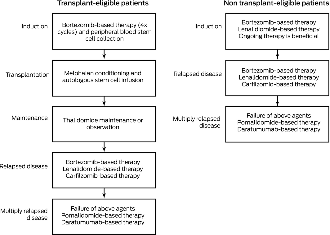
*Always consider clinical trials when available.
Box 4 – Revised International Staging System for myeloma*
|
Stage |
Definition (all criteria must be met) |
5‐Year overall survival |
|||||||||||||
|
|
|||||||||||||||
|
Stage I |
|
82% |
|||||||||||||
|
Stage II |
|
62% |
|||||||||||||
|
Stage III |
|
40% |
|||||||||||||
|
|
|||||||||||||||
Competing interests
No relevant disclosures.
References
- Joshua DE. Multiple myeloma: the present and the future. Med J Aust 2005 Oct 3; 183: 344. https://www.mja.com.au/journal/2005/183/7/multiple-myeloma-present-and-future
- Rajkumar SV, Dimopoulos MA, Palumbo A, et al. International Myeloma Working Group updated criteria for the diagnosis of multiple myeloma. Lancet Oncol 2014; 15: e538–e548.
- Rajkumar SV. Multiple myeloma: 2016 update on diagnosis, risk‐stratification, and management. Am J Hematol 2016; 91: 719–734.
- Kyle R, Gertz M, Witzig T, et al. Review of 1027 patients with newly diagnosed multiple myeloma. Mayo Clin Proc 2003; 78: 21–33.
- Landgren O, Gridley G, Turesson I, et al. Risk of monoclonal gammopathy of undetermined significance (MGUS) and subsequent multiple myeloma among African American and white veterans in the United States. Blood 2006; 107: 904–906.
- Kyle R, Therneau T, Rajkumar S, et al. A long‐term study of prognosis in monoclonal gammopathy of undetermined significance. N Engl J Med 2002; 346: 564–569.
- Kyle R, Therneau T, Rajkumar S, et al. Prevalence of monoclonal gammopathy of undetermined significance. N Engl J Med 2006; 354: 1362–1369.
- Blimark CH, Turesson I, Genell A, et al. Outcome and survival of myeloma patients diagnosed 2008–2015. Real‐world data on 4904 patients from the Swedish Myeloma Registry. Haematologica 2018; 103: 506–513.
- Libby E, Garcia D, Quintana D, et al. Disease‐specific survival for patients with multiple myeloma: significant improvements over time in all age groups. Leuk Lymphoma 2014; 55: 2850–2857.
- Chapman MA, Lawrence MS, Keats JJ, et al. Initial genome sequencing and analysis of multiple myeloma. Nature 2011; 471: 467–472.
- Treon SP, Xu L, Yang G, et al. MYD88 L265P somatic mutation in Waldenstrom's macroglobulinemia. N Engl J Med 2012; 367: 826–833.
- Walker BA, Mavrommatis K, Wardell CP, et al. Identification of novel mutational drivers reveals oncogene dependencies in multiple myeloma. Blood 2018; 132: 587–597.
- Lohr JG, Stojanov P, Carter SL, et al. Widespread genetic heterogeneity in multiple myeloma: implications for targeted therapy. Cancer Cell 2014; 25: 91–101.
- Bolli N, Biancon G, Moarii M, et al. Analysis of the genomic landscape of multiple myeloma highlights novel prognostic markers and disease subgroups. Leukemia 2018; 32: 2604–2616.
- Bahlis NJ. Darwinian evolution and tiding clones in multiple myeloma. Blood 2012; 120: 927–928.
- Egan JB, Shi CX, Tembe W, et al. Whole‐genome sequencing of multiple myeloma from diagnosis to plasma cell leukemia reveals genomic initiating events, evolution, and clonal tides. Blood 2012; 120: 1060–1066.
- Keats JJ, Chesi M, Egan JB, et al. Clonal competition with alternating dominance in multiple myeloma. Blood 2012; 120: 1067–1076.
- Miller A, Asmann Y, Cattaneo L, et al. High somatic mutation and neoantigen burden are correlated with decreased progression‐free survival in multiple myeloma. Blood Cancer J 2017; 7: e612.
- Benson DM. Checkpoint inhibition in myeloma. Hematology Am Soc Hematol Educ Program 2016; 2016: 528–533.
- Chng WJ, Dispenzieri A, Chim CS, et al. IMWG consensus on risk stratification in multiple myeloma. Leukemia 2014; 28: 269–277.
- Palumbo A, Avet‐Loiseau H, Oliva S, et al. Revised International Staging System for multiple myeloma: a report from International Myeloma Working Group. J Clin Oncol 2015; 33: 2863–2869.
- Palumbo A, Avet‐Loiseau H, Oliva S, et al. Revised international staging system for multiple myeloma: a report from International Myeloma Working Group. J Clin Oncol 2015; 33: 2863–2869.
- Zhan F, Huang Y, Colla S, et al. The molecular classification of multiple myeloma. Blood 2006; 108: 2020–2028.
- Mateos MV, San Miguel JF. Smoldering multiple myeloma: when to observe and when to treat? Am Soc Clin Oncol Educ Book 2015: e484–e492.
- Rajkumar SV, Landgren O, Mateos MV. Smoldering multiple myeloma. Blood 2015; 125: 3069–3075.
- Mateos MV, Hernandez MT, Giraldo P, et al. Lenalidomide plus dexamethasone for high‐risk smoldering multiple myeloma. N Engl J Med 2013; 369: 438–447.
- Terpos E. Multiple myeloma: clinical updates from the American Society of Hematology Annual Meeting, 2017. Clin Lymphoma Myeloma Leuk 2018; 18: 321–334.
- Joshua D, Suen H, Brown R, et al. The T cell in myeloma. Clin Lymphoma Myeloma Leuk 2016; 16: 537–542.
- Kyle RA, Gertz MA, Witzig TE, et al. Review of 1027 patients with newly diagnosed multiple myeloma. Mayo Clin Proc 2003; 78: 21–33.
- Martinez‐Lopez J, Lahuerta JJ, Pepin F, et al. Prognostic value of deep sequencing method for minimal residual disease detection in multiple myeloma. Blood 2014; 123: 3073–3079.
- Paiva B, Puig N, Garcia‐Sanz R, San Miguel JF. Is this the time to introduce minimal residual disease in multiple myeloma clinical practice? Clin Cancer Res 2015; 21: 2001–2008.
- Rawstron AC, Gregory WM, de Tute RM, et al. Minimal residual disease in myeloma by flow cytometry: independent prediction of survival benefit per log reduction. Blood 2015; 125: 1932–1935.
- Moreau P, Zamagni E. MRD in multiple myeloma: more questions than answers? Blood Cancer J 2017; 7: 639.
- Mateos MV, Dimopoulos MA, Cavo M, et al. Daratumumab plus bortezomib, melphalan, and prednisone for untreated myeloma. N Engl J Med 2018; 378: 518–528.
- Dimopoulos MA, Lonial S, Betts KA, et al. Elotuzumab plus lenalidomide and dexamethasone in relapsed/refractory multiple myeloma: extended 4‐year follow‐up and analysis of relative progression‐free survival from the randomized ELOQUENT‐2 trial. Cancer 2018; 124: 4032–4043.
- Richardson PG, Attal M, Campana F, et al. Isatuximab plus pomalidomide/dexamethasone versus pomalidomide/dexamethasone in relapsed/refractory multiple myeloma: ICARIA Phase III study design. Future Oncol 2018; 14: 1035–1047.
- Hipp S, Tai YT, Blanset D, et al. A novel BCMA/CD3 bispecific T‐cell engager for the treatment of multiple myeloma induces selective lysis in vitro and in vivo. Leukemia 2017; 31: 1743–1751.
- Suen H, Bryant C, Hart D, Joshua D. Novel T cell therapies in multiple myeloma. Ann Hematol Oncol 2016; 3: 1120.
- Bu DX, Singh R, Choi EE, et al. Pre‐clinical validation of B cell maturation antigen (BCMA) as a target for T cell immunotherapy of multiple myeloma. Oncotarget 2018; 9: 25764–25780.
- Rosenblatt J, Avivi I, Vasir B, et al. Vaccination with dendritic cell/tumor fusions following autologous stem cell transplant induces immunologic and clinical responses in multiple myeloma patients. Clin Cancer Res 2013; 19: 3640–3648.
- Larocca A, Dold SM, Zweegman S, et al. Patient‐centered practice in elderly myeloma patients: an overview and consensus from the European Myeloma Network (EMN). Leukemia 2018; 32: 1697–1712.
- Quach H, Joshua D, Ho J, et al. Treatment of patients with multiple myeloma who are eligible for stem cell transplantation: position statement of the Myeloma Foundation of Australia Medical and Scientific Advisory Group. Intern Med J 2015; 45: 94–105.
- Quach H, Joshua D, Ho J, et al. Treatment of patients with multiple myeloma who are not eligible for stem cell transplantation: position statement of the myeloma foundation of Australia Medical and Scientific Advisory Group. Intern Med J 2015; 45: 335–343.
- Quach H, Prince HM; Medical Scientific Advisory Group. Clinical practice guideline — multiple myeloma. Myeloma Australia, 2014. http://myeloma.org.au/wp-content/uploads/2017/10/MSAG-Clinical-Practice-Guideline-Myeloma-V4-March-2017.pdf (viewed Feb 2019).
- Gibson J, Ho PJ, Joshua D. Evolving transplant options for multiple myeloma: autologous and nonmyeloablative allogenic. Transplant Proc 2004; 36: 2501–2503.
- Mahajan S, Tandon N, Kumar S. The evolution of stem‐cell transplantation in multiple myeloma. Ther Adv Hematol 2018; 9: 123–133.
- Krishnan A, Pasquini MC, Logan B, et al. Autologous haemopoietic stem‐cell transplantation followed by allogeneic or autologous haemopoietic stem‐cell transplantation in patients with multiple myeloma (BMT CTN 0102): a phase 3 biological assignment trial. Lancet Oncol 2011; 12: 1195–1203.
- McCarthy PL, Holstein SA, Petrucci MT, et al. Lenalidomide maintenance after autologous stem‐cell transplantation in newly diagnosed multiple myeloma: a meta‐analysis. J Clin Oncol 2017; 35: 3279–3289.
- Facon T, Dimopoulos MA, Dispenzieri A, et al. Final analysis of survival outcomes in the phase 3 FIRST trial of up‐front treatment for multiple myeloma. Blood 2018; 131: 301–310.
- Moreau P, Masszi T, Grzasko N, et al. Oral ixazomib, lenalidomide, and dexamethasone for multiple myeloma. N Engl J Med 2016; 374: 1621–1634.
- Varga C, Maglio M, Ghobrial IM, Richardson PG. Current use of monoclonal antibodies in the treatment of multiple myeloma. Br J Haematol 2018; 181: 447–459.
- Zamagni E, Tacchetti P, Pantani L, Cavo M. Anti‐CD38 and anti‐SLAMF7: the future of myeloma immunotherapy. Expert Rev Hematol 2018; 11: 423–435.
- Kumar SK, Berdeja JG, Niesvizky R, et al. Safety and tolerability of ixazomib, an oral proteasome inhibitor, in combination with lenalidomide and dexamethasone in patients with previously untreated multiple myeloma: an open‐label phase 1/2 study. Lancet Oncol 2014; 15: 1503–1512.
- Kumar SK, Lee JH, Lahuerta JJ, et al. Risk of progression and survival in multiple myeloma relapsing after therapy with IMiDs and bortezomib: a multicenter international myeloma working group study. Leukemia 2012; 26: 149–157.
- Dimopoulos MA, Goldschmidt H, Niesvizky R, et al. Carfilzomib or bortezomib in relapsed or refractory multiple myeloma (ENDEAVOR): an interim overall survival analysis of an open‐label, randomised, phase 3 trial. Lancet Oncol 2017; 18: 1327–1337.
- Mateos MV, Goldschmidt H, San‐Miguel J, et al. Carfilzomib in relapsed or refractory multiple myeloma patients with early or late relapse following prior therapy: a subgroup analysis of the randomized phase 3 ASPIRE and ENDEAVOR trials. Hematol Oncol 2018; 36: 463–470.
- Stewart AK, Rajkumar SV, Dimopoulos MA, et al. Carfilzomib, lenalidomide, and dexamethasone for relapsed multiple myeloma. N Engl J Med 2015; 372: 142–152.
- Bringhen S, Mina R, Cafro AM, et al. Once‐weekly carfilzomib, pomalidomide, and low‐dose dexamethasone for relapsed/refractory myeloma: a phase I/II study. Leukemia 2018; 32: 1803–1807.
- Scott A, Weber N, Tiley C, et al. “Real‐world” Australian experience of pomalidomide for relapsed and refractory myeloma. Leuk Lymphoma 2018; 59: 1514–1516.
- Ailawadhi S, Mikhael JR, LaPlant BR, et al. Pomalidomide‐dexamethasone in refractory multiple myeloma: long‐term follow‐up of a multi‐cohort phase II clinical trial. Leukemia 2018; 32: 719–728.
- Henry DH, Costa L, Goldwasser F, et al. Randomized, double‐blind study of denosumab versus zoledronic acid in the treatment of bone metastases in patients with advanced cancer (excluding breast and prostate cancer) or multiple myeloma. J Clin Oncol 2011; 29: 1125–1132.
- Terpos E, Berenson J, Raje N, Roodman GD. Management of bone disease in multiple myeloma. Expert Rev Hematol 2014; 7: 113–125.
- Dimopoulos MA, Roussou M, Gavriatopoulou M, et al. Bortezomib‐based triplets are associated with a high probability of dialysis independence and rapid renal recovery in newly diagnosed myeloma patients with severe renal failure or those requiring dialysis. Am J Hematol 2016; 91: 499–502.
- Dimopoulos MA, Terpos E, Chanan‐Khan A, et al. Renal impairment in patients with multiple myeloma: a consensus statement on behalf of the International Myeloma Working Group. J Clin Oncol 2010; 28: 4976–4984.
- Scheid C, Sonneveld P, Schmidt‐Wolf IG, et al. Bortezomib before and after autologous stem cell transplantation overcomes the negative prognostic impact of renal impairment in newly diagnosed multiple myeloma: a subgroup analysis from the HOVON‐65/GMMG‐HD4 trial. Haematologica 2014; 99: 148–154.
- Bridoux F, Carron PL, Pegourie B, et al. Effect of high‐cutoff hemodialysis vs conventional hemodialysis on hemodialysis independence among patients with myeloma cast nephropathy: a randomized clinical trial. JAMA 2017; 318: 2099–2110.
- Vacca A, Melaccio A, Sportelli A, et al. Subcutaneous immunoglobulins in patients with multiple myeloma and secondary hypogammaglobulinemia: a randomized trial. Clin Immunol 2018; 191: 110–115.
- Australian Institute of Health and Welfare. Cancer in Australia 2017 (Cat. No. CAN 100; Cancer Series No. 101). Canberra: AIHW; 2017. https://www.aihw.gov.au/reports/cancer/cancer-in-australia-2017/contents/table-of-contents (viewed Feb 2019).
- Bergin K, Moore E, McQuilten Z, et al. Design and development of the Australian and New Zealand (ANZ) myeloma and related diseases registry. BMC Med Res Methodol 2016; 16: 151.
- Bergin K, Moore E, McQuilten Z, et al. Design and development of the Australian and New Zealand (ANZ) myeloma and related diseases registry. BMC Med Res Methodol 2016; 16: 151.
Provenance: Commissioned; externally peer reviewed.
Splenic injury in severe cases of the zoonoses Q fever and rickettsial infection: diagnostic challenges
Ashleigh Drury, Philippa Harrison, Aiveen Bannan
Health and economic benefits of improving pre‐hospital identification of stroke in Australian women: a modelling study
Thomas Gadsden, Lei Si, Emily R Atkins, Cheryl Carcel, Xia Wang, Stephen Jan, Mark Woodward, Laura E Downey
Updating the diagnosis and management of elevated serum ferritin levels in the era of routine ferritin testing of blood donors by Australian Red Cross Lifeblood
Gary D Zhang, James Chen, Daniel M Johnstone, Martin B Delatycki, Katie Allen, John K Olynyk
Early cardiovascular collapse after envenoming by snakes in Australia, 2005–2020: an observational study (ASP‐31)
Geoffrey K Isbister, Katherine Z Isoardi, Angela L Chiew, Shane Jenkins, Nicholas A Buckley
Updated recommendations for warfarin reversal in the setting of four‐factor prothrombin complex concentrate
Danielle Robinson, James McFadyen, Eileen Merriman, Chee Wee Tan, Ross Baker, Huyen Tran
Updating the diagnosis and management of iron deficiency in the era of routine ferritin testing of blood donors by Australian Red Cross Lifeblood
Gary D Zhang, Daniel Johnstone, Michael F Leahy, John K Olynyk