




Medical education
Snapshot
Volume 210 - Issue 8
Blindness and acute pancreatitis: Purtscher‐like retinopathy
Authors: Anthony Yao and Bob Wang
Med J Aust 2019; 210 (8): 351-351.e1. || doi: 10.5694/mja2.50135
Published online: 6 May 2019
Published online: 6 May 2019
A 30-year-old woman was admitted for pancreatitis
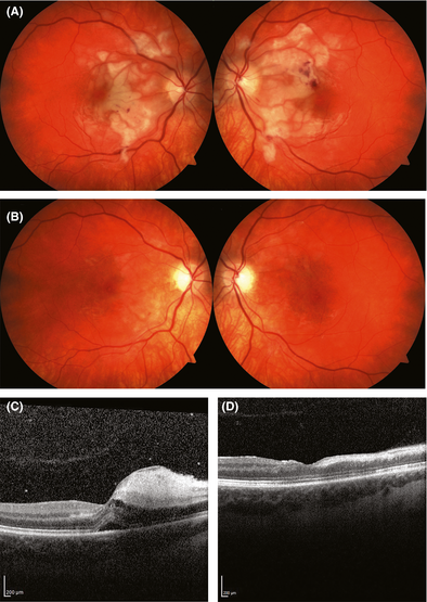
A 30‐year‐old woman was admitted for pancreatitis (peak lipase level, 1385 U/L; Ranson score, 2). Other than excess alcohol intake and previous smoking, she was well with no ocular history. Six hours after the onset of abdominal pain, there was rapid deterioration of central vision, with acuity dropping to 6/120 in both eyes. Fundoscopy demonstrated bilateral well defined areas of intraretinal whitening with perivascular sparing and cotton wool spots, consistent with Purtscher‐like retinopathy1 (Figure, A). Optical coherence tomography at acute presentation also demonstrated inner retinal swelling and disruption of layers (Figure, C). One‐month follow‐up showed resolution of the acute findings, with persistent mottled retinal pigment epithelium and retinal atrophy (Figure, B and D). Acuity remains < 6/60 in both eyes.
Competing interests
No relevant disclosures.
References
- Miguel AIM, Henriques F, Azevedo LFR, et al. Systematic review of Purtscher's and Purtscher‐like retinopathies. Eye 2013; 27: 1–13.
A Diagnostic Headache
Thomas Glynn, Michelle T. Leech, Stacy K. Goergen, Emily Lin, Nadeem Toodayan, Ralph Junckerstorff
Neisseria meningitidis: an uncommon cause of conjunctivitis
Chloe Story, Tina Marinelli, Simeon Crawford, Amrita Ronnachit
Dismantling barriers to research and clinical care for individuals with a vision impairment
Eden G Robertson, Kate Hetherington, Meredith Prain, Julia Hall, Leighton Boyd AM, Rosemary Boyd OAM, Emily Shepard, Hollie Feller, Sally Karandrews, Fleur O'Hare, Kanae Yamamoto, Matthew P Simunovic, Robyn V Jamieson, Alan Ma, Lauren Ayton AM, Anai Gonzalez‐Cordero
The New South Wales Pharmacy Trial for herpes zoster: on the nose?
Christian P Pappas, Timothy R Holmes, Minas T Coroneo
Skin fragility disorder misdiagnosed as child abuse: a cautionary tale
Heather G Mack, Fred K Chen, John R Grigg
Health economic aspects of inherited retinal diseases: looking for cost‐effective treatments
Benjamin Kamien, Rachael Heath Jeffery, Fred K Chen