




Medical education
Lessons from practice
Volume 209 - Issue 3
Brugada syndrome and undifferentiated syncope: use of an implantable loop recorder to document causation
Authors: Garry Hamilton, David O'Donnell and Hui-Chen Han
Med J Aust 2018; 209 (3): 113-114. || doi: 10.5694/mja17.01117
Published online: 6 August 2018
Published online: 6 August 2018
Implantable loop recorders allow for continuous cardiac rhythm monitoring in patients with undifferentiated syncope
Clinical record
A 32-year-old man was transferred to our tertiary hospital cardiology department after presenting to a regional Victorian hospital with syncope. He reported faintness while riding his off-road motorbike on a warm afternoon. While dismounting and removing his helmet, he developed palpitations and briefly lost consciousness. He reported that a similar episode had occurred one year earlier while he was getting out of the shower.
He had no past history of illicit drug use or significant alcohol intake. There was no family history of sudden death.
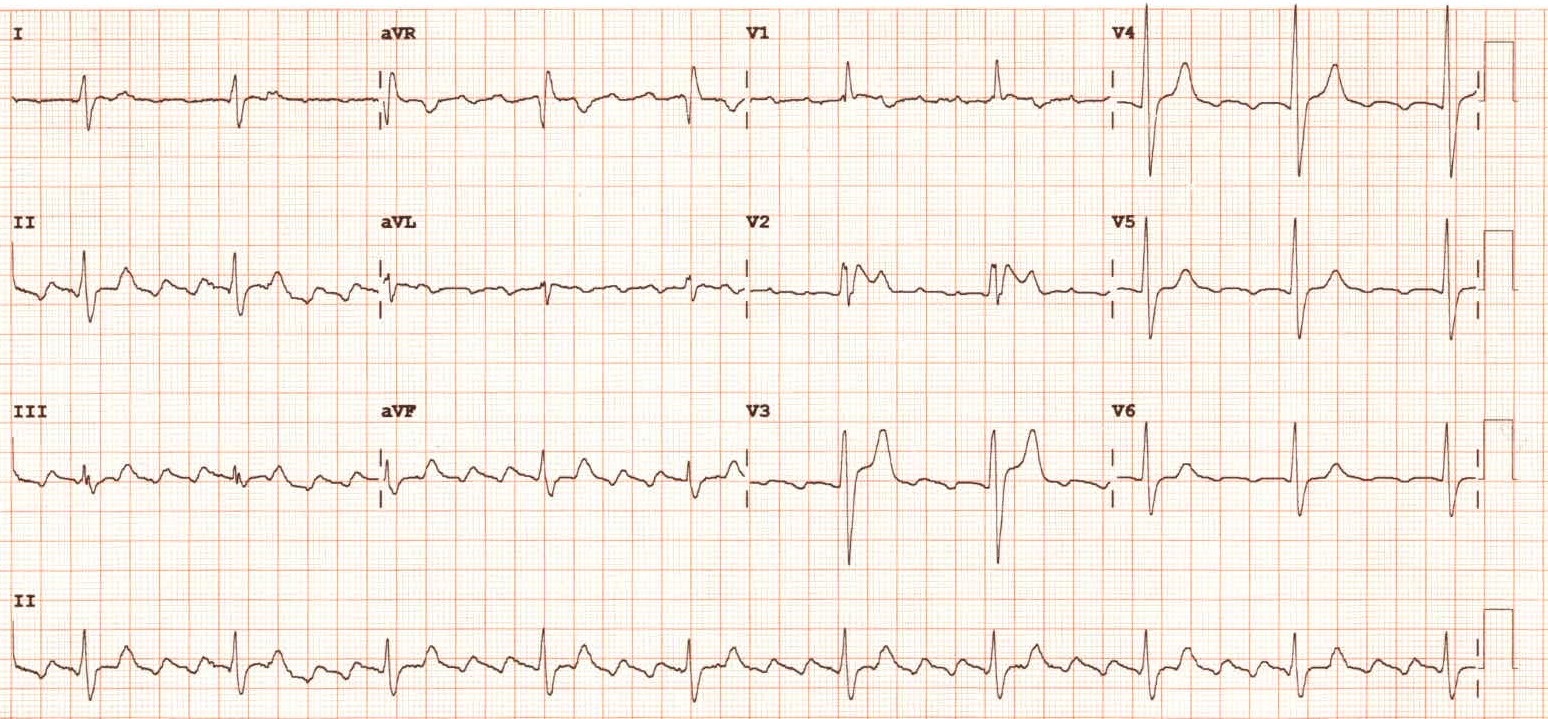
Electrocardiography (ECG) (Box 1) demonstrated atrial flutter with predominantly 4:1 conduction, right axis deviation, widened QRS complex (140 ms), dominant R wave in V1 and ≥ 2 mm ST segment elevation in V2 (saddleback appearance) without reciprocal changes. This was consistent with a type II Brugada pattern.
Basic investigations including a full blood count, electrolytes, renal and liver function, thyroid function, and high sensitivity troponin were all within normal limits.
The patient was commenced on metoprolol and rivaroxaban for his atrial flutter. Inpatient cardiac monitoring for 72 hours did not reveal any other rhythm disturbances. A transthoracic echocardiogram revealed normal cardiac size and function. Cardiac magnetic resonance imaging excluded underlying cardiac infiltration or scar as a substrate for ventricular arrhythmias.
He had no further syncopal episodes during his admission. Although his clinical history was suggestive of vasovagal syncope, he had a markedly abnormal ECG. An implantable loop recorder was inserted before discharge. Flecainide challenge testing (sodium channel blockade aimed at provoking ST-segment changes) to diagnose Brugada syndrome was planned once he had been adequately anti-coagulated for his atrial flutter.
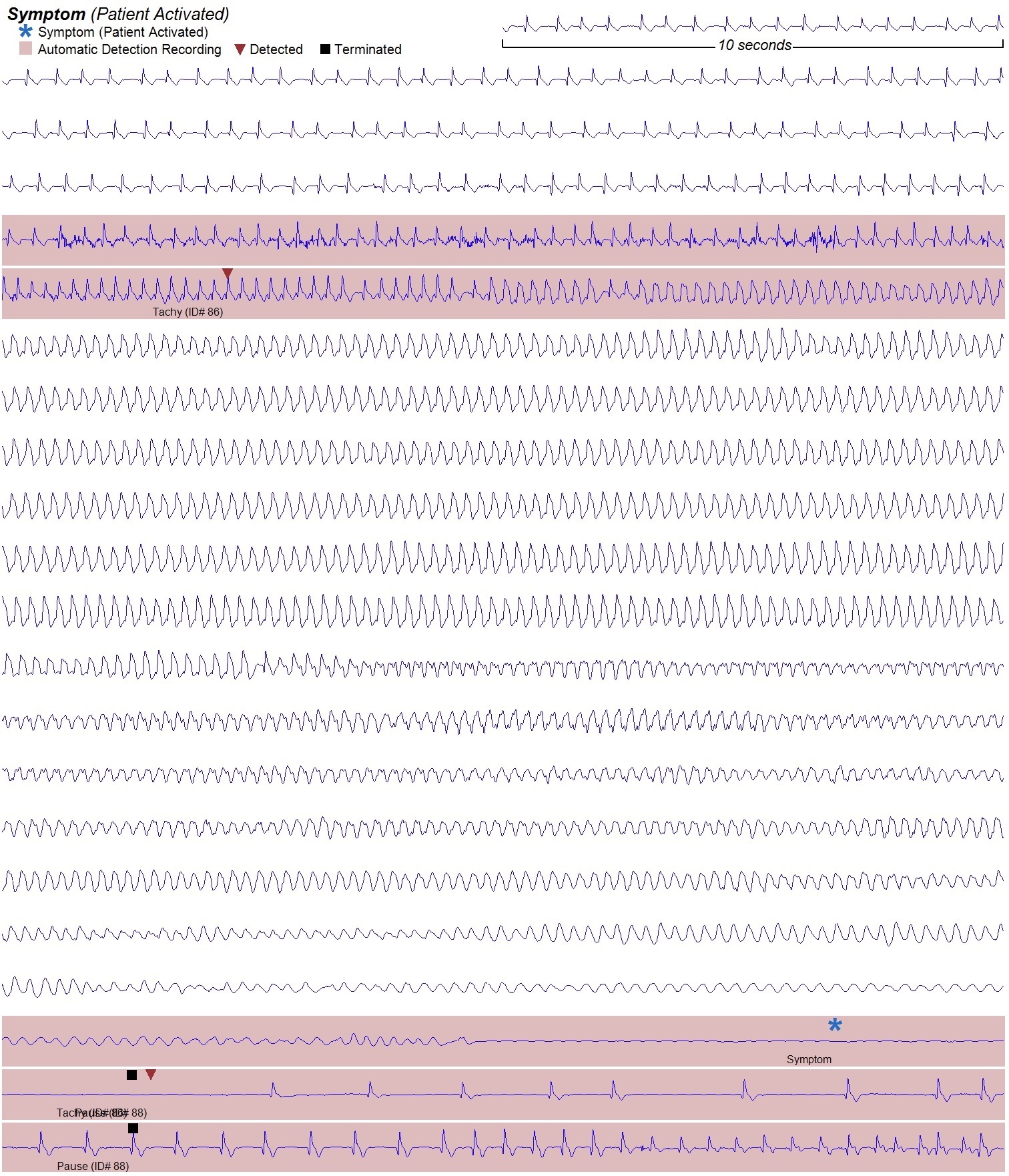
Four weeks later, he experienced another syncopal episode while climbing stairs. Similarly, this episode was preceded by pre-syncope and palpitations. Interrogation of his implantable loop recorder (Box 2) revealed ventricular tachycardia followed by ventricular fibrillation then 15 seconds of asystole, before spontaneous re-initiation of atrial flutter.
A diagnosis of Brugada syndrome was made based on ECG findings and documented ventricular arrhythmia. Subsequently, an implantable cardiac defibrillator (ICD) was inserted.
Two weeks after ICD insertion, the patient received an ICD shock. He was conscious but noticed palpitations. ICD interrogation demonstrated atrial flutter with 1:1 conduction degenerating into ventricular tachycardia, followed by anti-tachycardia pacing and an appropriate shock. It was determined that rapidly conducted atrial flutter was predisposing to ventricular tachycardia and he was commenced on sotalol. He has been well for a year now with no further arrhythmias.
Brugada syndrome is a disorder of cardiac repolarisation which predisposes patients to ventricular arrhythmias. The pathophysiology is thought to primarily involve abnormal sodium channel function under the influences of autonomic tone.1 Mutations in the SCN5A gene (resulting in sodium channel dysfunction) are the most commonly found genetic abnormality in Brugada syndrome, with autosomal dominant inheritance but incomplete penetrance.2
The estimated worldwide prevalence of Brugada pattern ECG changes is 0.23%, and they are most commonly seen in people of Asian descent, followed by people in Europe and the United States.2 A meta-analysis of 1545 patients with Brugada pattern ECG changes revealed an event rate (syncope, ICD shock or sudden death) of 10% at an average of 32 months follow-up.3 Males have a 5.5-fold increased risk of sudden cardiac death, and mean age for development of ventricular fibrillation is 41 ± 15 years.2
Brugada pattern ECG changes affecting leads V1–V2 are classified as either type I (coved) or type II (saddleback). These can fluctuate over time or under the influences of medications and physiological changes.4 Diagnosing Brugada syndrome requires the presence of characteristic ECG changes with additional clinical features (survived cardiac arrest, polymorphic ventricular tachycardia, history of non-vagal syncope, family history of sudden death or type I Brugada pattern ECG in relatives),4 although some suggest the diagnosis requires demonstration of type I ECG changes alone (either spontaneous or provoked).5
Investigations in patients with Brugada pattern ECG changes should include cardiac imaging to exclude structural heart disease, cardiac rhythm monitoring for ventricular arrhythmias, and pharmacological provocation testing in those who do not exhibit spontaneous type I ECG changes.5
The treatment of Brugada syndrome is aimed at preventing sudden death. Class I recommendations include lifestyle changes, such as avoiding aggravating drugs and excessive alcohol intake and immediate antipyretic treatment of fever. Those who survive cardiac arrest or demonstrate spontaneous sustained ventricular tachycardia should undergo ICD insertion.5
Our case highlights a rare, life-threatening cause of syncope and reinforces the need for thorough assessment of this presentation.Lessons from practice
Brugada syndrome is a disorder of cardiac sodium channels resulting in abnormal repolarisation on electrocardiography.
Mutations in the SCN5A gene are the most commonly found genetic abnormality in patients with Brugada syndrome.
Malignant ventricular arrhythmias in patients with Brugada syndrome are a rare but important cause of cardiac syncope.
Implantable loop recorders allow for continuous cardiac rhythm monitoring in patients with undifferentiated syncope.
Patients with Brugada syndrome and prior survived cardiac arrest or spontaneous sustained ventricular tachycardia are recommended to undergo implantable cardiac defibrillator insertion.
Competing interests
David O'Donnell has received lecture fees from Medtronic and St Jude Medical.
References
- Alings M, Wilde A. “Brugada” syndrome. Circulation 1999; 99: 666-673.
- Mizusawa Y, Wilde A. Brugada syndrome. Circ Arrhythm Electrophysiol 2012; 5: 606-616.
- Gehi AK, Duong TD, Metz LD, et al. Risk stratification of individuals with the Brugada electrocardiogram: a meta-analysis. J Cardiovasc Electrophysiol 2006; 17: 577-583.
- de Luna AB, Brugada J, Baranchuk A, et al. Current electrocardiographic criteria for diagnosis of Brugada pattern: a consensus report. J Electrocardiol 2012; 45: 433-442.
- Priori SG, Wilde AA, Horie M, et al. HRS/EHRA/APHRS expert consensus statement on the diagnosis and management of patients with inherited primary arrhythmia syndromes. Heart Rhythm 2013; 10: 1932-1963.
Provenance: Not commissioned; externally peer reviewed.
Rapid Growth in Diagnostic CT Knee Imaging in Australia: Implications for Imaging Stewardship
Hanwen Chen, Rachelle Buchbinder, Sean Docking, Ian A Harris
Disease‐specific survival for people with chronic myeloid leukaemia, South Australia, 1980–2020: a retrospective cohort study
Kerri Beckmann, Brendon J Kearney, Naranie Shanmuganathan, David Yeung, Tim Hughes
Technological advances in the search for a CURE for asthma
Sara Quon, Timothy M Johanson, Ridhima Wadhwa, Alen Faiz, Anthony Flynn, Gary P Anderson, Amanda J Cox, Nicholas P West, Michael P Menden, Rhys S Allan
Who gets asthma, and why?
Denby J Evans, Peter D Sly, Paul Foster, Chantal Donovan
The first Australian evidence‐based guidelines on male infertility
Darren J Katz, Liza O’Donnell, Robert I McLachlan, Tim J Moss, Clare V Boothroyd, Veena Jayadev, Sarah R Catford
Sex differences in management and outcomes of people with ST‐elevation myocardial infarction, New South Wales, 2011–2020: a retrospective cohort study
Samia Kazi, Simone Marschner, Haeri Min, Desi Quintans, James JH Chong, Ehsan Khan, David B Brieger, Clara K Chow