




Volume 207 - Issue 2
Iron stain following an intravenous iron infusion
Authors: Martin L Canning and Kenneth A Gilmore
Med J Aust 2017; 207 (2): 58. || doi: 10.5694/mja17.00040
Published online: 17 July 2017
Published online: 17 July 2017
Iron polymaltose was administered via rapid infusion
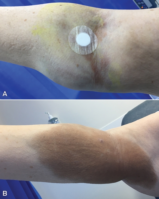
Iron polymaltose was administered via rapid infusion to a patient with iron deficiency anaemia. During administration, we observed staining and ceased the infusion (Figure, A; 30 minutes after cessation). Staining extended to a large part of the arm, and follow-up at 21 days revealed no change in size (Figure, B). Persistent discolouration of the skin is a risk associated with intramuscular and intravenous iron administration.1,2 It has been reported with multiple iron preparations,3-5 and one study found an incidence of 1.3%.5 Iron staining has been successfully treated with laser therapy, resulting in almost complete disappearance.3 Intravenous infusion sites should be monitored closely for swelling before and during administration to minimise the risk of extravasation causing staining.
Competing interests
No relevant disclosures.
References
- Australian Medicines Handbook [online]. Adelaide: Australian Medicines Handbook, 2017. https://amhonline.amh.net.au (accessed Mar 2017).
- Low MSY, Grigoriadis G. Iron deficiency and new insights into therapy. Med J Aust 2017; 207: 81-87.
- Hammami Ghorbel H, Lacour JP, Passeron T. Successful treatment with 532-nm Q-switched Nd:YAG laser of cutaneous siderosis following intravenous iron extravasation. J Eur Acad Dermatol Venereol 2015; 29: 818-819.
- Zuckerman MD, Greenston M. Extraordinary extravasation. N Z Med J 2014; 127: 100-101.
- Anker SD, Colet JC, Filippatos G, et al. Ferric carboxymaltose in patients with heart failure and iron deficiency. N Engl J Med 2009; 361: 2436-2448.
Intranasal Adrenaline Use for the Management of Insect Venom-Induced Anaphylaxis
Thomas J. Young, Wilhelmina R. F. Johnson, Braydon L. Pennington, Troy Wanandy, Wun Y. Lau, Thanh-Thao A. Le
Gabapentinoid Poisoning Presentations to Victorian Emergency Departments: A 15-Year Trend Analysis
Bishaal Tej Gurung, Amy McNeilage, Angus Skeen, Tina Lam, Joanna F. Dipnall, Jane Hayman, Suzanne Nielsen, Ting Xia
Still Treating Yesterday's Risk? Reconsidering Antiviral Use for Mild-to-Moderate COVID-19 Cases in a Broadly Immune Population
Hadar Mudrik-Zohar, Tim Cutfield, Susan Morpeth, Thomas Hills, Eamon Duffy, Laura J. Edwards, Allen C. Cheng, Steven Y. C. Tong
Birth prevalence, clinical sequelae, and management of congenital cytomegalovirus infections in Australia, 1999–2023: a national prospective study
Ece Egilmezer, Suzy M Teutsch, Carlos Nunez, Stuart T Hamilton, Adam W Bartlett, Pamela Palasanthiran, Elizabeth J Elliott, William D Rawlinson
Concurrent use of hormonal long‐acting reversible contraception by women of reproductive age dispensed teratogenic medications, Australia, 2013–2021
Aaron E Boyce, Tony Caccetta, Jo‐Ann See, Rosemary L Nixon AM
Concurrent use of hormonal long‐acting reversible contraception by women of reproductive age dispensed teratogenic medications, Australia, 2013–2021
Michelle KY Chen, Adrian Lim, Deshan F Sebaratnam