




Volume 207 - Issue 2
Iron deficiency and new insights into therapy
Authors: Michael SY Low and George Grigoriadis
Med J Aust 2017; 207 (2): 81-87. || doi: 10.5694/mja16.01304
Published online: 17 July 2017
Published online: 17 July 2017
Iron deficiency and iron deficiency anaemia remain prevalent in Australia
Summary
- Iron deficiency and iron deficiency anaemia remain prevalent in Australia.
- The groups at highest risk are pre-menopausal women, socially disadvantaged people and those of Indigenous background.
- Diagnosing iron deficiency using a full blood examination and iron studies can be difficult and can be further complicated by concomitant inflammation. Results of iron studies should always be interpreted as an overall picture rather than focusing on individual parameters. In difficult clinical scenarios, soluble transferrin receptor assays can be useful.
- Management of iron deficiency involves identification and treatment of the cause of iron deficiency, as well as effective iron replacement.
- Clinicians should always take a detailed history and perform a comprehensive physical examination of a patient with iron deficiency. Patients should be monitored even if a likely cause of iron deficiency is identified.
- Patients who fail to respond to iron replacement or maintain iron status should be referred for further investigation, including endoscopy to exclude internal bleeding.
- Both enteral and parenteral iron are effective at replacing iron. For most adult patients, we recommend trialling daily oral iron (30–100 mg of elemental iron) as the first-line therapy.
- Safety and efficacy of intravenous iron infusions have improved with the availability of a newer formulation, ferric carboxymaltose. Patients who fail to respond to oral iron replacement can be safely managed with intravenous iron.
- Blood transfusion for iron deficiency anaemia should be reserved for life-threatening situations and should always be followed by appropriate iron replacement.
Iron deficiency (ID) is a deficit in total body iron which, when severe, can lead to iron deficiency anaemia (IDA). ID is considered the most prevalent nutritional deficiency in the world.1,2 Recent estimates suggest that about a third of non-pregnant women worldwide are anaemic,1 with IDA comprising about half of all cases.2
In Australia, the burden of ID is harder to define, as only a few large scale studies exist. The prevalence of ID and IDA depends on the groups studied, but estimates for IDA range from 1% to 14% in toddlers and 10% to 55% in women.3 The highest rates of ID and IDA are seen in pre-menopausal women, socially disadvantaged people and those of Indigenous background.3-5 A recent study by the Australian Red Cross Blood Service found that, among new blood donors, 12% of female donors and 1.3% of male donors were iron deficient.6 The combined rate of anaemia (ie, haemoglobin level below normal) from all causes in new donors of both sexes was 5.6%.6 Whether these estimates represent the overall burden across Australian society is unclear, due to the strict age- and health-related criteria for potential blood donors in Australia. The Australian Health Survey in 2012 suggested that about 3% of Australia’s population were anaemic, with 71% of this attributable to IDA.5 It is therefore estimated that over a quarter of a million Australians live with IDA, and many more have ID without anaemia. Regardless of the exact number, ID and IDA continue to be major public health problems in Australia.
Here, we discuss the causes, effects, diagnosis and management of ID. For this narrative review, we searched PubMed for original papers and review articles published between 2000 and 2016, as well as using specialist society publications and guidelines, to formulate an evidence-based overview of ID and IDA.
Causes and effects of iron deficiency
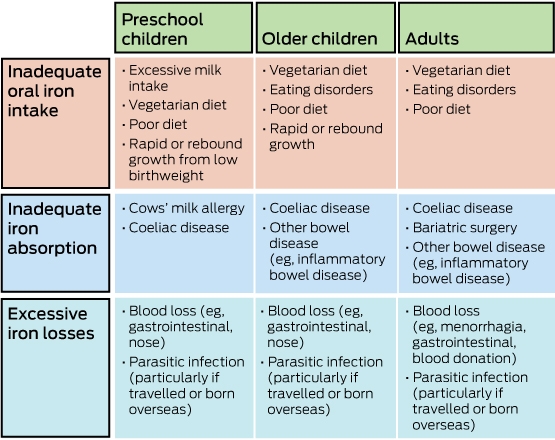
The causes of ID have been reviewed previously.3,7 In their most simplistic form, ID and IDA are a result of unbalanced loss of iron, most commonly from bleeding, without adequate replacement by intake of oral iron. Important causes of ID are shown in Box 1.
The effects of ID and IDA in children include impaired cognition and behavioural problems.5,8,9 In adults, they can lead to reduced productivity, tiredness and lethargy.10 In pregnant women, IDA has been associated with neonates of low birthweight, prematurity and maternal morbidity.11 Given these potentially severe consequences, timely diagnosis, investigation and treatment of ID and IDA are important for optimal patient care.
Diagnosis of iron deficiency
One of the major problems in diagnosing ID and IDA is difficulty in interpreting test results. Classically, patients with suspected IDA will have a full blood examination and iron studies, in addition to other pertinent tests. Iron studies include measurement of serum or plasma iron levels, transferrin levels, transferrin saturations (calculated from total iron binding capacity) and ferritin levels. Serum iron levels, when assessed alone, are usually unhelpful for investigating ID, but they are used to calculate other measures within the iron studies. Transferrin is the transport molecule for iron in blood, and transferrin saturation is a measure of how much iron is bound to transferrin. Ferritin is a measure of iron reserve and is the most reliable parameter in the iron studies for the diagnosis of ID.12
The World Health Organization defines ID as a serum or plasma ferritin level of < 12 µg/L in children under 5 years of age or < 15 µg/L in adults and children over 5 years of age.13 However, these cut-offs are not universally adopted, with the Royal College of Pathologists of Australasia recommending that a ferritin level < 30 µg/L be considered abnormal for adults.14 Even more confusingly, laboratories may use different cut-offs depending on the method of ferritin analysis and the population in which the assay was validated.14,15 These differing definitions for abnormally low ferritin levels make interpretation of results difficult. Furthermore, ferritin is an acute phase reactant and its levels can be raised in inflammatory states such as infection, autoimmune disease or alcoholic and non-alcoholic fatty liver disease. It is often taught that patients with ferritin levels greater than 100 µg/L are unlikely to be iron deficient, although there are exceptions.3
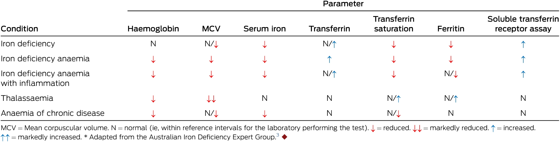
A summary of how to interpret the results of a full blood examination and iron studies, adapted from the Australian Iron Deficiency Expert Group, is shown in Box 2.3 We recommend that the results of iron studies be interpreted as a whole. For example, a borderline low ferritin level may still represent ID in an unwell patient, and suspicion should be raised if the transferrin and transferrin saturation levels indicate IDA, regardless of the ferritin level.
The two main differential diagnoses for IDA (which is typically a microcytic, hyopchromic anaemia) are thalassaemia and anaemia of chronic disease. Thalassaemia can usually be differentiated from IDA by the presence of a family history, a disproportionately low mean corpuscular volume compared with haemoglobin level, a red cell distribution width within reference intervals or only slightly raised16 and an adequate ferritin level. A haemoglobin electrophoresis test or high performance liquid chromatography to determine the presence of haemoglobinopathy can often be diagnostic, although a-thalassaemia is more difficult to detect and may require genetic testing for diagnosis.17 On the other hand, anaemia of chronic disease can be particularly difficult to distinguish from IDA. In situations where iron studies are compromised by the presence of inflammation, we use soluble transferrin receptor assays to differentiate between the two conditions, as soluble transferrin receptor levels are raised only in the setting of ID. Although raised soluble transferrin receptor levels have high sensitivity and specificity for ID,18 this method has limited use in clinical practice because of confusion about the best method of reporting (total level or as a ratio to ferritin), unfamiliarity among clinicians and lack of a standardised cut-off.18 Nevertheless, we feel that soluble transferrin receptor assays are useful to differentiate IDA from anaemia of chronic disease in difficult cases.
The gold standard for diagnosing IDA remains bone marrow biopsy, with analysis of particulate iron using a Perls’ Prussian blue stain (Box 3).12 Due to the invasiveness of this procedure and the ability to diagnose most patients with serum testing, we do not recommend this investigation even in difficult cases. In our clinical practice, if ID cannot be clearly differentiated from other conditions, a trial of iron replacement is often attempted, with close clinical monitoring. Nevertheless, bone marrow biopsies are sometimes performed in the diagnostic investigation of anaemia or other cytopenias to exclude primary bone marrow disease. When it is performed, the information available from a bone marrow biopsy should not be ignored. We recommend that a Perls’ stain be performed to analyse body iron stores in all patients who have a bone marrow biopsy for the investigation of anaemia.
Management of iron deficiency
The management of ID consists of investigation and treatment of the cause, iron replacement by either the enteral (oral) or parenteral (injectable) route, follow-up and further investigations when required.
Investigation of the cause of iron deficiency
Investigation of ID should include a detailed history, physical examination and appropriate biochemical and interventional investigations. Importantly, consideration should be given to a patient’s family history, age, sex and risk factors for causes.
An algorithm of how we investigate ID, adapted from the guidelines of the Gastroenterological Society of Australia and the Australian Iron Deficiency Expert Group, is shown in Box 4.3,7 Our algorithm is a general guideline and does not replace a clinician’s need to consider the patient’s unique situation and investigate when appropriate.
Iron replacement therapies
Oral iron
Daily oral iron is commonly prescribed as an initial intervention by clinicians treating ID and IDA and is recommended by the World Health Organization.19 However, oral iron replacement is not without its risks. While some risks, such as a reported increased rate of malaria,20-22 are less relevant in Australia, clinicians should be aware of the adverse effects and risks of treatment with oral iron.
The relative benefits of daily oral iron replacement in different at-risk populations have been analysed in a series of recent systematic reviews and meta-analyses.10,23-26 Evidence-based benefits and risks according to these studies are summarised in Box 5, which illustrates the quality of the evidence that daily oral iron replacement has an effect on the stated outcome, not the magnitude of its effect. In some age groups, insufficient numbers were studied for the stated outcomes, so although no effect could be demonstrated in the meta-analysis, it is possible there is an effect that has not been detected. For example, as very few studies reported cognitive outcomes in females older than 12 years, no benefit could be observed.10 It remains unclear if daily oral iron therapy has a benefit for cognitive outcomes in this population. Further, only females were included in the meta-analysis for people aged over 12 years, as ID is rare in males above this age and should be investigated as shown in Box 4. Nevertheless, it is likely that males will have similar benefit and risk profiles as females for oral iron replacement. Although these studies give a general overview of the effects of daily oral iron therapy in different age groups, the included studies encompassed areas with different endemic infections and socio-economic status than Australia. These differences should be considered when weighing up the risks and benefits of iron replacement in Australia. We feel that the data strongly support the use of daily oral iron replacement to treat ID and IDA for all ages, but clinicians should be aware of, and monitor for, possible adverse effects during therapy.
The type of iron, dose and duration of daily oral iron therapy were also analysed in these studies. Commonly available forms of oral iron in Australia are shown in Box 6. Our recommendations for choice of iron replacement therapy, based on these studies and previous recommendations, are shown in Box 7. Some evidence suggests that ferrous sulfate (compared with other forms of iron) and the addition of vitamin C may help further reduce rates of anaemia; however, we do not feel this evidence is sufficiently robust to specifically recommend these interventions.10 A reticulocyte response is expected after about 72 hours of treatment with daily oral iron, with improvements in haemoglobin levels of about 20 g/L every 3 weeks.3 If daily oral iron is not feasible because of adverse effects or problems with patient adherence, intermittent oral iron (once, twice or three times a week) has been shown to be effective.28 Studies have suggested that second-daily oral iron dosing may maximise iron absorption and reduce intestinal exposure to non-absorbed iron, which may reduce adverse effects;29 however, longer term studies to confirm these findings are required. A Cochrane review of intermittent oral iron replacement showed it improved haemoglobin levels and iron status, but not as effectively as daily dosing. Hence, our preference is for daily oral iron where possible.28
Parenteral iron
Parenteral iron is an alternative for patients who have major adverse effects from oral iron, are unwilling to take regular oral medication or are unable to absorb oral iron. In these cases, we recommend intravenous (IV) iron replacement (Box 7). We also recommend IV iron for adults with severe IDA (haemoglobin level < 80 g/L), as evidence suggests that IV iron corrects haemoglobin levels faster than oral iron.30-32 Intramuscular iron has previously been used, but we strongly discourage its use due to the safety of new IV iron formulations, pain associated with intramuscular injection and the possibility of permanent skin discolouration.3
Three IV iron formulations are available on the Pharmaceutical Benefits Scheme in Australia: iron sucrose, iron polymatose and ferric carboxymaltose. Several others are available overseas (eg, iron dextran, ferumoxytol and iron gluconate). Iron sucrose and iron polymatose are second-generation iron agents that have largely been superseded by the newer formulation, ferric carboxymaltose, as they cannot be given in high doses because of possible instability in the iron–carbohydrate complex and toxicity from non-transferrin-bound iron.33 Ferric carboxymaltose has an excellent safety profile, is easily administered and can be given at high doses.34 It is administered at a dose of up to 1000 mg, either undiluted via a slow IV push or diluted in 250 mL of 0.9% sodium chloride over at least 15 minutes.33,34 For most patients, one dose of ferric carboxymaltose is sufficient to return haemoglobin and ferritin levels to within normal reference intervals. However, when required, a repeat dose can be administered 7 days after the first dose. In trials and post-marketing experience of ferric carboxymaltose, anaphylactic or anaphylactoid reactions have been reported in only about 0.1% of patients.31,34,35 More common adverse reactions include headache, nausea, injection site reactions, hypertension, hypophosphataemia (usually asymptomatic) and flushing. Importantly, rates of hypotension appeared to be lower with ferric carboxymaltose than with other iron formulations.34
Blood transfusions
Although red cell transfusion is an effective treatment for IDA, we recommend it only in life-threatening circumstances. As a single unit of fractionated red blood cells (resuspended) is estimated to contain about 200 mg of elemental iron,36 it is an inefficient method of replacing iron. Additionally, transfusion of red blood cells is expensive and exposes patients to rare but potentially life-threatening complications, including fluid overload and immunological and infectious hazards.3,37 We therefore only recommend it for use in patients who have severe anaemia that is compromising organ function (eg, angina pectoris or cardiac failure).3 When blood transfusion is required, each transfused unit of red blood cells is expected to increase the haemoglobin level by 10 g/L, in the absence of bleeding. We recommend transfusing the minimal number of units of red blood cells to control symptoms. Transfusion should always be followed by appropriate iron replacement (usually with IV iron, to facilitate the most rapid correction of haemoglobin level30).
Follow-up and further investigations
All patients should be routinely followed up after investigation and treatment of an underlying cause. Optimal timing of follow-up is not clear, as no firm guidelines exist. In our clinic, we routinely review patients with severe IDA (haemoglobin level < 80 g/L) within 2 weeks to ensure an appropriate reticulocytosis (indicated by a raised reticulocyte count and/or percentage) and response in haemoglobin level. We generally review patients with mild to moderate IDA (haemoglobin level > 80 g/L) in 3–6 months to ensure they remain iron replete and that no new symptoms requiring further investigation have evolved. Other clinicians may feel that more regular monitoring is warranted; we feel shorter intervals for follow-up are reasonable.
Common reasons why patients fail to respond to iron replacement therapies include poor adherence, impaired iron absorption and ongoing blood loss.3 When investigating a non-responding patient, we recommend that clinicians consider non-adherence and all conditions shown in Box 2, as well as following the algorithm in Box 4. Where appropriate, further investigation should include endoscopy to exclude gastrointestinal bleeding sources, regardless of symptoms, age and demographic characteristics. Failure to adequately monitor patients and appropriately investigate can have devastating consequences.38
Special situations
Children
As ID and anaemia have been associated with reduced cognitive function and behavioural problems in children, it is important to ensure they maintain adequate iron levels.5,8,9 We recommend the use of oral iron replacement as the first-line intervention for children (Box 7).39 IV iron can be given to children and has been shown to be safe when administered under the supervision of those familiar with its use.40 We recommend that children under the age of 16 years who need IV iron replacement because oral iron replacement has failed should see a specialist paediatrician for further investigation and appropriate dosing before receiving an IV iron infusion. Dosing is particularly important in children, as 20 mg/kg of elemental iron can be enough to cause iron poisoning, which can be lethal.41 Clinicians prescribing iron replacement therapies to children should educate parents to ensure that overdosing (either accidental or intentional) does not occur.
Pregnancy
Iron requirements are increased during pregnancy because of increased maternal red cell mass, placental and fetal iron requirements and peripartum blood loss.11 Additionally, it is commonly believed that breastfeeding increases the burden of iron loss in the nursing mother, although robust evidence for this is lacking.42 Regardless, women are at particularly high risk of ID and IDA during the peripartum period. The Royal Australian and New Zealand College of Obstetricians and Gynaecologists recommends all women be screened using a full blood examination at the first antenatal (booking) visit, with further testing including iron studies when suspicion of ID is raised by the presence of anaemia or reduced mean corpuscular volume.43 Given that ID in pregnancy has been associated with adverse outcomes for infants,11,44 close monitoring and correction of ID is crucial for optimal perinatal care.
A systematic review found that daily oral iron therapy is effective at improving maternal iron status and haemoglobin levels, with no evidence of increased rates of adverse effects.45 Daily oral iron showed a trend towards increased birthweight and lower rates of preterm labour, although these outcomes were not statistically significant. Another systematic review found that, similar to the findings for non-pregnant women, intermittent oral iron therapy was effective at reducing rates of anaemia and improving iron status in pregnant women and is therefore an alternative for those who cannot take daily oral iron.46 IV iron is safe to use in pregnancy, with trials showing superior correction of haemoglobin level and iron status compared with oral iron, with minimal adverse effects.11,47 Given the potential (if unproven) benefits of correcting iron status in pregnant women and their infants, in our practice we have a lower threshold for IV iron administration in pregnant women.
Peri-operative care
Anaemia in the peri-operative setting is associated with increased morbidity and mortality.48 Additionally, blood loss associated with surgery increases iron requirements in the peri-operative period. Evidence suggests that pre-operative iron replacement therapy (either parenteral or enteral) can help reduce red cell transfusions and hospital length of stay.48,49 Post-operative use of ferric carboxymaltose has also been shown to be effective in improving post-operative haemoglobin levels and reducing red cell transfusions.50 While the role of iron replacement in the peri-operative setting is currently being further examined through a meta-analysis,51 we feel that oral or IV iron should always be considered in appropriate patients to help reduce surgical complications and red cell transfusions.
Conclusions
ID and IDA remain prevalent in Australia. Diagnosing ID can be difficult, particularly in the setting of inflammation, and the results of iron studies should always be considered as an overall picture rather than focusing on single parameters. Adequate management of ID involves investigation and treatment of the cause, as well as appropriate iron replacement with enteral or parenteral iron. All patients with ID should be monitored clinically to ensure that iron stores remain replete and that further investigation is performed when warranted.
Box 3 – Perls’ Prussian blue stain for iron on bone marrow biopsy samples
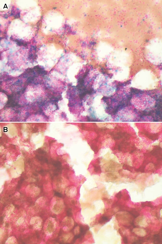
A: Bone marrow particle showing adequate particulate iron (iron reserves), indicated by the presence of blue iron deposits. B: Bone marrow particle of an iron-deficient patient with marked reduction in iron, indicated by the lack of blue deposits.
Box 4 – Algorithm for investigating iron deficiency*
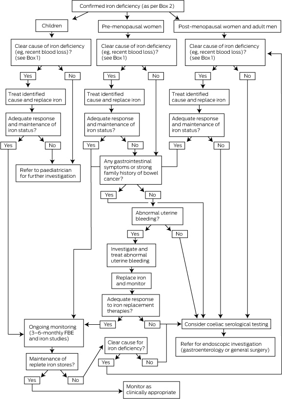
FBE = full blood examination. * Adapted from the guidelines of the Gastroenterological Society of Australia and the Australian Iron Deficiency Expert Group.3,7 Clinicians should consider referring patients with unexplained bleeding to a specialist haematologist for further investigation of possible bleeding disorders.
Box 5 – Benefits and adverse effects of daily oral iron replacement, by age group*
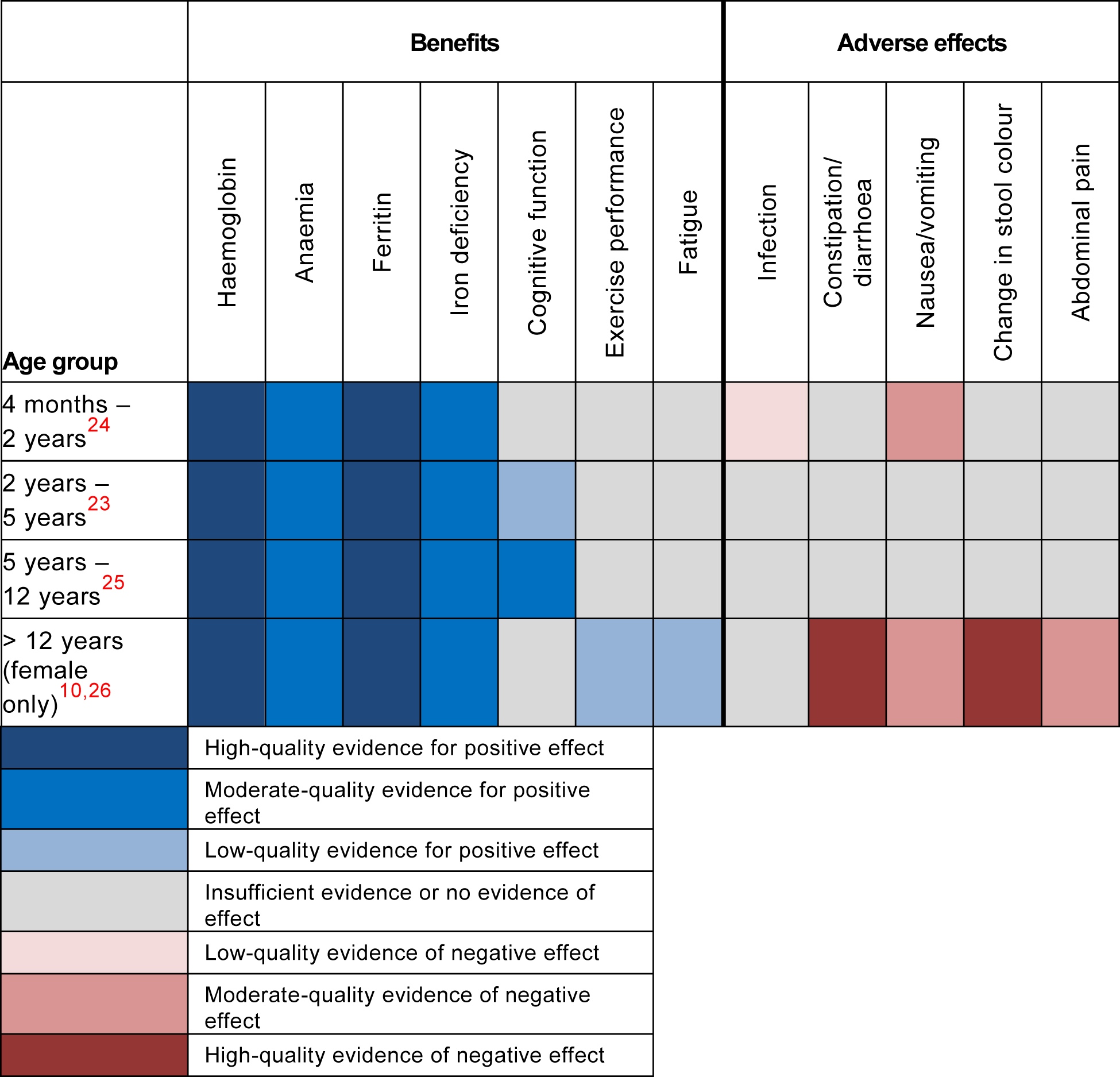
* Quality of evidence for an effect of daily oral iron replacement, as defined by the Cochrane Handbook for Systematic Reviews of Interventions using the Grades of Recommendation, Assessment, Development and Evaluation (GRADE) approach.27 The colours represent the quality of the evidence that daily oral iron replacement has an effect on the stated outcome, not the magnitude of its effect. The GRADE system also includes a “very low” quality category; none of the outcomes shown were classified in this category during meta-analysis.
Box 6 – Common oral iron formulations available in Australia
|
Type of oral iron |
Brand name examples (manufacturer) |
Dose of elemental iron |
Additional active ingredients* |
||||||||||||
|
|
|||||||||||||||
|
Recommended oral iron formulations that are commonly available |
|||||||||||||||
|
Ferrous sulfate |
Ferro-Gradumet (Abbott) |
105 mg |
Nil |
||||||||||||
|
|
Ferrograd C (Abbott) |
105 mg |
Vitamin C (500 mg) |
||||||||||||
|
|
FGF (Abbott) |
80 mg |
Folic acid (300 µg) |
||||||||||||
|
|
Fefol (Pharmacare) |
87 mg |
Folic acid (300 µg) |
||||||||||||
|
|
Ferro-Liquid (AFT Pharmaceuticals) |
30 mg/5 mL |
Nil |
||||||||||||
|
Ferrous fumarate |
Ferro-F-Tab (AFT Pharmaceuticals) |
100 mg |
Nil |
||||||||||||
|
|
Ferro-Tab (AFT Pharmaceuticals) |
67.5 mg |
Nil |
||||||||||||
|
Iron polymaltose |
Maltofer (Aspen) |
100 mg |
Nil |
||||||||||||
|
|
Maltofer Syrup (Aspen) |
50 mg/5 mL |
Nil |
||||||||||||
|
Other commonly available oral iron formulations (not recommended due to low dose of elemental iron)† |
|||||||||||||||
|
Ferrous fumarate |
Bio Iron (Blackmores) |
15 mg |
Vitamin C (100 mg) + folic acid (166.5 µg) + vitamin B12 (50 µg) |
||||||||||||
|
|
Iron Plus (Nature’s Own) |
5 mg |
Vitamin C (100 mg) + folic acid (95 µg) |
||||||||||||
|
Iron chelate |
Fab Iron (Care Pharmaceuticals) |
10 mg |
Nil |
||||||||||||
|
|
Iron Support (Clear Health) |
24 mg |
Vitamin C (150 mg) + vitamin B12 (100 µg) |
||||||||||||
|
|
Iron Max (Ethical Nutrients) |
24 mg |
Vitamin C (80 mg) + folic acid (500 µg) + vitamin B12 (500 µg) |
||||||||||||
|
|
Iron (Swisse) |
20 mg |
Vitamin C (16.52 mg) |
||||||||||||
|
Ferrous gluconate |
Liquid Iron (Nature’s Own) |
7.5 mg/5 mL |
Nil |
||||||||||||
|
|
Fab Iron Liquid (Care Pharmaceuticals) |
10 mg/mL |
Vitamin C (60 mg) + vitamin B12 (2.5 µg) |
||||||||||||
|
Iron glycinate |
Iron Plus (Cenovis) |
5 mg |
Vitamin C (100 mg) + vitamin B12 (50 µg) + folic acid (95 µg) |
||||||||||||
|
|
|||||||||||||||
|
* List of additional active ingredients is not complete and only includes those likely to affect iron metabolism. See product information for full details of other active ingredients. † List is not exhaustive and alternative branded oral iron replacement therapies may be available in different areas of Australia. |
|||||||||||||||
Box 7 – Preferred choice of iron replacement therapies*
|
Age |
Choice of therapy |
||||||||||||||
|
|
|||||||||||||||
|
Children |
|
||||||||||||||
|
First line |
2–6 mg/kg/day of ferrous sulfate oral solution for 3 months |
||||||||||||||
|
Second line |
Refer to paediatrician for further investigation and consideration of intravenous iron infusion |
||||||||||||||
|
Adults (> 16 years old) |
|||||||||||||||
|
First line |
Patients without adverse effects or relevant complications (see below) |
Daily oral iron at a dose of 30–100 mg of elemental iron a day for > 3 months (note: doses > 60 mg of elemental iron are associated with increased risk of gastrointestinal side effects) |
|||||||||||||
|
|
Patients with severe iron deficiency (haemoglobin level < 80 g/L) |
Consider intravenous ferric carboxymaltose 1000 mg (can be repeated 7 days later), followed by oral iron replacement |
|||||||||||||
|
|
Patients with gastrointestinal illness limiting iron absorption |
Consider intravenous ferric carboxymaltose 1000 mg (can be repeated 7 days later) |
|||||||||||||
|
Second line |
Patients with minor adverse effects† |
Consider intermittent oral iron (once, twice or three times a week) at a dose of 30–100 mg of elemental iron |
|||||||||||||
|
|
Patients with major adverse effects‡ or who are intolerant to oral iron |
Intravenous ferric carboxymaltose 1000 mg |
|||||||||||||
|
|
|||||||||||||||
|
* Our recommendations for choice of iron replacement therapy, based on recent studies10,23-26 and previous recommendations. † Common minor adverse effects include mild or moderate constipation or diarrhoea. We do not consider change in stool colour (see ) to be an adverse effect, although patients should be warned about this possibility. ‡ Major adverse effects include severe constipation or diarrhoea, nausea and vomiting. |
|||||||||||||||
Competing interests
No relevant disclosures.
Acknowledgements
Michael Low is employed by Monash Health and funded by a Royal Australasian College of Physicians (RACP) National Health and Medical Research Council (NHMRC) CRB Blackburn Scholarship. George Grigoriadis is employed by Monash Health and Alfred Health and funded by a Victorian Cancer Agency Clinical Research Fellowship. We thank Shahla Vilcassim (Monash Haematology) for her help in attaining the figure in .
References
- Stevens GA, Finucane MM, De-Regil LM, et al. Global, regional, and national trends in haemoglobin concentration and prevalence of total and severe anaemia in children and pregnant and non-pregnant women for 1995-2011: a systematic analysis of population-representative data. Lancet Glob Health 2013; 1: e16-e25.
- Stoltzfus R. Defining iron-deficiency anemia in public health terms: a time for reflection. J Nutr 2001; 131 (2S-2): 565S-567S.
- Pasricha SR, Flecknoe-Brown SC, Allen KJ, et al. Diagnosis and management of iron deficiency anaemia: a clinical update. Med J Aust 2010; 193: 525-532.
- Hopkins RM, Gracey MS, Hobbs RP, et al. The prevalence of hookworm infection, iron deficiency and anaemia in an Aboriginal community in north-west Australia. Med J Aust 1997; 166: 241-244.
- Callander EJ, Schofield DJ. Is there a mismatch between who gets iron supplementation and who needs it? A cross-sectional study of iron supplements, iron deficiency anaemia and socio-economic status in Australia. Br J Nutr 2016; 115: 703-708.
- Salvin HE, Pasricha SR, Marks DC, Speedy J. Iron deficiency in blood donors: a national cross-sectional study. Transfusion 2014; 54: 2434-2444.
- Digestive Health Foundation, Gastroenterological Society of Australia. Iron deficiency: clinical update. Melbourne: GESA, 2015. http://www.gesa.org.au/resources/clinical-guidelines-and-updates/iron-deficiency (accessed May 2017).
- Beard JL. Why iron deficiency is important in infant development. J Nutr 2008; 138: 2534-2536.
- World Health Organization. Iron deficiency anaemia: assessment, prevention and control. A guide for programme managers. Geneva: WHO, 2001. http://www.who.int/nutrition/publications/micronutrients/anaemia_iron_deficiency/WHO_NHD_01.3/en (accessed May 2017).
- Low MS, Speedy J, Styles CE, et al. Daily iron supplementation for improving anaemia, iron status and health in menstruating women. Cochrane Database Syst Rev 2016; (4): CD009747.
- Breymann C. Iron deficiency anemia in pregnancy. Semin Hematol 2015; 52: 339-347.
- Lopez A, Cacoub P, Macdougall IC, Peyrin-Biroulet L. Iron deficiency anaemia. Lancet 2016; 387: 907-916.
- Worwood M. Annex 2. Indicators of the iron status of populations: ferritin. In: Assessing the iron status of populations. 2nd ed. Geneva: World Health Organization and Centers for Disease Control and Prevention, 2007: 31-74.
- Garcia-Casal MN, Peña-Rosas JP, Pasricha SR. Rethinking ferritin cutoffs for iron deficiency and overload. Lancet Haematol 2014; 1: e92-e94.
- Peyrin-Biroulet L, Williet N, Cacoub P. Guidelines on the diagnosis and treatment of iron deficiency across indications: a systematic review. Am J Clin Nutr 2015; 102: 1585-1594.
- Dugdale AE. Diagnosis and management of iron deficiency anaemia: a clinical update. Med J Aust 2011; 194: 429.
- Tan YL, Kidson-Gerber G. Antenatal haemoglobinopathy screening in Australia. Med J Aust 2016; 204: 226-230.
- Infusino I, Braga F, Dolci A, Panteghini M. Soluble transferrin receptor (sTfR) and sTfR/log ferritin index for the diagnosis of iron-deficiency anemia. A meta-analysis. Am J Clin Pathol 2012; 138: 642-649.
- World Health Organization. Guideline: daily iron supplementation in adult women and adolescent girls. Geneva: WHO, 2016. http://www.who.int/nutrition/publications/micronutrients/guidelines/daily_iron_supp_womenandgirls/en (accessed May 2017).
- Pasricha SR, Drakesmith H. Iron deficiency anemia: problems in diagnosis and prevention at the population level. Hematol Oncol Clin North Am 2016; 30: 309-325.
- Sazawal S, Black RE, Ramsan M, et al. Effects of routine prophylactic supplementation with iron and folic acid on admission to hospital and mortality in preschool children in a high malaria transmission setting: community-based, randomised, placebo-controlled trial. Lancet 2006; 367: 133-143.
- Tielsch JM, Khatry SK, Stoltzfus RJ, et al. Effect of routine prophylactic supplementation with iron and folic acid on preschool child mortality in southern Nepal: community-based, cluster-randomised, placebo-controlled trial. Lancet 2006; 367: 144-152.
- Thompson J, Biggs BA, Pasricha SR. Effects of daily iron supplementation in 2- to 5-year-old children: systematic review and meta-analysis. Pediatrics 2013; 131: 739-753.
- Pasricha SR, Hayes E, Kalumba K, Biggs BA. Effect of daily iron supplementation on health in children aged 4–23 months: a systematic review and meta-analysis of randomised controlled trials. Lancet Glob Health 2013; 1: e77-e86.
- Low M, Farrell A, Biggs BA, Pasricha SR. Effects of daily iron supplementation in primary-school-aged children: systematic review and meta-analysis of randomized controlled trials. CMAJ 2013; 185: E791-E802.
- Pasricha SR, Low M, Thompson J, et al. Iron supplementation benefits physical performance in women of reproductive age: a systematic review and meta-analysis. J Nutr 2014; 144: 906-914.
- Higgins JPT, Green S, editors. Section 12.2. Assessing the quality of a body of evidence. In: Cochrane handbook for systematic reviews of interventions. Version 5.1.0. The Cochrane Collaboration, 2011. http://handbook.cochrane.org/chapter_12/12_2_assessing_the_quality_of_a_body_of_evidence.htm (accessed May 2017).
- Fernández-Gaxiola AC, De-Regil LM. Intermittent iron supplementation for reducing anaemia and its associated impairments in menstruating women. Cochrane Database Syst Rev 2011; (12): CD009218.
- Moretti D, Goede JS, Zeder C, et al. Oral iron supplements increase hepcidin and decrease iron absorption from daily or twice-daily doses in iron-depleted young women. Blood 2015; 126: 1981-1989.
- Rognoni C, Venturini S, Meregaglia M, et al. Efficacy and safety of ferric carboxymaltose and other formulations in iron-deficient patients: a systematic review and network meta-analysis of randomised controlled trials. Clin Drug Investig 2016; 36: 177-194.
- Onken JE, Bregman DB, Harrington RA, et al. A multicenter, randomized, active-controlled study to investigate the efficacy and safety of intravenous ferric carboxymaltose in patients with iron deficiency anemia. Transfusion 2014; 54: 306-315.
- Schatz U, Arneth B, Siegert G, et al. Iron deficiency and its management in patients undergoing lipoprotein apheresis. Comparison of two parenteral iron formulations. Atheroscler Suppl 2013; 14: 115-122.
- Chunilal S, Paricha S, Bennett A, et al. Safety of RAPid INJECTion of undiluted ferric carboxymaltose to patients with iron deficiency anaemia (RAPINJECT) [abstract]. Annual Scientific Meeting of the Haematology Society of Australia and New Zealand, 13–16 November 2016. Melbourne.
- Bregman DB, Goodnough LT. Experience with intravenous ferric carboxymaltose in patients with iron deficiency anemia. Ther Adv Hematol 2014; 5: 48-60.
- Onken JE, Bregman DB, Harrington RA, et al. Ferric carboxymaltose in patients with iron-deficiency anemia and impaired renal function: the REPAIR-IDA trial. Nephrol Dial Transplant 2014; 29: 833-842.
- Porter JB. Practical management of iron overload. Br J Haematol 2001; 115: 239-252.
- Australian Red Cross Blood Service. Classification & incidence of adverse events. http://www.transfusion.com.au/adverse_transfusion_reactions/classification_and_incidence (accessed May 2017).
- Anonymous. Iron deficiency anaemia in a young woman: a plea for early investigation. Med J Aust 2013; 198: 562.
- The Royal Children’s Hospital Melbourne. Iron preparations and therapy. Iron supplementation. http://www.rch.org.au/genmed/clinical_resources/Oral_Iron_Preparations (accessed May 2017).
- Laass MW, Straub S, Chainey S, et al. Effectiveness and safety of ferric carboxymaltose treatment in children and adolescents with inflammatory bowel disease and other gastrointestinal diseases. BMC Gastroenterol 2014; 14: 184.
- Chang TP, Rangan C. Iron poisoning: a literature-based review of epidemiology, diagnosis, and management. Pediatr Emerg Care 2011; 27: 978-985.
- Dewey KG. Impact of breastfeeding on maternal nutritional status. Adv Exp Med Biol 2004; 554: 91-100.
- Royal Australian and New Zealand College of Obstetricians and Gynaecologists. Routine antenatal assessment in the absence of pregnancy complications (C-OBS 03B). 2016. https://www.ranzcog.edu.au/Statements-Guidelines (accessed May 2017).
- Burke RM, Leon JS, Suchdev PS. Identification, prevention and treatment of iron deficiency during the first 1000 days. Nutrients 2014; 6: 4093-4114.
- Peña-Rosas JP, De-Regil LM, Garcia-Casal MN, Dowswell T. Daily oral iron supplementation during pregnancy. Cochrane Database Syst Rev 2015; (7): CD004736.
- Peña-Rosas JP, De-Regil LM, Gomez Malave H, et al. Intermittent oral iron supplementation during pregnancy. Cochrane Database Syst Rev 2015; (10): CD009997.
- Breymann C, Milman N, Mezzacasa A, et al. Ferric carboxymaltose vs. oral iron in the treatment of pregnant women with iron deficiency anemia: an international, open-label, randomized controlled trial (FER-ASAP). J Perinat Med 2016; doi: 10.1515/jpm-2016-0050 [Epub ahead of print].
- Hallet J, Hanif A, Callum J, et al. The impact of perioperative iron on the use of red blood cell transfusions in gastrointestinal surgery: a systematic review and meta-analysis. Transfus Med Rev 2014; 28: 205-211.
- Froessler B, Palm P, Weber I, et al. The important role for intravenous iron in perioperative patient blood management in major abdominal surgery: a randomized controlled trial. Ann Surg 2016; 264: 41-46.
- Khalafallah AA, Yan C, Al-Badri R, et al. Intravenous ferric carboxymaltose versus standard care in the management of postoperative anaemia: a prospective, open-label, randomised controlled trial. Lancet Haematol 2016; 3: e415-e425.
- Elhenawy AM, Meyer SR, Bagshaw SM, et al. Role of preoperative intravenous iron therapy to correct anemia before major surgery: study protocol for systematic review and meta-analysis. Syst Rev 2015; 4: 29.
Provenance: Commissioned; externally peer reviewed.
Splenic injury in severe cases of the zoonoses Q fever and rickettsial infection: diagnostic challenges
Ashleigh Drury, Philippa Harrison, Aiveen Bannan
Health and economic benefits of improving pre‐hospital identification of stroke in Australian women: a modelling study
Thomas Gadsden, Lei Si, Emily R Atkins, Cheryl Carcel, Xia Wang, Stephen Jan, Mark Woodward, Laura E Downey
Updating the diagnosis and management of elevated serum ferritin levels in the era of routine ferritin testing of blood donors by Australian Red Cross Lifeblood
Gary D Zhang, James Chen, Daniel M Johnstone, Martin B Delatycki, Katie Allen, John K Olynyk
Early cardiovascular collapse after envenoming by snakes in Australia, 2005–2020: an observational study (ASP‐31)
Geoffrey K Isbister, Katherine Z Isoardi, Angela L Chiew, Shane Jenkins, Nicholas A Buckley
Updated recommendations for warfarin reversal in the setting of four‐factor prothrombin complex concentrate
Danielle Robinson, James McFadyen, Eileen Merriman, Chee Wee Tan, Ross Baker, Huyen Tran
Updating the diagnosis and management of iron deficiency in the era of routine ferritin testing of blood donors by Australian Red Cross Lifeblood
Gary D Zhang, Daniel Johnstone, Michael F Leahy, John K Olynyk