




Volume 187 - Issue 1
Mycobacterium ulcerans infection in Brazil
Authors: Vitorino M dos Santos, Flávio L Noronha, Érica C Vicentina and Camila C Lima
Med J Aust 2007; 187 (1): 63-64.
Published online: 2 July 2007
Published online: 2 July 2007
To the Editor: Recent articles in the Journal referred to clinical characteristics of lesions caused by Mycobacterium ulcerans in Australia, and to recommendations and challenges in their management.1-3
Brazil may also be an endemic area of this devastating neglected but treatable disease. In developing countries, cases of Bairnsdale or Buruli ulcer (BU) can be misdiagnosed or underreported because neither the general public nor health care workers have sufficient knowledge about the disease, and because affected people usually have little contact with the health care system, or do not seek prompt treatment.4 Expensive tests like the polymerase chain reaction are not available to confirm all suspicious cases, and smears can give a low diagnostic yield; there are often minimal histopathological changes and absence of bacilli, particularly in patients with long-standing lesions previously treated with effective antimicrobial drugs.4
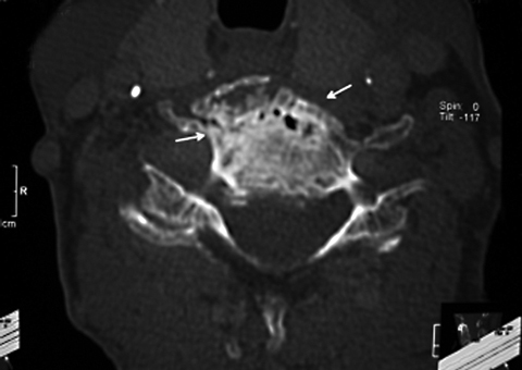
We report the case of a 65-year-old Brazilian woman with a 2-year history of BU in her extremities coexistent with osteomyelitis in the fourth cervical vertebra (Figure 1), and evidence of inadequate nutrition. Although she had received BCG vaccine as an infant, mycobacteria osteomyelitis developed in the site of an arthrodesis performed in 1998 to treat an accidental fracture.4,5
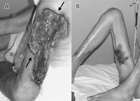
This patient had lived in a poor riverside rural area with a humid, hot climate. As in descriptions of Australian cases, our patient was much older than the age (5–15 years) at which most cases of M. ulcerans infection are reported in tropical and subtropical regions.1,2,4 Before her disease was characterised through positive cultures for M. ulcerans in samples from skin and bone lesions, the main differential diagnosis was ulcers resulting from fungal infection and leishmaniasis,4 conditions that are frequently seen in the region where she lived. The earlier skin lesions had appeared in May 2004 as papules and nodules, and evolved as painless, chronic, indolent ulcers with undermined edges.2,4 Despite treatment in another hospital that included surgery as well as medical therapy with rifamycin, aminoglycoside and quinolone antibiotics, the disease recurred. On admission to our hospital in August 2006, she had an extensive ulcer on her left arm in addition to scars on the right inner thigh (Figure 2).
After nearly 2 months of hospitalisation, the patient was discharged to continue antimicrobial therapy with outpatient follow-up. Despite this, the lesions are healing very slowly.
References
- Asiedu K, Wansbrough-Jones M. Mycobacterium ulcerans infection (Buruli or Bairnsdale ulcer): challenges in developing management strategies. Med J Aust 2006; 186: 55-56.
- Johnson PDR, Hayman JA, Quek TY, et al. Consensus recommendations for the diagnosis, treatment and control of Mycobacterium ulcerans infection (Bairnsdale or Buruli ulcer) in Victoria, Australia. Med J Aust 2007; 186: 64-68. 0_CBBCHJFE
- Lavender CJ, Senanayake SN, Fyfe JAM, et al. First case of Mycobacterium ulcerans disease (Bairnsdale or Buruli ulcer) acquired in New South Wales. Med J Aust 2006; 186: 62-63. 0_CBBHCGJF
- van der Werf TS, Stienstra Y, Johnson RC, et al. Mycobacterium ulcerans disease. Bull World Health Organ 2005; 83: 785-791. 0_CBBBAEDD
- Portaels F, Aguiar J, Debacker M, et al. Mycobacterium bovis BCG vaccination as prophylaxis against Mycobacterium ulcerans osteomyelitis in Buruli ulcer disease. Infect Immun 2004; 72: 62-65. 0_i1091856
Shifting focus to adolescent wellbeing and inclusive participation in the digital age
Allyson R Todd, Elena Wang, Stephanie R Partridge
The clinical presentation, investigation, and management of women diagnosed with endometriosis in Australian general practices, 2011–2021: an open cohort study
Danielle Mazza, Kailash Thapaliya, Sharinne B Crawford, Alissia Hui, Maryam Moradi, Luke E Grzeskowiak
The impact of the Breast Screen NSW transition from film to digital mammography, 2002–2016: a linked population health data analysis
Rachel Farber, Nehmat Houssami, Katy J L Bell
Non‐technical errors associated with deaths in surgical care, Australia, 2012–2019, by surgical specialty (Australian and New Zealand Audit of Surgical Mortality): a retrospective cohort study
Jesse Ey, Victoria Kollias, Octavia Lee, Kelly Hou, Matheesha Herath, John B North, Ellie Treloar, Suzanne Edwards, Martin Bruening, Adam J Wells, Guy J Maddern
The CURE Asthma roadmap
Gary P Anderson, Anthony Flynn, Phil G Bardin, John D Blakey, Shyamali C Dharmage, Paul Foster, Peter G Gibson, Adam Jaffe, Alan James, Christine R Jenkins, Sundram Sivamalai, Peter D Sly, Guy B Marks, Vanessa M McDonald, Judy Wetttenhall
Evidence from Australian cohort studies about asthma trajectories and transitions across the life course: a narrative review
Caroline Lodge, Xin Dai, Ingrid A. Laing, Michael P Menden, Anthony Flynn, Gary P Anderson, Sarath Ranganathan, Shyamali C Dharmage