




Volume 179 - Issue 10
What mole is that?
Author: Rodney D Sinclair
Med J Aust 2003 || doi: 10.5694/j.1326-5377.2003.tb05666.x
Published online: 11 March 2002
Published online: 11 March 2002
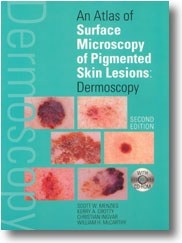
An atlas of surface microscopy of pigmented skin lesions: dermoscopy.
Scott W Menzies, Kerry A Crotty, Christian Ingvar, William H McCarthy. Sydney: McGraw-Hill, 2003 (vii + 175pp). ISBN 0 07 471102 4.
This book on surface microscopy focuses specifically on the diagnosis of pigmented skin lesions based upon their morphology and, in particular, their morphology when examined with a hand held surface microscope. The authors have data to show that general practitioners achieve a 39% improvement in the sensitivity for the diagnosis of melanoma when using this Atlas. This cannot be specifically interpreted as meaning that fewer melanomas are missed when using this book, but it may translate to fewer benign naevi being excised unnecessarily. Nevertheless, any book that improves diagnostic accuracy has to be welcomed, and this book certainly does that.
Examination of pigmented lesions using surface microscopy is a double-edged sword. To the uninitiated, hand held microscopes are confusing and may actually diminish diagnostic accuracy. As such, no general practitioner should pick up a hand held surface microscope without first having read this book.
This excellent book is simply written and beautifully illustrated. It is the best of its type in the discipline and the first edition has been widely used by dermatologists and dermatology trainees. The steps to diagnosis are clear and constructive, and the quiz on the CD-ROM that accompanies the book is also very useful.
At $94.95, including a CD-ROM, it is excellent value and I recommend it to all general practitioners contemplating the use of a hand held surface microscope.
Rodney D SinclairDermatologistMelbourne, VIC
Invasive melanoma - clinical view (a) and surface microscopy (b). Reproduced with permission.
Diagnosing Acute Kava Dermopathy: A Case Report of a Characteristic Cutaneous Eruption
Ali Abid, Nicholas Allen, Abeer Hagelamin, Christopher Henderson, Artiene Tatian
Localised Herpes Simplex Following Midline Laparotomy
Jessica S. Bulluss, Paul Chee, Matthew J. Verheyden
No silver lining with health misinformation: argyria caused by intentional silver consumption
Luke Collins, Logesh Palanikumar, Stephen Bacchi
Lingual Raynaud phenomenon
Michael Taggart, Mina John
Melasma in the male: a less well recognised entity
Tim Aung, Rowland Noakes
Age group‐specific changes in keratinocyte cancer treatment rates in Australia, 2012–2021: a retrospective cohort study based on MBS claims data
Catherine M Olsen, Nirmala Pandeya, Rachel E Neale, David C Whiteman