




Medical education
Snapshot
Volume 218 - Issue 3
Wong‐type dermatomyositis
Authors: Li Chai and Ze‐Hu Liu
Med J Aust 2023; 218 (3): 113-113. || doi: 10.5694/mja2.51802
Published online: 20 February 2023
Published online: 20 February 2023
A previously healthy 27-year-old woman presented to the dermatology department following a 3-month history of an itchy erythema
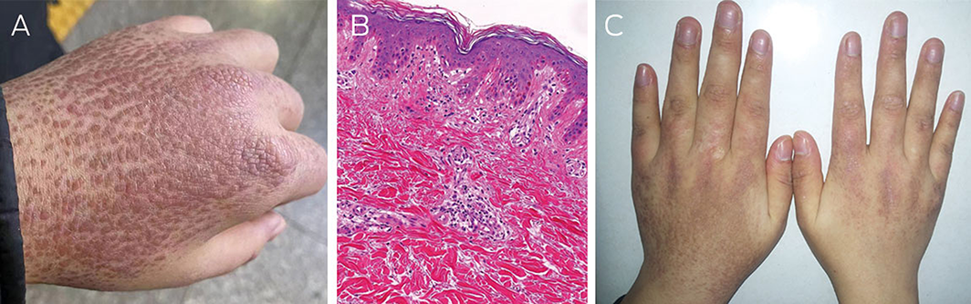
A previously healthy 27‐year‐old woman presented to the dermatology department following a 3‐month history of an itchy erythema initially involving the face and which later progressed to her trunk and upper limbs. Physical examination revealed multiple hyperkeratotic follicular papules on the hands (Figure A). Laboratory examination revealed elevated levels of creatine kinase (487 U/L; reference interval, 40–200 U/L). Autoimmune antibody tests were negative. Electromyography revealed a myogenic pattern, and a myositis panel was positive for transcription intermediary factor 1‐γ (TIF1‐γ) antibody. The histopathological features revealed follicular hyperkeratosis and superficial lymphocytic perivascular and vacuolar interface alterations (Figure B [haematoxylin‐eosin stain, original magnification ×100]). A series of comprehensive malignancy workup was negative. The patient was diagnosed with Wong‐type dermatomyositis, a rare variant of dermatomyositis, characterised by pinkish, hyperkeratotic follicular papules with intervening areas of unaffected skin known as “islands of sparing”.1 To our knowledge, only 30 cases have been reported in the literature.2 The patient was successfully treated with systemic methylprednisolone (initially 40mg per day, subsequently transitioned to 10mg per day) and tacrolimus (initially 0.5mg twice a day, subsequently transitioned to 1mg twice a day) (Figure C).
Competing interests
No relevant disclosures.
References
- Wong KO. Dermatomyositis: a clinical investigation of twenty‐three cases in Hong Kong. Br J Dermatol 1969; 81: 544‐547.
- Bax CE, Grinnell M, Concha JSS, Werth VP. Wong‐type dermatomyositis in an African American patient. Arthritis Rheumatol 2021; 73: 630.
Rhabdomyolysis After COVID-19
Navya Kataria, Abel Devadass, Lana Sundac, Laura De Souza, Nabeel Sheikh, Samantha Ng, Scott Campbell, Yishen Wang, Ross S. Francis
Technological advances in the search for a CURE for asthma
Sara Quon, Timothy M Johanson, Ridhima Wadhwa, Alen Faiz, Anthony Flynn, Gary P Anderson, Amanda J Cox, Nicholas P West, Michael P Menden, Rhys S Allan
The CURE Asthma roadmap
Gary P Anderson, Anthony Flynn, Phil G Bardin, John D Blakey, Shyamali C Dharmage, Paul Foster, Peter G Gibson, Adam Jaffe, Alan James, Christine R Jenkins, Sundram Sivamalai, Peter D Sly, Guy B Marks, Vanessa M McDonald, Judy Wetttenhall
The first live term birth following uterus transplantation in Australia
Rebecca Deans, Brigitte Gerstl, Antonia W Shand, Sarah Lyons, Aaron Budden, Helen L Barrett, Grant Luxton, Mangalee Fernando, Kenneth Yong, Karen Keung, Kaushalya Arulpragasam, Henry Pleass, King Man Wan, Eva Kehag, Jana‐Emily Pittman, Mianna Lotz, Maria Fenn, Erin Nesbitt‐Hawes, Lily Byun, Katrina Tang, Mats Brannstrom, Jason Abbott
The public health impacts of mining in Australia
Javier Cortes‐Ramirez, Ruby N Michael, Leisa‐Maree Toms, Melissa Haswell
Lingual Raynaud phenomenon
Michael Taggart, Mina John