




Medical education
Lessons from practice
Volume 216 - Issue 11
Strongyloides hyperinfection: a preventable complication of immunosuppression
Authors: Robert G Stolz, Mark R Dowling, Belinda B Lin, Kylie Mason and Stephen Muhi
Med J Aust 2022; 216 (11): 561-562. || doi: 10.5694/mja2.51561
Published online: 20 June 2022
Published online: 20 June 2022
A 70-year-old man of non-English speaking background migrated from Vietnam to Melbourne, Victoria, in 2007.
Clinical record
A 70‐year‐old man of non‐English speaking background migrated from Vietnam to Melbourne, Victoria, in 2007. In October 2019, he was diagnosed with plasmablastic lymphoma and commenced chemotherapy with rituximab, etoposide, prednisolone, vincristine, cyclophosphamide and doxorubicin. Before chemotherapy, he was screened for tuberculosis using QuantiFERON‐TB Gold (QIAGEN) and for blood‐borne viruses using serology. Twelve days after his third cycle of chemotherapy, he developed pain in the left iliac fossa, anorexia, rigors and a dry cough.
On presentation, he was febrile (39°C) and in septic shock. He required intensive care for inotropic support and was prescribed piperacillin–tazobactam following blood culture collection. Investigations revealed pancytopenia, with neutropenia 0.2×10⁹/L (reference interval [RI], 2.0–8.0×10⁹/L) and an eosinophil count of 0.1×10⁹ (RI, 0.0–0.5×10⁹/L). Inflammatory markers were elevated, with C‐reactive protein 237mg/L (RI, <5mg/L). Blood cultures did not reveal bacteraemia. Computed tomography of the abdomen and pelvis demonstrated multifocal colonic inflammation.
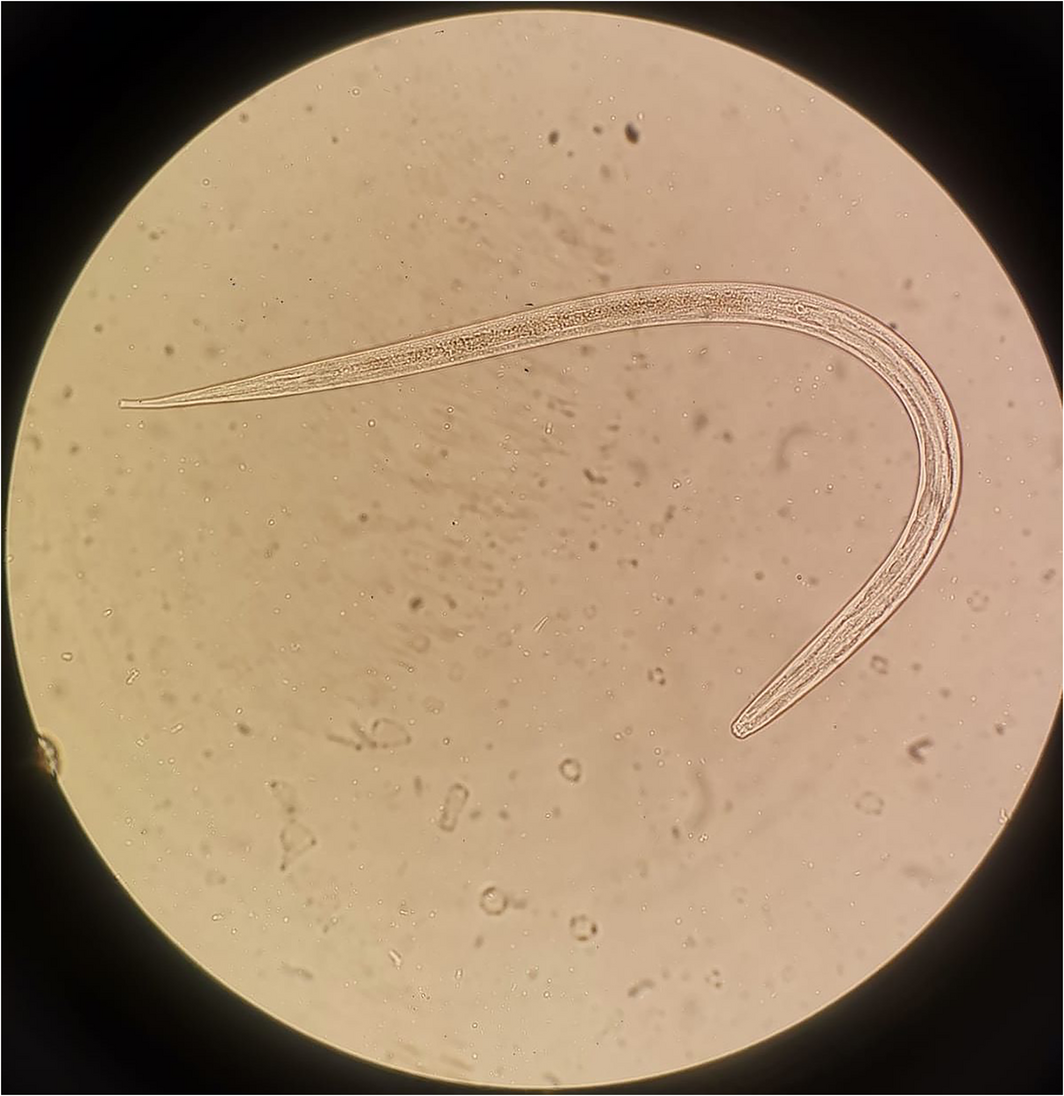
He then developed non‐bloody diarrhoea, and general faecal microscopy and culture revealed multiple parasites consistent with filariform Strongyloides stercoralis (Box). Harada‐Mori culture was examined after 24 hours using light microscopy demonstrating characteristic thrashing motility (, video). S. stercoralis serology was negative. He commenced daily oral ivermectin for 14days, with stool examination at day 13 of treatment confirming clearance. Clinically, he responded rapidly to ivermectin, and continued chemotherapy with outpatient observation. One year after treatment he remains in remission.
Discussion
S. stercoralis is an intestinal parasitic roundworm with global prevalence which can lead to lifelong infection by way of its ability to autoinfect humans. At‐risk groups within Australia include migrants and returned travellers from tropical and subtropical regions, as well as Indigenous people from rural and remote areas of northern Australia.1 Immunocompetent people with chronic strongyloidiasis are often asymptomatic or exhibit mild respiratory or gastrointestinal symptoms with no serious sequelae.2
Patients undergoing immunosuppressive treatment are at risk of developing Strongyloides hyperinfection and dissemination due to the rapid proliferation of larvae, resulting in sepsis and multiorgan dysfunction. Exposure to high dose glucocorticoids is the strongest risk factor, with hyperinfection seen in patients receiving as little as 6days of high dose prednisolone.2 Therefore, patients receiving glucocorticoid‐containing regimes for haematological malignancies are at particularly high risk.
In immunocompromised hosts, the case fatality rate of hyperinfection is at least 60%.3 Lung infiltration results in cough, with chest infiltrates on imaging, which may progress to diffuse alveolar haemorrhage; gastrointestinal symptoms vary from diffuse bloody diarrhoea to paralytic ileus.2 Translocation of enteric bacteria accompanying larvae that penetrate through the intestinal wall can cause sepsis. Despite septic shock, we did not isolate an enteric organism in blood cultures.
Sensitivity of S. stercoralis serology for chronic infection is only 83–89%.2 Therefore, all patients currently living in endemic regions should receive ivermectin before immunosuppression, with ivermectin dosing schedules depending on serostatus.4 Patients who continue to live in or visit endemic regions should receive ongoing prophylactic ivermectin during immunosuppression, without further investigation if asymptomatic.4 Moreover, in patients who are already immunosuppressed, sensitivity of serology is further reduced, highlighting the importance of screening all patients who were born in, resided in or visited endemic regions before treatment.4
If hyperinfection is suspected, empiric treatment should include ivermectin together with broad spectrum coverage for enteric bacteria.2 Microscopy is particularly helpful in patients with hyperinfection, who are more likely to have visible larvae on direct inspection of faeces or sputum.3 Culture‐based techniques, such as the Harada‐Mori or agar plate culture, are more sensitive (72–89%) compared with microscopy alone but should not delay empirical treatment if hyperinfection is suspected.5 Due to the intermittent release of larvae, submission of multiple samples is recommended. Eighty per cent of patients with hyperinfection will not have eosinophilia at presentation, with concomitant corticosteroid therapy blunting eosinophilic responses, making eosinophilia an unreliable marker of disease.3
Although case reports show large variation in treatment regimens for hyperinfection, the accepted treatment of choice is ivermectin. Despite ivermectin being generally well tolerated, case reports of encephalopathy due to P‐glycoprotein polymorphisms have been reported.6 Albendazole monotherapy is likely inferior to ivermectin or mebendazole.3
A recent audit of screening for latent strongyloidiasis at the Royal Melbourne Hospital in patients undergoing immunosuppression demonstrated a screening rate of 38%.7 Moreover, patients from non‐English speaking backgrounds and older patients were 2.5 times less likely to be screened, despite clinician‐reported awareness of latency of infection. This case further highlights the importance of screening and pre‐emptive treatment, with vulnerable patients at particular risk of this neglected tropical disease.
Lessons from practice
- Patients who have previously lived in or visited Strongyloides endemic regions should be serologically screened for latent strongyloidiasis before significant immunosuppression, and receive pre‐emptive ivermectin if seropositive.
- Patients who currently live in or visit Strongyloides endemic regions should receive ivermectin before immunosuppression and ongoing prophylactic ivermectin during immunosuppression, regardless of serostatus.
- Maintain a high index of suspicion for Strongyloides hyperinfection in immunosuppressed patients from high risk groups with sepsis; direct examination of faeces or sputum under microscopy provides rapid diagnosis, and faecal culture techniques may increase diagnostic yield.
- There remain numerous barriers to screening patients for latent strongyloidiasis, and we encourage clinicians to develop protocolised screening tools to allow for more consistent screening of patients before immunosuppression.
Competing interests
No relevant disclosures.
References
- Miller A, Smith ML, Judd JA, Speare R. Strongyloides stercoralis: systematic review of barriers to controlling strongyloidiasis for Australian indigenous communities. PLoS Negl Trop Dis 2014; 8: e3141.
- Greaves D, Coggle S, Pollard C, et al. Strongyloides stercoralis infection. BMJ 2013; 347: f4610.
- Buonfrate D, Requena‐Mendez A, Angheben A, et al. Severe strongyloidiasis: a systematic review of case reports. BMC Infect Dis 2013; 13: 78.
- eTG Complete. Strongyloides stercoralis prophylaxis in immunocompromised adults without HIV infection. West Melbourne (Australia): Therapeutic Guidelines Limited; 2019. https://www.tg.org.au/ (viewed June 2021).
- Polanco L, Gutiérrez L, Cardona Arias J. [Diagnosis of Strongyloides stercoralis infection. Meta‐analysis on evaluation of conventional parasitological methods (1980–2013)] [Spanish]. Rev Esp Salud Publica 2014; 88: 581‐600.
- Baudou E, Lespine A, Durrieu G, et al. Serious ivermectin toxicity and human ABCB1 nonsense mutations. N Engl J Med 2020; 383: 787‐789.
- Muhi S, Ko DK, McGuinness SL, et al. A mixed‐method analysis of screening for Strongyloides stercoralis prior to immunosuppression: a problem of limited bandwidth? Intern Med J 2020; doi: [Epub ahead of print].
Provenance: Not commissioned; externally peer reviewed.
Costs of Delivering the Hep B PAST Model of Care in Remote Australia: A Retrospective Pre–Post Study
Hoa Nguyen, Anh Le Tuan Nguyen, Paula Binks, Melita McKinnon, Emily Vintour-Cesar, Karen Wills, Nicola Stephens, Benjamin Cowie, Joshua S. Davis, Yuejen Zhao, Peter Nihill, Julie A. Campbell, Andrew J. Palmer, Jane Davies, Barbara de Graaff
Buruli Ulcer Acquired From Ringtail Possum Rescue
Daniel P. O'Brien, Naomi Clarke, Sara Bonnici, Mohammad A. Hussain
Q Fever Visceral Abscesses After Contact With Native Australian Marsupials
Samantha J. Laws, Enzo Binotto, James Stewart, Jennifer Ho, Trent Yarwood, Josh Hanson, Simon Smith
Hospital-Admitted Injection-Related Infections Among Incarcerated People Who Inject Drugs in Australia: A Retrospective Cohort Study
Andrew Palmer, Matthew Carter, Jeremy Yeo, Cecilia Shim, Jason Connor, Jeremy Hayllar, Gerald Holtmann, Naomi Moy, Elliott G. Playford, Naomi Runnegar, Paul J. Clark
The Live Attenuated Influenza Vaccine in Australia: An Additional Tool for Influenza Prevention
Cyra Patel, Alexis Pillsbury, Tran Nguyen, Xia Wang, Helen E. Quinn, Clayton K. Chiu, Allen C. Cheng, Katie L. Flanagan, Zhicheng Wang
Reflex Testing for Hepatitis D Infection: A Unique Opportunity to Reduce Hepatitis D-Related Chronic Liver Disease Deaths in Australia
Jessica Howell, Lauren Andersson, Miriam T. Levy, James O'Beirne, Leon Adams, Katharine Irvine, Avik Majumdar, Golo Ahlenstiel, Kathy Jackson, Krispin Hajkowicz, Joseph Doyle, Jane Davies, Sarah Cherian, Wayne Dimech, Alexander J. Thompson