




Medical education
Lessons from practice
Volume 216 - Issue 11
Kerion: a great mimicker
Authors: Abhinav Rajkumar and Philip N Britton
Med J Aust 2022; 216 (11): 563-567. || doi: 10.5694/mja2.51588
Published online: 20 June 2022
Published online: 20 June 2022
A previously well 9-year-old boy presented to the outpatients’ clinic with a tender, swollen occipital scalp lesion progressing over one month’s duration
Clinical record
A previously well 9‐year‐old boy presented to the outpatients’ clinic with a tender, swollen occipital scalp lesion progressing over one month’s duration; erythema, scaling, focal alopecia and occipital lymphadenopathy were noted (Box 1, A). He was systemically well with no fever. There was no response to 2weeks’ oral cephalexin. He was admitted for surgical drainage; scalp scrapings and surgical aspirates were sent for bacterial and fungal cultures. He was discharged the following day with 2weeks’ oral cephalexin and topical scalp miconazole.
Three weeks later, the surgical site showed a persistent skin defect, alopecia and scaling (Box 1, B). Scalp scrapings grew Trichophyton tonsurans with no fungal or bacterial growth from surgical aspirates. Kerion was diagnosed clinically, supported by scalp scraping results, and oral terbinafine was commenced for one month. After 3weeks of terbinafine, the skin defect persisted, with suboptimal healing and ongoing scaling (Box 1, C); therapy was switched to second line weekly oral fluconazole. After one month’s fluconazole, the skin defect had healed, with no scaling; early hair regrowth was seen (Box 1, D).
Discussion
Tinea capitis is a fungal dermatophyte infection of the scalp and hair follicles most commonly seen in pre‐pubescent children and is more prevalent in boys than in girls.1,2 Dermatophytes can inhabit animal skin (zoophilic; eg, Microsporum canis), human skin (anthropophilic; eg, T. tonsurans) or soil (geophilic; eg, Nannizzia gypsea).2 Recent phylogenetic insights have prompted reclassification of several dermatophyte genera.3 Trichophyton and Microsporum genera most commonly cause tinea capitis worldwide, with causative species varying by geographical location.2 In New South Wales, Australia, T. tonsurans and M. canis are most common,4 with risk factors for tinea capitis including animal exposure, scalp shaving, overcrowding and suboptimal hygiene.1 Tinea capitis is common in specific refugee5 and Indigenous communities6 in Australia.
Mild forms of tinea capitis present with black dots, representing broken hair follicles, and scaly scalp patches.1 In contrast, kerion (Greek for “honeycomb”) is severe tinea capitis with tender, erythematous, boggy pustules with overlying crusting, and itchy, scaling dermatophytid reactions developing over several weeks.7 Focal alopecia and occipital or cervical lymphadenopathy are common.7 Kerion results from intense host‐derived inflammatory responses, as dermatophytes do not directly cause tissue damage or invade tissues in immunocompetent hosts. Zoophilic dermatophytes (particularly M. canis) and endothrix dermatophytes that preferentially infect deep hair shafts (particularly T. tonsurans) more commonly cause progression to kerion, as they induce more pronounced, deep‐seated dermal inflammatory responses.7,8,9
Clinical features of kerion can cause diagnostic confusion, with frequent misdiagnosis as bacterial scalp abscesses and other conditions (Box 2).7 Diagnosis is primarily clinical but is aided by additional diagnostic tests (Box 3).7 Direct microscopy and fungal cultures on scalp scrapings help support the diagnosis, although a third of clinically diagnosed kerion may be fungal culture negative.9 Nucleic acid tests now available in Australia can rapidly identify dermatophytes in culture negative cases.10 Bacterial cultures are unnecessary when signs of local superinfection are absent.9
The treatment of tinea capitis, including kerion, involves oral antifungal therapy; topical antifungal therapies do not effectively penetrate hair shafts and follicles.11 Randomised controlled trials indicate that prolonged courses of oral griseofulvin, terbinafine or fluconazole are effective against all forms of tinea capitis, including kerion (Box 4); shorter durations are associated with relapse, and treatment should continue until lesion resolution.11 Bacterial superinfection is uncommon, and concomitant antibacterial therapy in uncomplicated cases is not beneficial.9 Lack of clinical improvement after 4weeks of appropriate antifungal therapy should prompt use of second line agents.11 Thrice weekly treatment with a sporicidal shampoo, such as selenium sulphide, helps eradicate fungal conidiospores and arthroconidia, which can transmit infection to contacts and cause reinfection.1
Limiting dermatophyte transmission is important, as studies suggest secondary attack rates of up to 30% in tinea capitis in classroom settings.12 Asymptomatic household contacts require thrice weekly treatment with sporicidal shampoo, with prompt treatment of infected individuals.1 Veterinary examination and treatment of infected pets is important in cases with zoophilic dermatophytoses.1 Towels, pillows, bed sheets and hair trimmers of index cases should not be shared and should be sterilised.1
Case series indicate that delayed, inappropriate treatment of kerion can lead to non‐reversible alopecia.9 In immunocompetent, systemically well patients, case reports suggest surgical drainage of kerion offers little additional benefit, with procedures themselves possibly contributing to scarring.13,14 Concomitant oral corticosteroid therapy in kerion remains controversial; studies suggest corticosteroids may reduce symptoms, but do not reduce the rates of irreversible alopecia.15 When kerion is promptly and appropriately treated, case reports indicate irreversible alopecia can be avoided, with positive treatment outcomes.9
Lessons from practice
- Kerion is a severe form of tinea capitis caused by inflammatory reactions to dermatophyte scalp infections. It can be easily confused with bacterial scalp abscess and various other conditions. If the child is unwell, immunocompromised or sustained penetrating traumatic scalp injury, serious bacterial or invasive fungal scalp infection requires consideration.
- Diagnosis is primarily clinical, but is aided by dermoscopy, microscopy and fungal culture of scalp scrapings. Bacterial superinfection is uncommon; bacterial cultures and antibacterial therapy is unnecessary in most cases. Prolonged courses of oral griseofulvin, terbinafine or fluconazole are needed; shorter durations are associated with relapse, and topical antifungal therapy is insufficient. Infected individuals should use sporicidal shampoo to minimise transmission.
- Asymptomatic household contacts require use of sporicidal shampoo, and infected household members (and infected pets in zoophilic infections) require examination and treatment. Infected bedding and hair trimmers should be sterilised and not shared.
- Delayed or inappropriate treatment of kerion can result in persistent inflammation and irreversible alopecia. Oral steroids may reduce pain but have not been shown to reduce alopecia. Routine surgical drainage in uncomplicated cases is not shown to be beneficial and may instead contribute to further scarring.
Box 1 – (A) Appearance of scalp lesions on initial surgical outpatient clinic review before surgical drainage;* (B) appearance on clinic review after incision and drainage and treatment with oral antibiotics and topical antifungals,† (C) after one month’s treatment with oral terbinafine,‡ and (D) after one month of treatment with weekly fluconazole§
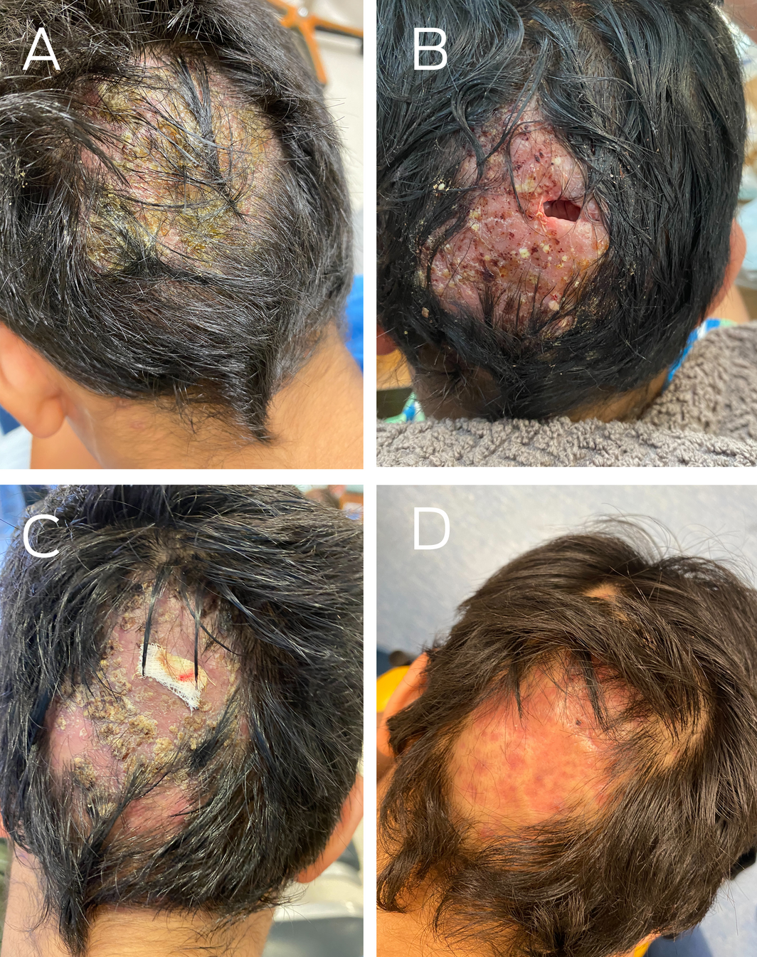
* Crusted, pustular lesions with associated scaling and focal hair loss. † Poorly healing skin defect at surgical site, with persistent scaly lesions and hair loss. ‡ Some suboptimal healing has occurred. The skin defect at the surgical site (underneath piece of gauze) has not fully healed, with ongoing scaling and persistent hair loss. § Skin defect has closed and healed and scaling has resolved, with some early regrowth of hair seen.
Box 2 – Clinical features seen in kerion compared with common differential diagnoses7,16
|
Clinical features |
Condition |
||||||||||||||
|
Kerion |
Bacterial scalp abscess* |
Bacterial folliculitis |
Dissecting folliculitis† |
Folliculitis decalvans‡ |
Invasive fungal scalp infection§ |
Seborrheic dermatitis¶ |
Autoimmune causes of alopecia** |
Contact dermatitis |
Eczema or psoriasis†† |
Neoplastic scalp diseases‡‡ |
|||||
|
|
|||||||||||||||
|
Scaly skin lesions |
Yes |
No |
No |
No |
No |
Yes |
Yes§§ |
Sometimes¶¶ |
Yes |
Yes |
Sometimes seen in LCH |
||||
|
Pustules with overlying crusting |
Yes |
Yes |
Yes |
Yes |
Yes |
Yes |
No |
No |
No |
No |
No |
||||
|
Tender, boggy lesions |
Yes |
Yes |
No |
Yes |
Yes |
Yes |
No |
No |
No |
No |
No |
||||
|
Localised cervical or occipital lymphadenopathy |
Yes |
Yes |
No |
Yes |
Yes |
Yes |
No |
No |
No |
No |
Yes |
||||
|
Focal or diffuse |
Focal |
Focal |
Focal |
Diffuse |
Diffuse |
Focal |
Diffuse |
Focal |
Focal |
Focal |
Focal/diffuse*** |
||||
|
Significant alopecia |
Yes |
No |
No |
Yes |
Yes |
Yes |
No |
Yes |
No |
No |
Yes |
||||
|
Response to antibacterial therapy |
No |
Yes |
Yes |
Yes |
Yes |
No |
No |
No |
No |
No |
No |
||||
|
Response to antifungal therapy |
Yes |
No |
No |
No |
No |
Sometimes††† |
Yes‡‡‡ |
No |
No |
No |
No |
||||
|
|
|||||||||||||||
|
LCH = Langerhans cell histiocytosis. * Rarely seen except in individuals with immune deficiency or secondary infection following preceding traumatic or penetrating scalp injury. † Inflammatory responses to blocked follicles, associated with folliculitis in other body regions, associated with alopecia, more common in adults. ‡ Inflammatory responses to skin bacteria by‐products in susceptible individuals, associated with alopecia, more common in adults. § In penetrating scalp injuries and in immunocompromised hosts, invasive fungal scalp infection requires consideration, often needing urgent investigation and treatment. In chronic mucocutaneous candidiasis, recurrent Candida scalp infections due to immune dysregulation and interleukin pathway defects can be associated with alopecia, but invasive disease is uncommon. ¶ Irritant reaction to products from scalp colonisation with non‐dermatophyte Malassezia fungi. ** Autoimmune disorders can be primary (alopecia areata) or systemic (lupus erythematosus, dermatomyositis). †† Psoriasis and atopic dermatitis are often associated with scaly plaques at other body sites. ‡‡ Uncommon; LCH can present with a scaly scalp mass. §§ Often greasy. ¶¶ Sometimes seen in dermatomyositis. *** In LCH. ††† If agent active against fungus. ‡‡‡ Topical antifungal shampoo. |
|||||||||||||||
Box 3 – Investigations that can help support the diagnosis of kerion7,10
|
Diagnostic investigation |
Finding |
||||||||||||||
|
|
|||||||||||||||
|
Appearance under ultraviolet light |
|
||||||||||||||
|
Dermoscopic examination |
|
||||||||||||||
|
Microscopy of skin scrapings or plucked hairs with added potassium hydroxide (dissolves keratinocytes) |
|
||||||||||||||
|
Fungal culture of skin scrapings, sterile brushed specimen or plucked hairs |
|
||||||||||||||
|
Histology |
|
||||||||||||||
|
Nucleic acid based diagnostic methods |
|
||||||||||||||
|
|
|||||||||||||||
|
|
|||||||||||||||
Box 4 – Treatment regimens for kerion and tinea capitis11
|
Medication |
Mechanism |
Indication |
Suggested durations of therapy and monitoring |
Side effects, monitoring and interactions |
|||||||||||
|
|
|||||||||||||||
|
Oral griseofulvin |
|
|
|
|
|||||||||||
|
Oral terbinafine |
|
|
|
|
|||||||||||
|
Oral azoles (fluconazole, itraconazole)* |
|
|
|
|
|||||||||||
|
|
|||||||||||||||
|
PPI = proton pump inhibitor. * Voriconazole is rarely used as it has a similar activity compared with other azoles but a higher chance of adverse reactions. |
|||||||||||||||
Competing interests
No relevant disclosures.
References
- Kelly BP. Superficial fungal infections. Pediatr Rev 2012; 33: e22‐e37.
- Rodríguez‐Cerdeira C, Martínez‐Herrera E, Szepietowski JC, et al. A systematic review of worldwide data on tinea capitis: analysis of the last 20years. J Eur Acad Dermatol Venereol 2021; 35: 844‐883.
- de Hoog GS, Dukik K, Monod M, et al. Toward a novel multilocus phylogenetic taxonomy for the dermatophytes. Mycopathologia 2017; 182: 5‐31.
- Rogers M, Muir D, Pritchard R. Increasing importance of Trichophyton tonsurans in childhood tinea in New South Wales. The pattern of childhood tinea in New South Wales, Australia 1979–1988: the emergence of Trichophyton tonsurans as an important pathogen in tinea capitis in white children. Australas J Dermatol 1993; 34: 5‐8.
- Mutch RC, Cherian S, Nemba K et al. Tertiary paediatric refugee health clinic in Western Australia: analysis of the first 1026 children. J Paediatr Child Health 2012; 48: 582‐587.
- Kaminski GW, Green AC. Tinea capitis in aboriginal children at Maningrida, Northern Territory, Australia. A variant of Microsporum canis. Australas J Dermatol 1977; 18: 88‐97.
- John AM, Schwartz RA, Janniger CK. The kerion: an angry tinea capitis. Int J Dermatol 2018; 57: 3‐9.
- Aste N, Pau M, Biggio P. Kerion Celsi: a clinical epidemiological study. Mycoses 1998; 41: 169‐173.
- Proudfoot LE, Higgins EM, Morris‐Jones R. A retrospective study of the management of pediatric kerion in Trichophyton tonsurans infection. Pediatr Dermatol 2011; 28: 655‐657.
- Ross IL, Weldhagen GF, Kidd SE. Detection and identification of dermatophyte fungi in clinical samples using a commercial multiplex tandem PCR assay. Pathology 2020; 52: 473‐477.
- Fuller LC, Barton RC, Mohd Mustapa MF, et al. British Association of Dermatologists’ guidelines for the management of tinea capitis 2014. Br J Dermatol 2014; 171: 454‐463.
- Fox PB, Rush‐Munro FM. Tinea capitis due to Trichophyton sulphureum. N Z Med J 1953; 52: 488‐493.
- von Laer Tschudin L, Laffitte E, Baudraz‐Rosselet F, et al. Tinea capitis: no incision nor excision. J Pediatr Surg 2007; 42: E33‐E36.
- Gibbon KL, Goldsmith P, Salisbury JA, Bewley AP. Unnecessary surgical treatment of fungal kerions in children. BMJ 2000; 320: 696‐697.
- Hussain I, Muzaffar F, Rashid T, et al. A randomized, comparative trial of treatment of kerion celsi with griseofulvin plus oral prednisolone vs. griseofulvin alone. Med Mycol 1999; 37: 97‐99.
- Errichetti E, Stinco G. Dermoscopy in general dermatology: a practical overview. Dermatol Ther (Heidelb) 2016; 6: 471‐507.
Provenance: Not commissioned; externally peer reviewed.
Costs of Delivering the Hep B PAST Model of Care in Remote Australia: A Retrospective Pre–Post Study
Hoa Nguyen, Anh Le Tuan Nguyen, Paula Binks, Melita McKinnon, Emily Vintour-Cesar, Karen Wills, Nicola Stephens, Benjamin Cowie, Joshua S. Davis, Yuejen Zhao, Peter Nihill, Julie A. Campbell, Andrew J. Palmer, Jane Davies, Barbara de Graaff
Buruli Ulcer Acquired From Ringtail Possum Rescue
Daniel P. O'Brien, Naomi Clarke, Sara Bonnici, Mohammad A. Hussain
Q Fever Visceral Abscesses After Contact With Native Australian Marsupials
Samantha J. Laws, Enzo Binotto, James Stewart, Jennifer Ho, Trent Yarwood, Josh Hanson, Simon Smith
Hospital-Admitted Injection-Related Infections Among Incarcerated People Who Inject Drugs in Australia: A Retrospective Cohort Study
Andrew Palmer, Matthew Carter, Jeremy Yeo, Cecilia Shim, Jason Connor, Jeremy Hayllar, Gerald Holtmann, Naomi Moy, Elliott G. Playford, Naomi Runnegar, Paul J. Clark
The Live Attenuated Influenza Vaccine in Australia: An Additional Tool for Influenza Prevention
Cyra Patel, Alexis Pillsbury, Tran Nguyen, Xia Wang, Helen E. Quinn, Clayton K. Chiu, Allen C. Cheng, Katie L. Flanagan, Zhicheng Wang
Reflex Testing for Hepatitis D Infection: A Unique Opportunity to Reduce Hepatitis D-Related Chronic Liver Disease Deaths in Australia
Jessica Howell, Lauren Andersson, Miriam T. Levy, James O'Beirne, Leon Adams, Katharine Irvine, Avik Majumdar, Golo Ahlenstiel, Kathy Jackson, Krispin Hajkowicz, Joseph Doyle, Jane Davies, Sarah Cherian, Wayne Dimech, Alexander J. Thompson