




Medical education
Lessons from practice
Volume 215 - Issue 7
An unusual case of non‐infective endocarditis in undiagnosed antiphospholipid syndrome
Authors: Jessica V Yao, Subodh B Joshi, John G Morgan and Melissa GY Lee
Med J Aust 2021; 215 (7): 311-312.e1. || doi: 10.5694/mja2.51246
Published online: 4 October 2021
Published online: 4 October 2021
A 57-year-old man presented with left facial droop and dysarthria due to a multiterritory ischaemic stroke affecting the right posterior and middle cerebral arteries
Clinical record
A 57‐year‐old man presented with left facial droop and dysarthria due to a multiterritory ischaemic stroke affecting the right posterior and middle cerebral arteries. His previous history included a segmental pulmonary embolus in the setting of pneumonia. He was not taking any regular medications and denied recreational drug use. There was no relevant family history. He was haemodynamically stable, afebrile and euvolaemic. White cell count was normal at 7.8 × 10⁹/L (reference interval [RI], 4–12 × 109/L). C‐reactive protein was mildly elevated at 9 mg/L (RI, < 5 mg/L). On day 1, transthoracic echocardiogram revealed a vegetation on the anterior mitral valve leaflet tip with moderate to severe mitral regurgitation, normal left atrial size and biventricular size and systolic function. Three sets of blood cultures were taken before commencing intravenous ceftriaxone and flucloxacillin. He was transferred to our hospital for surgical consideration.
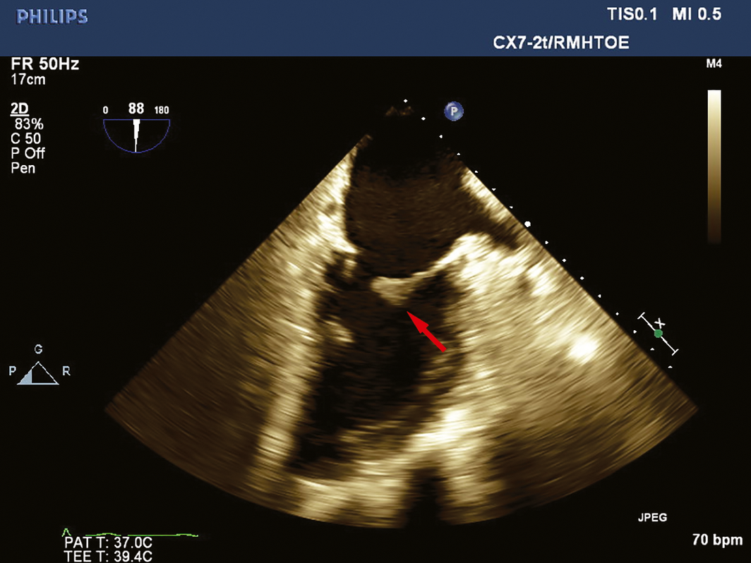
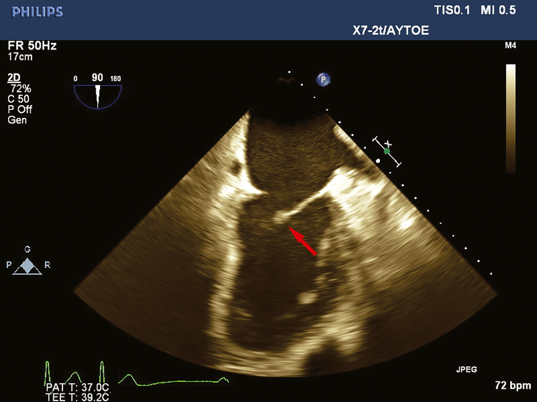
On day 3, transoesophageal echocardiogram confirmed a 2 cm independently mobile lesion on the anterior mitral valve leaflet tip with severe central mitral regurgitation (Box 1). Extended 27‐day blood cultures remained negative. Routine coagulation studies revealed an elevated activated partial thromboplastin time (aPTT) of 77 seconds (RI, 23–36 seconds) with normal international normalised ratio (INR). aPTT did not correct with mixing studies. Elevated dilute Russell viper venom time screen ratio of 2.63 (RI, < 1.2) confirmed presence of lupus anticoagulant. Thrombophilia screen confirmed high anticardiolipin IgG > 2024 (RI, < 20) and β2‐glycoprotein 1 antibody > 6100 (RI, < 20) suggestive of antiphospholipid syndrome. There were no clinical features of systemic lupus erythematosus and autoimmune screen was normal. Computed tomography of the chest, abdomen, and pelvis demonstrated no embolic events or malignancy. A diagnosis of non‐infective endocarditis (NIE) due to presumed antiphospholipid syndrome was made. A multidisciplinary consensus opinion involving cardiology, cardiothoracic surgery, infectious diseases, haematology, and stroke teams was made for non‐surgical management with warfarin (target INR, 2–3). Antibiotics were ceased. He was discharged home on day 10. Anticardiolipin IgG and β2‐glycoprotein 1 antibody remained elevated at 12 weeks, confirming antiphospholipid syndrome. Repeat transoesophageal echocardiogram at 3 weeks demonstrated significantly reduced mitral valve lesion size with only mild mitral regurgitation (Box 2). The patient continues on lifelong warfarin with no residual stroke symptoms.
Discussion
While NIE is rarely diagnosed clinically,1 in an autopsy study, NIE was three times more common than infective endocarditis.2 NIE is associated with high morbidity and mortality mainly due to under‐recognition. Causes include mechanical stress, hypercoagulable states such as malignancy, and autoimmune diseases including antiphospholipid syndrome.2,3 Turbulent blood flow in hypercoagulable states is hypothesised to induce endothelial cell injury leading to platelet and fibrin deposition interweaving to form vegetations,3 most commonly affecting the aortic and mitral valves.3,4 Systemic embolisation affects 40% of patients with NIE.4
Differentiating NIE from infective endocarditis can be challenging. Clinically, infective endocarditis usually manifests with fevers and weight loss, whereas NIE commonly presents as recurrent systemic embolisation.1 Patients with NIE often have features suggestive of their underlying condition such as constitutional symptoms of occult malignancy, arthritis suggestive of systemic lupus erythematosus, or recurrent thromboses indicating antiphospholipid syndrome. Stroke is more common in NIE compared with infective endocarditis (33% v 13%),3 with NIE vegetations more likely to detach given minimal inflammation or adherence to the valve.4
Close scrutiny of laboratory markers is important, as abnormalities may indicate particular NIE‐associated conditions. On echocardiogram, NIE lesions are typically 0.1–2 cm in diameter, broad‐based, sessile and irregular.1,3 They usually attach to the leaflet closure line and move with the leaflets, compared with independently mobile infective endocarditis vegetations. Significant valvular regurgitation is rare in NIE.1 Our case describes NIE in the setting of antiphospholipid syndrome. Despite unusual NIE echocardiographic features of an independently mobile mitral valve mass with severe mitral regurgitation, surgery was ultimately avoided with anticoagulation. Antiphospholipid syndrome is an autoimmune disease characterised by venous and arterial thromboses and recurrent miscarriages. Clinical features and laboratory criteria are crucial to diagnosis. The latter includes the identification of antiphospholipid antibodies or lupus anticoagulant on two occasions at least 12 weeks apart to minimise false positives. NIE occurs in up to 15% of patients with antiphospholipid syndrome.5
Mainstay NIE management involves treatment of the underlying condition and consideration of therapeutic anticoagulation to prevent recurrent embolisation.3 While warfarin is typically used in autoimmune‐associated NIE,5 heparin and enoxaparin are superior in malignancy‐related NIE. There is no consensus for use of direct oral anticoagulants or corticosteroids.3 Surgery is seldom necessary and reserved for severe valvular dysfunction, large vegetations, or recurrent embolism despite anticoagulation.
The diagnosis of NIE remains challenging based on clinical and echocardiographic features alone. Targeted history taking and scrutiny of investigations, including cultures, inflammatory markers, and coagulation studies, are paramount and may prevent unnecessary surgery. Our case exemplifies the value of a multidisciplinary approach to endocarditis.
Lessons from practice
- While uncommon, non‐infective causes should be considered in all patients with endocarditis.
- Non‐infective endocarditis can be difficult to distinguish from infective endocarditis based on clinical and echocardiographic features alone, and targeted history taking and scrutiny of blood cultures, inflammatory markers, and coagulation studies are necessary.
- The mainstay treatment of non‐infective endocarditis is treatment of the underlying cause. Surgery is seldom required.
Competing interests
No relevant disclosures.
References
- Lee V, Gilbert JD, Byard RW. Marantic endocarditis — a not so benign entity. J Forensic Leg Med 2012; 19: 312–315.
- Bussani R, De‐Giorgio F, Pesel G, et al. Overview and comparison of infectious endocarditis and non‐infectious endocarditis. a review of 814 autoptic cases. In Vivo 2019; 33: 1565–1572.
- Liu J, Frishman WH. Nonbacterial thrombotic endocarditis: pathogenesis, diagnosis, and management. Cardiol Rev 2016; 24: 244–247.
- Joshi SB, Richards MJ, Holt DQ, et al. Marantic endocarditis presenting as recurrent arterial embolization. Int J Cardiol 2009; 132: e14–16.
- Lenz CJ, Mankad R, Klarich K, et al. Antiphospholipid syndrome and the relationship between laboratory assay positivity and prevalence of non‐bacterial thrombotic endocarditis: a retrospective cohort study. J Thromb Haemost 2020; 18: 1408–1414.
Provenance: Not commissioned; externally peer reviewed.
Fatty Liver Disease in Australia: A Narrative Review on the Epidemiology, Natural History, Prognostication and Management in People With Metabolic Dysfunction
Karl Vaz, Daniel Clayton-Chubb, William W. Kemp, Stuart K. Roberts, Ammar Majeed
Sex Differences in Management and Outcomes of People With ST-Elevation Myocardial Infarction, New South Wales, 2011–2020: A Retrospective Cohort Study
Adam C. Livori, Jedidiah I. Morton, Dianna J. Magliano, Jonathan E. Shaw
Rethinking Rheumatic Fever Diagnosis: Progress, Pitfalls and the Path Towards a Definitive Test
Anna P. Ralph, Rachel Webb, Tom Parks, Nicole J. Moreland, Asha C. Bowen, Benjamin Jones, Emma Ndagire, Andrea Z. Beaton
Sex Differences in Management and Outcomes of People With ST-Elevation Myocardial Infarction, New South Wales, 2011–2020: A Retrospective Cohort Study
Samia Kazi, Simone Marschner, Clara K. Chow
Distribution of Cardiovascular Disease Risk Based on the Updated 2023 Guideline-Recommended Australian Cardiovascular Disease Risk Algorithm and Comparison With the 2012 Algorithm: An Observational Study
Nina Lazarevic, Meghana Bhat, Grace Joshy, Danielle C. Butler, Mark Woodward, Anushka Patel, Rod T. Jackson, Garry Jennings, Rosemary Wyber, Ellie Paige, Emily Banks
They Brought the Hospital to Me
Hugh N. Burrill