




Medical education
Lessons from practice
Volume 214 - Issue 8
Epipericardial fat necrosis: chest pain in a young pregnant woman
Authors: Rohan V Navani, Claudia Ashkar and Harry Gibbs
Med J Aust 2021; 214 (8): 355-356.e1. || doi: 10.5694/mja2.51007
Published online: 3 May 2021
Published online: 3 May 2021
A 28-year-old woman at 16 weeks’ gestation presented to the emergency department with a 2-day history of severe, worsening left- sided pleuritic
chest pain
Clinical record
A 28‐year‐old woman at 16 weeks’ gestation presented to the emergency department with a 2‐day history of severe, worsening left‐sided pleuritic chest pain. She denied fever, dyspnoea and other symptoms. There was no history of chest trauma. The pain was exacerbated by movement and relieved by lying in the right recumbent position. She had two previous uncomplicated pregnancies with no cardiac or other medical history.
Physical examination demonstrated regular heart rate 70 beats per minute, blood pressure 100/60 mmHg, respiratory rate 18 breaths per minute, and oxygen saturation 95% on room air. The left anterior ribs were tender to palpation at the costochondral margin. There was no pericardial friction rub and the lungs were clear to auscultation. There was mild left‐sided calf tenderness to deep palpation but no unilateral calf swelling or erythema.
Investigations revealed a white blood cell count of 11.38 × 10⁹/L (reference interval [RI], 3.9–12.7 × 10⁹/L), neutrophilia (70%), C‐reactive protein (CRP) 13 mg/L (RI, < 5 mg/L), undetectable troponin, and D‐dimer 0.44 µg/mL (RI, < 0.50 µg/mL). An electrocardiogram showed sinus rhythm. Chest x‐ray was unremarkable. Left lower limb Doppler ultrasound ruled out venous thrombosis.
The patient was admitted to short stay for observation with a presumed diagnosis of costochondritis and a differential diagnosis of pulmonary embolism. Despite analgesia and supportive care, she continued to experience significant ongoing chest pain and it was decided to exclude pulmonary embolism.
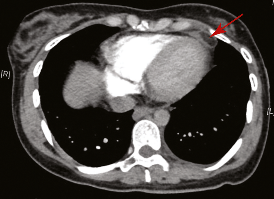
Ventilation–perfusion (V/Q) scanning is the preferred imaging modality for pulmonary embolism in pregnancy at our institution but was limited during the coronavirus disease 2019 (COVID‐19) pandemic due to the risk of respiratory secretion aerosolisation. Computed tomography pulmonary angiography (CTPA), which was performed after discussion of its risks and benefits, did not demonstrate pulmonary embolism. However, there was a region of focal inflammatory fat stranding in the pericardium adjacent to the left ventricular apex (Box), consistent with epipericardial fat necrosis. This radiological finding, in combination with her clinical presentation and biochemical evidence of inflammation (raised white blood cell count with predominant neutrophilia), led to the diagnosis of epipericardial fat necrosis. While the CRP was only mildly elevated, in combination with the other findings, it supported the diagnosis.
She was subsequently discharged with analgesia. Six weeks later, her pain had resolved and cardiac magnetic resonance imaging demonstrated reduced prominence of the pericardial fat adjacent to the left ventricular anterior apical wall with no signs of pericardial fat oedema. She had not experienced any pregnancy‐related complications.
Discussion
Epipericardial fat necrosis is an underdiagnosed cause of pleuritic chest pain. In a retrospective series, epipericardial fat necrosis was found in up to 2.6% of patients undergoing chest computed tomography (CT) scanning compared with 0.3% for whom fat necrosis was the provisional diagnosis.1
The exact aetiology of epipericardial fat necrosis remains unknown. Hypotheses include acute torsion of a vascular pedicle, pre‐existing structural abnormality of the adipose tissue that makes it vulnerable to trauma, and abrupt changes in intravascular pressure associated with the Valsalva manoeuvre leading to haemorrhage in the loosely attached adipose tissue.2
The diagnosis is suggested by the triad of acute pleuritic chest pain with characteristic CT findings of an encapsulated fatty lesion with inflammatory changes and thickening of the adjacent pericardium.3 Clinically, the patient often has isolated pleuritic chest pain, with an elevated CRP being the only laboratory abnormality, reflecting inflammation around the necrotic fat, as occurred with our patient.2 We found no other cases associated with pregnancy in our literature review.
CTPA is the recommended imaging modality for suspected pulmonary embolism due to its high diagnostic accuracy and frequent identification of an alternative diagnosis.4 V/Q scanning, however, is preferred for suspected pulmonary embolism in pregnancy due to a lower breast radiation dose.4 In this case, CTPA was performed as there was reduced availability of V/Q scanning due to the COVID‐19 pandemic. The diagnosis of epipericardial fat necrosis would not have been made if a V/Q scan had been performed. We suggest that CTPA should be considered in pregnancy if an alternate thoracic pathology may be present.
Before epipericardial fat necrosis was a recognised entity, the mass demonstrated on CT in these patients was often presumed to be a tumour and surgically excised, as was done for 19 published cases between 1957 and 1999.5 It is now appreciated that epipericardial fat necrosis is a benign, self‐limiting condition with no need for surgical resection. The clinical approach is conservative, using non‐steroidal anti‐inflammatory analgesia with a follow‐up CT scan in 4–8 weeks to ensure a reduction of lesion size and resolving inflammatory changes.3
We highlight the importance of this uncommon, benign condition. Furthermore, we emphasise the utility of a CT scan in contrast to a V/Q scan in a patient with undifferentiated pleuritic chest pain.
Lessons from practice
- Epipericardial fat necrosis is an uncommon cause of pleuritic chest pain.
- The diagnosis of epipericardial fat necrosis can be established by a computed tomography scan, which typically demonstrates a characteristic, ovoid, fatty lesion in the anterior mediastinum with associated inflammatory fat stranding and/or an adjacent thickened pericardium.
- Compared with a ventilation–perfusion scan, computed tomography pulmonary angiography has the advantage of providing alternative diagnoses in patients with suspected pulmonary embolism.
Competing interests
No relevant disclosures.
References
- Giassi Kde S, Costa AN, Bachion GH, et al. Epipericardial fat necrosis: an underdiagnosed condition. Br J Radiol 2014; 87: 20140118.
- Gayer G. Mediastinal (epipericardial) fat necrosis: an overlooked and little known cause of acute chest pain mimicking acute coronary syndrome. Semin Ultrasound CT MR 2017; 38: 629–633.
- Pineda V, Caceres J, Andreu J, et al. Epipericardial fat necrosis: radiologic diagnosis and follow‐up. AJR Am J Roentgenol 2005; 185: 1234–1236.
- Tran HA, Gibbs H, Merriman E, et al. New guidelines from the Thrombosis and Haemostasis Society of Australia and New Zealand for the diagnosis and management of venous thromboembolism. Med J Aust 2019; 210: 227–235. https://www.mja.com.au/journal/2019/210/5/new-guidelines-thrombosis-and-haemostasis-society-australia-and-new-zealand
- Inoue S, Fujino S, Tezuka N, et al. Encapsulated pericardial fat necrosis treated by video‐assisted thoracic surgery: report of a case. Surg Today 2000; 30: 739–743.
Provenance: Not commissioned; externally peer reviewed.
Fatty Liver Disease in Australia: A Narrative Review on the Epidemiology, Natural History, Prognostication and Management in People With Metabolic Dysfunction
Karl Vaz, Daniel Clayton-Chubb, William W. Kemp, Stuart K. Roberts, Ammar Majeed
Sex Differences in Management and Outcomes of People With ST-Elevation Myocardial Infarction, New South Wales, 2011–2020: A Retrospective Cohort Study
Adam C. Livori, Jedidiah I. Morton, Dianna J. Magliano, Jonathan E. Shaw
Rethinking Rheumatic Fever Diagnosis: Progress, Pitfalls and the Path Towards a Definitive Test
Anna P. Ralph, Rachel Webb, Tom Parks, Nicole J. Moreland, Asha C. Bowen, Benjamin Jones, Emma Ndagire, Andrea Z. Beaton
Sex Differences in Management and Outcomes of People With ST-Elevation Myocardial Infarction, New South Wales, 2011–2020: A Retrospective Cohort Study
Samia Kazi, Simone Marschner, Clara K. Chow
Distribution of Cardiovascular Disease Risk Based on the Updated 2023 Guideline-Recommended Australian Cardiovascular Disease Risk Algorithm and Comparison With the 2012 Algorithm: An Observational Study
Nina Lazarevic, Meghana Bhat, Grace Joshy, Danielle C. Butler, Mark Woodward, Anushka Patel, Rod T. Jackson, Garry Jennings, Rosemary Wyber, Ellie Paige, Emily Banks
They Brought the Hospital to Me
Hugh N. Burrill