




Volume 210 - Issue 5
New guidelines from the Thrombosis and Haemostasis Society of Australia and New Zealand for the diagnosis and management of venous thromboembolism
Authors: Huyen A Tran, Harry Gibbs, Eileen Merriman, Jennifer L Curnow, Laura Young, Ashwini Bennett, Chee Wee Tan, Sanjeev D Chunilal, Chris M Ward, Ross Baker and Harshal Nandurkar
Med J Aust 2019; 210 (5): 227-235. || doi: 10.5694/mja2.50004
Published online: 11 February 2019
Published online: 11 February 2019
Correction(s) for this article:
Erratum | Published online: 17 February 2020
The THANZ guideline for the diagnosis and management of VTE has been developed by the VTE Working Group based on up-to-date evidence and using an evidence-based approach. The guideline aims to promote optimal management of VTE. The extended version of the guideline can be found on the THANZ website (www.thanz.org.au/resources/thanz-guidelines).
Abstract
Introduction: Venous thromboembolism (VTE), including deep vein thrombosis (DVT) and pulmonary embolism (PE), is the third most common cardiovascular disease and, globally, more than an estimated 10 million people have it yearly. It is a chronic and recurrent disease. The symptoms of VTE are non‐specific and the diagnosis should actively be sought once considered. The mainstay of VTE treatment is anticoagulation, with few patients requiring additional intervention.
A working group of experts in the area recently completed an evidence‐based guideline for the diagnosis and management of DVT and PE on behalf of the Thrombosis and Haemostasis Society of Australia and New Zealand (www.thanz.org.au/resources/thanz-guidelines).
Main recommendations:
- The diagnosis of VTE should be established with imaging; it may be excluded by the use of clinical prediction rules combined with D‐dimer testing.
- Proximal DVT or PE caused by a major surgery or trauma that is no longer present should be treated with anticoagulant therapy for 3 months.
- Proximal DVT or PE that is unprovoked or associated with a transient risk factor (non‐surgical) should be treated with anticoagulant therapy for 3–6 months.
- Proximal DVT or PE that is recurrent (two or more) and provoked by active cancer or antiphospholipid syndrome should receive extended anticoagulation.
- Distal DVT caused by a major provoking factor that is no longer present should be treated with anticoagulant therapy for 6 weeks.
- For patients continuing with extended anticoagulant therapy, either therapeutic or low dose direct oral anticoagulants can be prescribed and is preferred over warfarin in the absence of contraindications.
- Routine thrombophilia testing is not indicated.
- Thrombolysis or a suitable alternative is indicated for massive (haemodynamically unstable) PE.
Changes in management as a result of the guideline: Most patients with acute VTE should be treated with a factor Xa inhibitor and be assessed for extended anticoagulation.
Venous thromboembolism (VTE), including deep vein thrombosis (DVT) and pulmonary embolism (PE), is the third most common cardiovascular disease, with an annual incidence of more than 10 million people globally.1 In Australia, at least 17 000 people develop VTE each year (annual incidence, 0.83 per 1000 population).2 The lifetime risk of VTE is 8%, with 1% of people aged over 80 years experiencing their first VTE. This disease is a major cause of health‐related economic loss for the patient and the community (estimated to be $1.7 billion for Australia in 2008).3 It is a chronic and frequently recurrent disease.
VTE can be fatal if untreated; long term morbidity includes post‐thrombotic syndrome (PTS) and pulmonary hypertension. Symptoms of VTE are non‐specific, and the diagnosis should actively be sought once considered. A diagnosis of VTE has an impact on subsequent pregnancies, oestrogen use, surgery, life insurance and, occasionally, long‐haul travel.
This guideline summary outlines the recommendations for the diagnosis and management of VTE on behalf the Thrombosis and Haemostasis Society of Australia and New Zealand (THANZ) (Box 1).
Methods
The VTE Writing Group was established within THANZ and it comprised experts in the field of thromboembolic disorders in Australia and New Zealand. All members undertook a detailed literature review and critically appraised existing evidence on the diagnosis and treatment of VTE. Drafts of evidence‐based recommendations, practice points and background manuscript were developed. We then conducted a 2‐day face to face meeting on 25–26 February 2018 to draft the guideline. Further revisions were made via emails or face to face meetings. The summary recommendations follow the National Health and Medical Research Council levels of evidence (www.mja.com.au/sites/default/files/NHMRC.levels.of.evidence.2008-09.pdf) and the Grading of Recommendations Assessment, Development and Evaluation (GRADE) system (www.gradeworkinggroup.org) to determine the strength of the recommendations.
Risk factors for venous thromboembolism
There are inherited and acquired VTE risk factors. Multiple risk factors often coexist in an individual, each contributing to the overall VTE risk. While hereditary thrombophilia is associated with an increased VTE risk, there is little clinical benefit of testing for this condition, as its utility in decision making regarding anticoagulation is low.4
It is important to delineate whether a VTE event was provoked or unprovoked. Provoking factors can be further classified as surgical (recent major surgery) or non‐surgical and transient or persistent (Box 2). Such clinical categorisation is important as it has an impact on the risk of VTE recurrence and duration of anticoagulation (Box 3).5 VTE occurring within 2 months of a transient provoking risk factor has one‐half the risk of recurrent VTE after stopping anticoagulant therapy compared with patients with no transient risk factor.6
It is important to consider occult malignancy in unprovoked VTE, as up to 10% of such patients are diagnosed with cancer in the year after a VTE diagnosis.7 Clinical assessment should include a thorough clinical examination and age‐appropriate screening for malignancy (Box 4). In addition, abdominopelvic computed tomography scan does not identify more early stage cancers or improve outcome.7
Diagnosis and treatment of pulmonary embolism and deep vein thrombosis
Clinical presentations of VTE are non‐specific, and only about 20% of patients with clinically suspected VTE have it objectively confirmed.10 A misdiagnosis of VTE has significant implications, including needless cessation of effective hormonal contraception in women and unnecessary ante‐ and post‐partum injections of low molecular weight heparin, and in older patients, anticoagulation is associated with higher rates of major and fatal bleeding.11 Conversely, failure to diagnose VTE can result in fatal PE. Therefore, objective testing is required to establish the diagnosis of VTE.
The need for imaging can be determined by the use of clinical prediction rules combined with D‐dimer testing, avoiding unnecessary radiological investigations that expose patients to radiation and potential nephrotoxic contrast dyes, which are costly and inconvenient.
Clinical prediction rules
The most validated prediction rules for VTE are the Wells and Geneva scores12 (Box 5). However, they alone cannot safely exclude VTE and must be used in combination with D‐dimer testing.13,15GRADE: Strong; Evidence: High. These algorithms are designed for outpatient or emergency department assessment and are not applicable to hospitalised patients.
The pulmonary embolism rule‐out criteria (PERC) is a scoring system for excluding PE.14 It is only applicable to patients younger than 50 years of age and when the estimated rate of PE is low (< 15%).16 This rate is seen in most Australian and New Zealand emergency departments and, therefore, PERC can be applied. If used in this way, additional investigations can be avoided in some patients. Patients with a positive PERC score should be assessed further for PE.
D‐dimer assay
D‐dimer levels are increased in VTE but also in many other conditions, including malignancy, inflammation, infection, trauma and pregnancy.17
A negative D‐dimer result is a useful rule‐out test when combined with an unlikely (non‐high) clinical probability, avoiding imaging in many cases (Box 6 and Box 7).12 A positive D‐dimer result alone is not diagnostic of VTE and requires further radiological investigation.
Imaging
Lower extremity duplex ultrasound
Duplex ultrasound is accurate for diagnosing and excluding DVT. In Australia and New Zealand, the entire deep venous system, from ankle to groin, is evaluated with duplex ultrasound. In general, a negative single whole leg ultrasound excludes DVT and anticoagulation can be withheld (Box 6).18
Diagnosing recurrent ipsilateral DVT is challenging, as incomplete resolution of thrombus occurs in up to 30–50% of patients after DVT.19 For this reason, many clinicians perform a single repeat duplex ultrasound after 3–6 months if anticoagulation is to be ceased (Box 4). GRADE: Strong; Evidence: Low. These images may be compared with future imaging in the event of new symptoms suspicious for DVT. Multiple repeat scans in the absence of symptoms are unhelpful and do not alter management. GRADE: Strong; Evidence: Low.
Computed tomography pulmonary angiography
Computed tomography pulmonary angiography (CTPA) is the preferred imaging modality for suspected PE due to its accuracy.8GRADE: Strong; Evidence, High. However, CTPA involves significant exposure to ionising radiation (3–5 mSv) and requires iodinated contrast, which can cause nephrotoxicity (up to 14%) and allergic reactions (< 1%).9
Ventilation–perfusion scanning
Ventilation–perfusion (VQ) scanning does not require radiocontrast, and so is suitable for patients with renal impairment. A normal VQ scan effectively excludes PE, and a high probability scan is diagnostic.20 However, 27–55% of patients have non‐diagnostic lung scans; these patients require testing with serial ultrasound of the legs or CTPA to exclude PE (Box 7).21 In pregnant women, given the absence of contrast combined with studies showing that the proportion of diagnostic VQ scans is high, VQ scan is the preferred diagnostic investigation.22GRADE: Strong; Evidence: Low.
Treatment of venous thromboembolism
The spectrum of VTE ranges from distal DVT, which may not require anticoagulation, through proximal DVT to potentially life‐threatening PE requiring additional invasive strategies. The treatment for DVT depends on its anatomical extent: in proximal DVT, thrombus is present in the popliteal (and its trifurcation) or a more proximal vein; in distal DVT, thrombus only occurs in the tibial, peroneal, gastrocnemius and soleal veins.23
Anticoagulation is indicated in most cases of VTE because it is highly effective in preventing thrombus extension or recurrence by at least 80%.5
Anticoagulant therapy for deep vein thrombosis and pulmonary embolism
Direct oral anticoagulants (DOACs) and warfarin are equally effective and can be prescribed to most patients. GRADE: Strong; Evidence: High.5,24 DOACs do not require routine monitoring, have no known food interactions and few drug interactions, and are favoured in most instances. However, DOACs should not be used during pregnancy or breastfeeding, in which case low molecular weight heparin is indicated.5 Edoxaban and rivaroxaban have been shown to be as efficacious as dalteparin in cancer‐related thrombosis, but are associated with an increased risk for major bleeding or clinically relevant non‐major bleeding (CRNMB) and, therefore, can be considered when appropriate.25,26
Oral factor Xa inhibitors (eg, apixaban, rivaroxaban) are preferred to dabigatran or warfarin to treat proximal DVT and PE because they do not require parenteral anticoagulation for initiation (Box 8). GRADE: Strong; Evidence: High.5
Duration of anticoagulation
Proximal deep vein thrombosis and pulmonary embolism
All patients with proximal DVT and PE should receive anticoagulant therapy for at least 3 months. GRADE: Strong; Evidence: Strong. Patients whose proximal DVT or PE were provoked by major surgery or major trauma can cease anticoagulation at this time.5
Distal deep vein thrombosis
Uncertainty exists about the value of anticoagulation for distal DVT. In general, anticoagulation is used for proximal DVT and PE, but serial duplex ultrasound (two duplex ultrasound scans over 2 weeks) is reasonable (GRADE: Strong; Evidence: Moderate), especially if the risk of bleeding is increased. Most distal DVT can be treated for 6–12 weeks. GRADE: Strong; Evidence: Moderate.30
Extended anticoagulation for deep vein thrombosis and pulmonary embolism (beyond 3–6 months)
For patients whose events were unprovoked or associated with transient risk factors (non‐surgical), decide whether to stop or to continue with extended anticoagulant therapy after 3 months of anticoagulation. Continuing therapy for longer than 3 months reduces the risk of VTE recurrence during therapy by at least 80% but is associated with a major bleeding risk of < 1% per year. Once anticoagulant therapy is stopped, the risk of recurrence is the same as for patients who cease treatment after 3–6months when followed up over time.27
The decision to stop or extend anticoagulation beyond 3 months is challenging and depends on the balance between the risks of bleeding and VTE recurrence (see below). Clinical “equipoise” is common and patient preference is important. Box 9 recommends the duration of anticoagulation for different types of VTE based on recurrence rates (Box 8) and risk factors for recurrence (Box 10) while considering patient preference.
Among patients for whom it has been decided to extend anticoagulant therapy, consider low intensity anticoagulation GRADE Strong; Evidence: High. The risk of major bleeding and CRNMB on therapeutic anticoagulation (DOACs or warfarin) varies from 2% to 3% per year34 but is less in patients who have completed 6 months of oral anticoagulants without bleeding.5 Apixaban 2.5 mg twice daily is as efficacious as 5.0 mg twice daily for preventing VTE recurrence beyond 6 months, with no difference in major bleeding and a trend to have less CRNMB.28 Likewise, rivaroxaban 10 mg once daily is as efficacious as 20 mg once daily with a trend to have less major bleeding and CRNMB.29
Aspirin (100 mg daily) reduces the rate of VTE recurrence to a much lesser extent than oral anticoagulants but is associated with similar rates of bleeding to rivaroxaban 10 mg daily.29,35 Therefore, aspirin should be avoided, unless anticoagulation cannot be used. GRADE: Strong; Evidence: High.
Predictors of venous thromboembolism recurrence
Many risk factors for VTE recurrence have been identified, although few have a major effect (Box 10). Most clinical decisions can be made by assessing the following predictors of recurrence: unprovoked, non‐surgical provoking factor and persistent risk factors versus provoked by surgery; PE and proximal DVT versus distal DVT;36 prior VTE;5 and male sex.31
Bleeding risk
The strongest predictor of bleeding is active or recent (< 30 days) bleeding, which usually contraindicates anticoagulant therapy.37 Other predictors of bleeding include prior history of bleeding (especially while receiving anticoagulation), a potential bleeding lesion (eg, peptic ulceration), recent surgery (within 14 days), severe kidney disease, and active cancer. The decision as to when bleeding risk outweighs the benefit of anticoagulation may be difficult and is often subjective. Importantly, among patients with no bleeding for whom recent therapeutic anticoagulation has been prescribed, the subsequent risk of major bleeding is very low (0.8–1.6% per year), particularly with low intensity DOACs, and similar to patients who are not taking anticoagulants.5,28
Thrombophilia testing
The presence of an inherited thrombophilia does not influence initial anticoagulant treatment. Only rare deficiencies of natural inhibitors (antithrombin, protein C or S) significantly increase the risk of recurrent VTE to warrant extended anticoagulation, and uncertainty remains with homozygous or other compound heterozygosity states.32,38 Neither factor V Leiden nor prothrombin gene mutation heterozygosity change treatment duration or advice for family members and should not be routinely sought (Box 4).33
It is reasonable to test for antiphospholipid syndrome in patients with unprovoked VTE. GRADE: Strong; Evidence: Moderate.
Complications of venous thromboembolism
Chronic thromboembolic pulmonary hypertension (CTEPH) is form of pre‐capillary pulmonary hypertension which results after pulmonary obstruction with thrombus and organised fibrous tissue, accompanied by pulmonary arteriopathy. The incidence of CTEPH after acute PE is 3.4%.39 Patients typically report persisting dyspnoea despite anticoagulation over the subsequent 2 years. VQ scan and echocardiography should be performed if CTEPH is suspected (Box 4). If untreated, CTEPH portends a poor prognosis, with a 5‐year survival rate of 30%. Treatment ranges from medical therapies aiming to vasodilate the pulmonary vasculature to pulmonary endarterectomy.40GRADE: Low; Evidence: Moderate.
PTS is characterised by clinical features (eg, swelling, discomfort, hyperpigmentation and lipodermatosclerosis) in a limb with previous DVT.41 It occurs in one in three patients following DVT.42 Radiological findings are insufficient to diagnose PTS; the Villalta scale is the most commonly used clinical scale.43
Anticoagulation does not prevent PTS.44 Thrombolysis relieves venous outflow obstruction and has been used to prevent PTS in extensive DVT, although clinical trial results have been mixed.45,46 Clinical trials evaluating elastic compression stockings among patients with proximal DVT have reported conflicting findings in reducing PTS incidence.47 Hence, elastic compression stockings may be useful only to control symptoms of leg swelling and pain. GRADE: Strong; Evidence: Moderate.
Invasive strategies for venous thromboembolism management
Invasive treatment modalities for acute removal of thrombosis have been investigated, with the goals of rapidly relieving acute right ventricular pressure overload in PE and thereby improving survival or rapidly relieving venous obstruction to prevent vein dysfunction and PTS and reduce VTE recurrence.
The following strategies have been investigated with variable results: systemic administration of thrombolytic agents; catheter‐directed thrombolysis, which uses lower thrombolytic doses with or without the addition of mechanical clot disruption; and acute surgical thrombectomy.46,48,49,50
These therapies have a limited role in management of acute VTE (Box 11).
Inferior vena cava filter insertion may prevent PE in patients with acute VTE and who have an absolute contraindication to anticoagulation, such as active bleeding, but are not recommended in patients treated with anticoagulants for acute VTE.51GRADE: Strong; Evidence: Strong.
Conclusion
The THANZ guideline for the diagnosis and management of VTE has been developed by the VTE Working Group based on up‐to‐date evidence and using an evidence‐based approach. The guideline aims to promote optimal management of VTE. The extended version of the guideline can be found on the THANZ website (www.thanz.org.au/resources/thanz-guidelines).
Box 1 – Thrombosis and Haemostasis Society of Australia and New Zealand guidelines: evidence‐based recommendations for the diagnosis and management of pulmonary embolism (PE) and deep vein thrombosis (DVT)
|
Interventions |
GRADE* |
Evidence† |
|||||||||||||
|
|
|||||||||||||||
|
Diagnosis of PE and DVT |
A non‐high pre‐test probability (Wells or Geneva score) combined with a negative D‐dimer result safely excludes VTE without imaging |
Strong |
High |
||||||||||||
|
A single negative complete ultrasound is sufficient to exclude DVT |
Strong |
High |
|||||||||||||
|
PE can be excluded without D‐dimer or radiological testing in selected patients if the PE rule‐out criteria (negative PERC rule) are met |
Strong |
Moderate |
|||||||||||||
|
A normal VQ scan or a negative technically adequate CTPA excludes PE and anticoagulation can be safely withheld |
Strong |
High |
|||||||||||||
|
Treatment of VTE |
Distal DVT caused by a major provoking factor that is no longer present requires OACs for 6 weeks |
Strong |
Moderate |
||||||||||||
|
Distal DVT that has been unprovoked or with persisting risk factors requires OACs for 3 months |
Strong |
Moderate |
|||||||||||||
|
Proximal DVT or PE caused by major surgery or trauma that is no longer present requires OACs for 3 months |
Strong |
High |
|||||||||||||
|
Proximal DVT or PE that is unprovoked or associated with a transient (non‐surgical) risk factor requires OACs for 3–6 months |
Strong |
High |
|||||||||||||
|
For DVT or PE that is provoked by active cancer, treat with therapeutic LMWH for at least 6 months |
Strong |
High |
|||||||||||||
|
For patients continuing with extended anticoagulation, either therapeutic or low dose DOAC is preferred over warfarin in the absence of contraindications |
Strong |
High |
|||||||||||||
|
Aspirin should be avoided unless anticoagulation cannot be used |
Strong |
High |
|||||||||||||
|
Thrombophilia testing |
Patients with VTE provoked by surgery or major trauma should not be screened for hereditary thrombophilia |
Strong |
High |
||||||||||||
|
Additional Interventions |
|||||||||||||||
|
PE |
For patients with massive PE (sustained hypotension) and a low risk of bleeding, administer thrombolytic therapy or an alternative (eg, surgical embolectomy or catheter‐based therapy) depending on local availability |
Strong |
Moderate |
||||||||||||
|
IVC filter insertion may prevent PE in patients with acute VTE and an absolute contraindication to anticoagulation, such as active bleeding, but are not recommended in patients treated with anticoagulants for acute VTE |
Strong |
High |
|||||||||||||
|
DVT |
CDT may be considered in selected patients with extensive proximal DVT (involves common iliac veins) and low bleeding risk |
Strong |
Low |
||||||||||||
|
Elastic compression stockings may be useful only to control symptoms of leg swelling and pain following DVT |
Strong |
Moderate |
|||||||||||||
|
|
|||||||||||||||
|
CDT = catheter‐directed thrombolysis. CTPA = computed tomography pulmonary angiography. DOAC = direct oral anticoagulant. IVC = inferior vena cava. LMWH = low molecular weight heparin. OACs = oral anticoagulants. PERC = pulmonary embolism rule‐out criteria. VQ = ventilation–perfusion. * Grading of Recommendations Assessment, Development and Evaluation; www.gradeworkinggroup.org. † www.mja.com.au/sites/default/files/NHMRC.levels.of.evidence.2008-09.pdf. ◆ |
|||||||||||||||
Box 2 – Examples of non‐surgical transient, or persistent provoking factors for venous thromboembolism (VTE)5
|
Type of VTE |
Examples |
||||||||||||||
|
|
|||||||||||||||
|
Non‐surgical transient |
|
||||||||||||||
|
Persistent provoking |
|
||||||||||||||
|
|
|||||||||||||||
|
|
|||||||||||||||
Box 3 – Types of venous thromboembolism (VTE) and associated VTE recurrence rates5
|
Type of VTE |
Recurrence rate at one year after stopping anticoagulation |
Recurrence rate at 5 years after stopping anticoagulation |
|||||||||||||
|
|
|||||||||||||||
|
First VTE provoked by major surgery or major trauma |
1% |
3% |
|||||||||||||
|
First VTE provoked by transient risk factor (non‐surgical) |
5% |
15% |
|||||||||||||
|
Provoked VTE with persistent risk factors (eg, active cancer) |
15% |
45% |
|||||||||||||
|
First unprovoked distal DVT |
5% |
15% |
|||||||||||||
|
First unprovoked proximal DVT or PE |
10% |
30% |
|||||||||||||
|
Second episode of unprovoked VTE |
15% |
45% |
|||||||||||||
|
|
|||||||||||||||
|
DVT = deep vein thrombosis. PE = pulmonary embolism. ◆ |
|||||||||||||||
Box 4 – Good practice points for the diagnosis and management of pulmonary embolism (PE) and deep vein thrombosis (DVT)
|
Clinical setting |
Practice point |
||||||||||||||
|
|
|||||||||||||||
|
Diagnosis of PE and DVT |
|
||||||||||||||
|
Subsegmental PE |
|
||||||||||||||
|
Incidental PE in patients with cancer |
|
||||||||||||||
|
Thrombophilia testing |
|
||||||||||||||
|
Complications of VTE |
|||||||||||||||
|
Chronic thromboembolic pulmonary hypertension |
|
||||||||||||||
|
Invasive strategies — PE |
|
||||||||||||||
|
|
|||||||||||||||
|
CTPA = computed tomography pulmonary angiography. CUS = compression ultrasonography. DVT = deep vein thrombosis. IVC = inferior vena cava. LMWH = low molecular weight heparin. PE = pulmonary embolism. TTE = transthoracic echocardiography. VQ = ventilation–perfusion. ◆ |
|||||||||||||||
Box 5 – Clinical prediction rules for pulmonary embolism (PE)12,13,14 and deep vein thrombosis (DVT)15
|
Simplified Geneva score for PE |
Simplified Wells score for PE |
Simplified Wells score for DVT |
PERC rule* |
||||||||||||
|
|
|||||||||||||||
|
Age > 65 years |
1 |
Clinical sign and symptoms of DVT |
1 |
Active cancer (receiving treatment within past 6 months or palliative treatment) |
1 |
Age > 50 years |
1 |
||||||||
|
Surgery or fracture previous 4 weeks |
1 |
Immobility/surgery previous 4 weeks |
1 |
Paralysis, paresis or recent plaster immobilisation of the lower extremity |
1 |
Recent trauma/surgery |
1 |
||||||||
|
Previous VTE |
1 |
Previous VTE |
1 |
Recently bedridden for 3 days or more, major surgery within 3 months requiring general or regional anaesthesia |
1 |
Prior VTE |
1 |
||||||||
|
Haemoptysis |
1 |
Haemoptysis |
1 |
Localised tenderness along distribution of the deep venous system |
1 |
Haemoptysis |
1 |
||||||||
|
Active cancer |
1 |
Malignancy |
1 |
Entire leg swollen |
1 |
Oestrogen use |
1 |
||||||||
|
Unilateral leg pain |
1 |
Alternative diagnosis less likely than PE |
1 |
Calf swelling at least 3 cm larger than the asymptomatic side |
1 |
Arterial oxygen < 94% |
1 |
||||||||
|
Heart rate |
|
Heart rate > 100 beats/min |
1 |
Pitting oedema of the symptomatic leg |
1 |
Heart rate > 100 beats/min |
1 |
||||||||
|
75–94 beats/min |
1 |
|
|
|
|
|
|
||||||||
|
> 95 beats/min |
2 |
|
|
|
|
|
|
||||||||
|
Pain on lower leg deep vein palpation or unilateral oedema |
1 |
|
|
Previous documented DVT |
1 |
Unilateral leg swelling |
1 |
||||||||
|
|
|
|
|
Collateral superficial veins (non‐varicose) |
1 |
|
|
||||||||
|
|
|
|
|
Alternative diagnosis at least as likely as deep vein thrombosis |
−2 |
PERC negative |
0 |
||||||||
|
|
|
|
|
|
|
PERC positive |
≥ 1 |
||||||||
|
Low |
0–1 |
|
|
|
|
|
|
||||||||
|
Moderate |
2–4 |
|
|
|
|
|
|
||||||||
|
High |
≥ 5 |
|
|
|
|
|
|
||||||||
|
Unlikely |
0–2 |
Unlikely |
0–1 |
Unlikely |
< 2 |
|
|
||||||||
|
Likely |
≥ 3 |
Likely |
≥ 2 |
Likely |
≥ 2 |
|
|
||||||||
|
|
|||||||||||||||
|
PERC = pulmonary embolism rule‐out criteria. VTE = venous thromboembolism. * The estimated rate of PE must be low (< 15%). ◆ |
|||||||||||||||
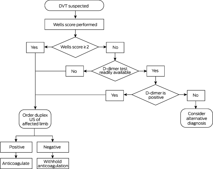
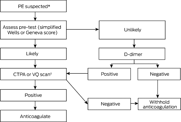
Box 8 – Anticoagulant options for acute venous thromboembolism (VTE)5,27,28,29
|
Anticoagulant |
Initiation dose |
Maintenance dose |
|||||||||||||
|
|
|||||||||||||||
|
Apixaban* |
|
|
|||||||||||||
|
Rivaroxaban† |
|
|
|||||||||||||
|
Dabigatran‡ |
|
|
|||||||||||||
|
Warfarin |
|
|
|||||||||||||
|
LMWH§ |
|
|
|||||||||||||
|
|
|||||||||||||||
|
CrCl = creatinine clearance. INR = international normalised ratio. LMWH = low molecular weight heparin. * Requires CrCl ≥ 25 mL/min; reimbursed for VTE only in Australia. † Requires CrCl ≥ 30 mL/min; reimbursed for VTE in Australia and New Zealand. ‡ Reimbursed for VTE only in New Zealand. § If LMWH is required for a patient with CrCl ≤ 30 mL/min, seek expert advice. Twice‐daily dosing of dalteparin and enoxaparin may be preferred for patients at high risk of bleeding, such as patients who are older, are at extremes of weight (eg, ≥ 150 kg) or have a malignancy. ◆ |
|||||||||||||||
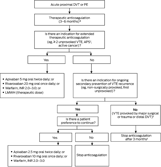
Box 10 – Risk factors for recurrent venous thromboembolism (VTE)5,30,31,32,33
|
|
Risk factor |
||||||||||||||
|
|
|||||||||||||||
|
Strong risk factors for recurrence |
|
||||||||||||||
|
Moderate risk factors for recurrence |
|
||||||||||||||
|
Factors that have little or no effect on recurrence |
|
||||||||||||||
|
|
|||||||||||||||
|
|
|||||||||||||||
Box 11 – Role of additional interventions in venous thromboembolism (VTE)
|
|
Definition |
Intervention |
GRADE* |
Level of evidence† |
|||||||||||
|
|
|||||||||||||||
|
Pulmonary embolism |
|
|
|
|
|||||||||||
|
Massive |
Sustained hypotension (systolic BP < 90mmHg for 15 min or requiring inotropic support or pulselessness or sustained HR < 40 beats/min with signs/symptoms of shock) |
Thrombolysis or alternative based on local expertise and availability (eg, surgical embolectomy, catheter‐based intervention, ECMO) |
Strong |
Moderate |
|||||||||||
|
Submassive |
Systolic BP > 90 mmHg and RV dysfunction or myocardial necrosis defined by:
|
Anticoagulation |
Strong |
Moderate |
|||||||||||
|
Consider lysis or other invasive therapy if very high thrombus burden, poor cardiorespiratory reserve and low bleeding risk |
Low |
Moderate |
|||||||||||||
|
Haemodynamically stable |
|
Anticoagulation |
Strong |
High |
|||||||||||
|
Deep vein thrombosis |
|
|
|
|
|||||||||||
|
Iliofemoral |
Thrombus involving at least the common iliac vein |
Consider pharmaco‐mechanical thrombus dissolution (phlegmasia cerulea dolens) |
Strong |
Low |
|||||||||||
|
Other (non‐iliofemoral) |
|
Anticoagulation |
Strong |
High |
|||||||||||
|
|
|||||||||||||||
|
BNP = brain natriuretic peptide. BP = blood pressure. CT = computed tomography. HR = heart rate. ECMO = extracorporeal membrane oxygenation. HR = heart rate. NT‐proBNP = N‐terminal pro‐brain natriuretic peptide. RV = right ventricle. * Grading of Recommendations Assessment, Development and Evaluation; www.gradeworkinggroup.org. † www.mja.com.au/sites/default/files/NHMRC.levels.of.evidence.2008-09.pdf. ◆ |
|||||||||||||||
Competing interests
No relevant disclosures.
References
- Raskob GE, Angchaisuksiri P, Blanco AN, et al. Thrombosis: a major contributor to global disease burden. SeminThromb Hemost 2014; 40: 724–735.
- Ho WK, Hankey GJ, Eikelboom JW. The incidence of venous thromboembolism: a prospective, community‐based study in Perth, Western Australia. Med J Aust 2008; 189: 144–147. https://www.mja.com.au/journal/2008/189/3/incidence-venous-thromboembolism-prospective-community-based-study-perth-western
- Fletcher J, Baker R, Fisher C, et al. The burden of venous thromboembolism in Australia. Access Economics, 2008. https://www.safetyandquality.gov.au/wp-content/uploads/2018/10/Access-Economics_The-burden-of-VTE-in-Australia_2008.pdf (viewed Jan 2019).
- Connors JM. Thrombophilia Testing and Venous Thrombosis. N Engl J Med 2017; 377: 1177–1187.
- Kearon C, Akl EA, Ornelas J, et al. Antithrombotic Therapy for VTE Disease: CHEST Guideline. Chest 2016; 149: 315–352.
- Kearon C, Ageno W, Cannegieter SC, et al. Categorization of patients as having provoked or unprovoked venous thromboembolism: guidance from the SSC of ISTH. J Thromb Haemost 2016; 14: 1480–1483.
- van Es N, Le Gal G, Otten HM, et al. Screening for occult cancer in patients with unprovoked venous thromboembolism: a systematic review and meta‐analysis of individual patient data. Ann Intern Med 2017; 167: 410–417.
- Mos IC, Klok FA, Kroft LJ, et al. Safety of ruling out acute pulmonary embolism by normal computed tomography pulmonary angiography in patients with an indication for computed tomography: systematic review and meta‐analysis. J Thromb Haemost 2009; 7: 1491–1498.
- Mitchell AM, Jones AE, Tumlin JA, Kline JA. Prospective study of the incidence of contrast‐induced nephropathy among patients evaluated for pulmonary embolism by contrast‐enhanced computed tomography. Acad Emerg Med 2012; 19: 618–625.
- Di Nisio M, van Es N, Buller HR. Deep vein thrombosis and pulmonary embolism. Lancet 2016; 388: 3060–3073.
- Linkins LA, Choi PT, Douketis JD. Clinical impact of bleeding in patients taking oral anticoagulant therapy for venous thromboembolism: a meta‐analysis. Ann Intern Med 2003; 139: 893–900.
- Righini M, Robert‐Ebadi H, Le Gal G. Diagnosis of acute pulmonary embolism. J Thromb Haemost 2017; 15: 1251–1261.
- Wells P, Anderson D, Rodger M, et al. Derivation of a simple clinical model to categorize patients probability of pulmonary embolism: increasing the models utility with the SimpliRED D‐dimer. Thromb Haemost 2000; 83: 416–420.
- Kline JA, Mitchell AM, Kabrhel C, et al. Clinical criteria to prevent unnecessary diagnostic testing in emergency department patients with suspected pulmonary embolism. J Thromb Haemost 2004; 2: 1247–1255.
- Wells PS, Owen C, Doucette S, Fergusson D, Tran H. Does this patient have deep vein thrombosis? JAMA 2006; 295: 199–207.
- Freund Y, Cachanado M, Aubry A, et al. Effect of the Pulmonary Embolism Rule‐Out Criteria on Subsequent Thromboembolic Events Among Low‐Risk Emergency Department Patients: the PROPER randomized clinical trial. JAMA 2018; 319: 559–566.
- Righini M, Perrier A, De Moerloose P, Bounameaux H. D‐Dimer for venous thromboembolism diagnosis: 20 years later. J Thromb Haemost 2008; 6: 1059–1071.
- Bernardi E, Camporese G, Buller HR, et al. Serial 2‐point ultrasonography plus D‐dimer vs whole‐leg color‐coded Doppler ultrasonography for diagnosing suspected symptomatic deep vein thrombosis: a randomized controlled trial. JAMA 2008; 300: 1653–1659.
- Carrier M, Rodger MA, Wells PS, et al. Residual vein obstruction to predict the risk of recurrent venous thromboembolism in patients with deep vein thrombosis: a systematic review and meta‐analysis. J Thromb Haemost 2011; 9: 1119–1125.
- Sostman HD, Stein PD, Gottschalk A, et al. Acute pulmonary embolism: sensitivity and specificity of ventilation‐perfusion scintigraphy in PIOPED II study. Radiology 2008; 246: 941–946.
- Anderson DR, Kahn SR, Rodger MA, et al. Computed tomographic pulmonary angiography vs ventilation‐perfusion lung scanning in patients with suspected pulmonary embolism: a randomized controlled trial. JAMA 2007; 298: 2743–2753.
- Cahill AG, Stout MJ, Macones GA, Bhalla S. Diagnosing pulmonary embolism in pregnancy using computed‐tomographic angiography or ventilation–perfusion. Obstet Gynecol 2009; 114: 124–129.
- Palareti G, Schellong S. Isolated distal deep vein thrombosis: what we know and what we are doing. J Thromb Haemost 2012; 10: 11–19.
- Pengo V, Denas G, Zoppellaro G, et al. Rivaroxaban vs warfarin in high‐risk patients with antiphospholipid syndrome. Blood 2018; 132: 1365–1371.
- Young AM, Marshall A, Thirlwall J, et al. Comparison of an oral factor xa inhibitor with low molecular weight heparin in patients with cancer with venous thromboembolism: results of a randomized trial (SELECT‐D). J Clin Oncol 2018; 36: 2017–2023.
- Raskob GE, van Es N, Verhamme P, et al. Edoxaban for the treatment of cancer‐associated venous thromboembolism. N Engl J Med 2018; 378: 615–624.
- Agnelli G, Prandoni P, Santamaria MG, et al. Three months versus one year of oral anticoagulant therapy for idiopathic deep venous thrombosis. Warfarin Optimal Duration Italian Trial Investigators. N Engl J Med 2001; 345: 165–169.
- Agnelli G, Buller HR, Cohen A, et al. Apixaban for extended treatment of venous thromboembolism. N Engl J Med 2013; 368: 699–710.
- Weitz JI, Lensing AWA, Prins MH, et al. Rivaroxaban or aspirin for extended treatment of venous thromboembolism. N Engl J Med 2017; 376: 1211–1222.
- Boutitie F, Pinede L, Schulman S, et al. Influence of preceding length of anticoagulant treatment and initial presentation of venous thromboembolism on risk of recurrence after stopping treatment: analysis of individual participants’ data from seven trials. BMJ 2011; 342: d3036.
- McRae S, Tran H, Schulman S, et al. Effect of patient's sex on risk of recurrent venous thromboembolism: a meta‐analysis. Lancet 2006; 368: 371–378.
- Coppens M, Reijnders JH, Middeldorp S, et al. Testing for inherited thrombophilia does not reduce the recurrence of venous thrombosis. J Thromb Haemost 2008; 6: 1474–1477.
- Segal JB, Brotman DJ, Necochea AJ, et al. Predictive value of factor V Leiden and prothrombin G20210A in adults with venous thromboembolism and in family members of those with a mutation: a systematic review. JAMA 2009; 301: 2472–2485.
- Ageno W, Mantovani LG, Haas S, et al. Safety and effectiveness of oral rivaroxaban versus standard anticoagulation for the treatment of symptomatic deep‐vein thrombosis (XALIA): an international, prospective, non‐interventional study. Lancet Haematol 2016; 3: e12–e21.
- Brighton TA, Eikelboom JW, Mann K, et al. Low‐dose aspirin for preventing recurrent venous thromboembolism. N Engl J Med 2012; 367: 1979–1987.
- Barco S, Corti M, Trinchero A, et al. Survival and recurrent venous thromboembolism in patients with first proximal or isolated distal deep vein thrombosis and no pulmonary embolism. J Thromb Haemost 2017; 15: 1436–1442.
- Nieto JA, Bruscas MJ, Ruiz‐Ribo D, et al. Acute venous thromboembolism in patients with recent major bleeding. The influence of the site of bleeding and the time elapsed on outcome. J Thromb Haemost 2006; 4: 2367–2372.
- De Stefano V, Simioni P, Rossi E, et al. The risk of recurrent venous thromboembolism in patients with inherited deficiency of natural anticoagulants antithrombin, protein C and protein S. Haematologica 2006; 91: 695–698.
- Hoeper MM, Humbert M, Souza R, et al. A global view of pulmonary hypertension. Lancet Respir Med 2016; 4: 306–322.
- Prior DL, Adams H, Williams TJ. Update on pharmacotherapy for pulmonary hypertension. Med J Aust 2016; 205: 271–276. https://www.mja.com.au/journal/2016/205/6/update-pharmacotherapy-pulmonary-hypertension
- Jain A, Cifu AS. Prevention, diagnosis, and treatment of postthrombotic syndrome. JAMA 2016; 315: 1048–1049.
- Kahn SR, Comerota AJ, Cushman M, et al. The postthrombotic syndrome: evidence‐based prevention, diagnosis, and treatment strategies: a scientific statement from the American Heart Association. Circulation 2014; 130: 1636–1661.
- Villalta S, Bagatella P, Piccioli A et al. Assessment of the validity and reproducibility of a clinical scale for the post‐thrombotic syndrome [abstract]. Haemostasis 1994; 24: 158a.
- Morling JR, Yeoh SE, Kolbach DN. Rutosides for treatment of post‐thrombotic syndrome. Cochrane Database Syst Rev 2015; (9): CD005625.
- Enden T, Haig Y, Klow NE, et al. Long‐term outcome after additional catheter‐directed thrombolysis versus standard treatment for acute iliofemoral deep vein thrombosis (the CaVenT study): a randomised controlled trial. Lancet 2012; 379: 31–38.
- Vedantham S, Goldhaber SZ, Julian JA, et al. Pharmacomechanical catheter‐directed thrombolysis for deep‐vein thrombosis. N Engl J Med 2017; 377: 2240–2252.
- Kahn SR, Shapiro S, Wells PS, et al. Compression stockings to prevent post‐thrombotic syndrome: a randomised placebo‐controlled trial. Lancet 2014; 383: 880–888.
- Meyer G, Vicaut E, Danays T, et al. Fibrinolysis for patients with intermediate‐risk pulmonary embolism. N Engl J Med 2014; 370: 1402–1411.
- Wan S, Quinlan DJ, Agnelli G, Eikelboom JW. Thrombolysis compared with heparin for the initial treatment of pulmonary embolism: a meta‐analysis of the randomized controlled trials. Circulation 2004; 110: 744–749.
- Sharifi M, Bay C, Skrocki L, et al; “MOPETT” Investigators. Moderate pulmonary embolism treated with thrombolysis (from the “MOPETT” trial). Am J Cardiol 2013; 111: 273–277.
- Decousus H, Leizorovicz A, Parent F, et al. A clinical trial of vena caval filters in the prevention of pulmonary embolism in patients with proximal deep‐vein thrombosis. Prévention du Risque d'Embolie Pulmonaire par Interruption Cave Study Group. N Engl J Med 1998; 338: 409–415.
Linked content
-
MJA Podcast: Associate Professor Huyen Tran
-
InSight+: Quiet revolution in treatment of venous thromboembolism
Provenance: Not commissioned; externally peer reviewed.
Splenic injury in severe cases of the zoonoses Q fever and rickettsial infection: diagnostic challenges
Ashleigh Drury, Philippa Harrison, Aiveen Bannan
Health and economic benefits of improving pre‐hospital identification of stroke in Australian women: a modelling study
Thomas Gadsden, Lei Si, Emily R Atkins, Cheryl Carcel, Xia Wang, Stephen Jan, Mark Woodward, Laura E Downey
Updating the diagnosis and management of elevated serum ferritin levels in the era of routine ferritin testing of blood donors by Australian Red Cross Lifeblood
Gary D Zhang, James Chen, Daniel M Johnstone, Martin B Delatycki, Katie Allen, John K Olynyk
Early cardiovascular collapse after envenoming by snakes in Australia, 2005–2020: an observational study (ASP‐31)
Geoffrey K Isbister, Katherine Z Isoardi, Angela L Chiew, Shane Jenkins, Nicholas A Buckley
Updated recommendations for warfarin reversal in the setting of four‐factor prothrombin complex concentrate
Danielle Robinson, James McFadyen, Eileen Merriman, Chee Wee Tan, Ross Baker, Huyen Tran
Updating the diagnosis and management of iron deficiency in the era of routine ferritin testing of blood donors by Australian Red Cross Lifeblood
Gary D Zhang, Daniel Johnstone, Michael F Leahy, John K Olynyk