




Medical education
Lessons from practice
Volume 213 - Issue 8
Polyneuritis cranialis from varicella zoster virus reactivation
Authors: Jesse A Schnall, Sadid F Khan, Luigi Zolio, Jason C Ray and Adam WJ Jenney
Med J Aust 2020; 213 (8): 352-353.e1. || doi: 10.5694/mja2.50791
Published online: 19 October 2020
Published online: 19 October 2020
A 68-year-old man with COPD, type 2 diabetes mellitus and stage II chronic kidney disease, presented to hospital with a 24-hour history of right- sided facial paralysis and 3 days of horizontal diplopia
Clinical record
A 68‐year‐old man with chronic obstructive pulmonary disease, type 2 diabetes mellitus and stage II chronic kidney disease, presented to hospital with a 24‐hour history of right‐sided facial paralysis and 3 days of horizontal diplopia. He had received 4 days of oral ciprofloxacin and oral prednisolone (37.5 mg) for 2 weeks of progressive right‐sided facial swelling with otalgia, initially thought to be otitis externa.
Examination revealed right facial cranial neuropathies. He had a sixth cranial nerve palsy and lower motor neuron seventh cranial nerve palsy. Audiometry confirmed eighth cranial nerve sensorineural involvement without vestibular dysfunction. Ninth and tenth cranial nerve palsies manifested as an absent gag reflex, asymmetric palatal movement and right vocal cord paralysis.
The patient was systemically well without fevers and had normal mentation. Examination of the remaining cranial nerves, cerebellar function, and upper and lower limbs was normal. Examination of the right external auditory meatus showed small, painful papules within the external auditory canal, with normal examination on the left. There was palpable, tender right submandibular swelling and lymphadenopathy without warmth or erythema.
Initially thought to have malignant otitis externa with base of skull osteomyelitis, he was commenced on intravenous piperacillin/tazobactam. Corticosteroids were discontinued. Brain magnetic resonance imaging (MRI) (with contrast) was normal. Both a computed tomography scan of the base of skull and a gallium bone scan revealed no malignant otitis externa or skull base osteomyelitis.
Varicella zoster virus (VZV) DNA was detected by polymerase chain reaction (PCR) from a swab of the lesions within the external auditory canal. Bacterial cultures revealed no significant growth. An antibody test for human immunodeficiency virus (HIV) — for which herpes zoster is an indicator condition — was negative. Further testing of immune status was not justified in the setting of known diabetes mellitus and steroid use. Antibiotic therapy was ceased and a 3‐week course of intravenous acyclovir was commenced. The right external auditory canal vesicles dried and crusted (Box 1). The patient had no previous history of primary VZV infection or shingles vaccination.
The diagnosis was revised to polyneuritis cranialis of right cranial nerves VI to X due to VZV reactivation. A lumbar puncture was not performed, as VZV DNA was found on the external auditory canal swab. Oral prednisolone commencing at 60 mg daily was slowly weaned over 3 weeks.
After 4 months, there has been negligible neurological recovery. The patient underwent a right tarsorrhaphy due to poor eyelid closure, and a thyroplasty for persisting right vocal cord paralysis (Box 1). Poor swallowing required the placement of a percutaneous endoscopic jejunostomy feeding tube.
Discussion
Polyneuritis cranialis is a clinical pattern of mononeuritis multiplex restricted to cranial nerves and of various aetiologies (Box 2). Differentiation of cranial nerve pathologies requires detailed history, examination and investigation. Time course can differentiate hyperacute (seconds to hours) vascular or traumatic causes from acute (days to weeks) inflammatory or infective aetiologies and subacute (months to years) malignant or infiltrative disease processes. Systemic features such as fever and constitutional symptoms may indicate inflammatory or infectious causes. A history of recent illness or environmental, toxin or medication exposure influences the probability of each differential diagnosis. Assessment of headache can differentiate diseases causing raised intracranial pressure or cranial nerves inflammation. Certain signs may suggest specific diagnoses, such as cranial nerve III, IV, V1, V2 and VI palsies in cavernous sinus pathology. Neuroimaging and cerebrospinal fluid analysis should be considered in all patients.
The lifetime risk of VZV reactivation is about 50%.2 Cranial nerve syndromes from VZV commonly involve the trigeminal nerve, presenting as herpes zoster ophthalmicus, or the facial nerve, presenting as Ramsay Hunt syndrome.3,4 Polyneuritis cranialis is a rare and severe manifestation of VZV that may be considered as part of a spectrum of complicated VZV infections that includes Ramsay Hunt syndrome.3,4,5 Seventh and eighth cranial nerve involvement and the presence of a characteristic vesicular “shingles” rash may distinguish VZV reactivation from conditions such as oculopharyngeal Guillain–Barré syndrome, yet skin lesions are only present if the involved nerve has a cutaneous distribution.3,4
MRI is important to investigate brainstem involvement and exclude other causes of peripheral cranial nerve palsies (Box 2). Enhancement of the cranial nerves is non‐specific, and does not exclude VZV reactivation when absent, yet may correlate with pathological findings in the acute phase of VZV reactivation.4 Cerebrospinal fluid analysis may show an elevated, predominantly lymphocytic cell count, elevated protein, VZV antibodies and detectable VZV on PCR.3,4,5
There is a paucity of data to guide therapy for polyneuritis cranialis. Suggested treatment involves a multiweek course of intravenous acyclovir and systemic corticosteroids. Longer courses have not shown superiority to shorter ones when long term recovery is the outcome measure.5
Prognosis is variable and complete recovery is rare.4,5 VZV vaccination is likely to be an important preventive consideration in an ageing population.2
Lessons from practice
• Differentiation of cranial nerve lesions relies on accurate discernment of the anatomical location, time course, and associated features of the pathology, aided by neuroimaging and cerebrospinal fluid analysis.
• The time course of illness can help differentiate hyperacute (vascular or trauma) from acute (inflammatory or infective) and subacute disease processes (malignant or infiltrative).
• Headache with “red flags” may suggest raised intracranial pressure or cranial nerves inflammation, while the presence of systemic features raises suspicion of inflammatory or infectious causes. Any recent illness or environmental, toxin or chemotherapy exposure should be considered when differentiating possible causes.
• The presence of skin vesicles with positive polymerase chain reaction (PCR) can distinguish varicella zoster virus (VZV) reactivation from other differentials. There is limited evidence to guide management and prognostication of severe VZV reactivations, with typical treatment involving a combined multiweek course of intravenous acyclovir and corticosteroids. Full recovery is rare.
Box 1 – Crusted varicella zoster virus rash in right external acoustic meatus at 2 weeks after admission (A). Persistent right facial nerve palsy and abducens nerve palsy at 3 weeks after admission (B)*
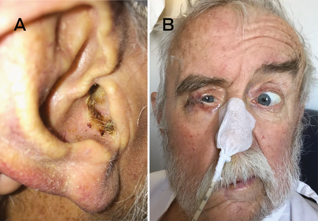
* Note that the patient's partially opened right tarsorrhaphy did not prevent abduction.
Box 2 – Differential diagnosis of multiple cranial nerve palsies*;1
|
|
|||||||||||||||
|
Infectious |
|
||||||||||||||
|
Inflammatory |
|
||||||||||||||
|
Neoplastic |
|
||||||||||||||
|
Vascular |
|
||||||||||||||
|
Bone disease |
|
||||||||||||||
|
|
|||||||||||||||
|
ANCA = antineutrophil cytoplasmic antibody; HIV = human immunodeficiency virus. * Central nervous system pathology involving the brainstem causing multiple cranial nerve palsies requires consideration, but is usually associated with concurrent sensorimotor deficits in the limbs and/or cerebellar dysfunction. † Via complication of vasculitis/mononeuritis multiplex. |
|||||||||||||||
Competing interests
No relevant disclosures.
References
- Carroll CG, Campbell WW. Multiple cranial neuropathies. Sem Neurol 2009; 29: 53–65.
- Zoster (Herpes Zoster): Australian Government Department of Health: Australian Immunisation Handbook; 2018. https://immunisationhandbook.health.gov.au/vaccine-preventable-diseases/zoster-herpes-zoster (viewed Jan 2020).
- Tateno F, Sakakibara R, Kishi M, et al. Zoster lower cranial neuropathy: A case report and a review of literature. Am J Case Rep 2011; 12: 137–139.
- Nisa L, Landis BN, Giger R, Leuchter I. Pharyngolaryngeal involvement by varicella‐zoster virus. J Voice 2013; 27: 636–641.
- Kim YH, Chang MY, Jung HH, et al. Prognosis of Ramsay Hunt syndrome presenting as cranial polyneuropathy. Laryngoscope 2010; 120: 2270–2276.
Provenance: Not commissioned; externally peer reviewed.
Costs of Delivering the Hep B PAST Model of Care in Remote Australia: A Retrospective Pre–Post Study
Hoa Nguyen, Anh Le Tuan Nguyen, Paula Binks, Melita McKinnon, Emily Vintour-Cesar, Karen Wills, Nicola Stephens, Benjamin Cowie, Joshua S. Davis, Yuejen Zhao, Peter Nihill, Julie A. Campbell, Andrew J. Palmer, Jane Davies, Barbara de Graaff
Buruli Ulcer Acquired From Ringtail Possum Rescue
Daniel P. O'Brien, Naomi Clarke, Sara Bonnici, Mohammad A. Hussain
Q Fever Visceral Abscesses After Contact With Native Australian Marsupials
Samantha J. Laws, Enzo Binotto, James Stewart, Jennifer Ho, Trent Yarwood, Josh Hanson, Simon Smith
Hospital-Admitted Injection-Related Infections Among Incarcerated People Who Inject Drugs in Australia: A Retrospective Cohort Study
Andrew Palmer, Matthew Carter, Jeremy Yeo, Cecilia Shim, Jason Connor, Jeremy Hayllar, Gerald Holtmann, Naomi Moy, Elliott G. Playford, Naomi Runnegar, Paul J. Clark
The Live Attenuated Influenza Vaccine in Australia: An Additional Tool for Influenza Prevention
Cyra Patel, Alexis Pillsbury, Tran Nguyen, Xia Wang, Helen E. Quinn, Clayton K. Chiu, Allen C. Cheng, Katie L. Flanagan, Zhicheng Wang
Reflex Testing for Hepatitis D Infection: A Unique Opportunity to Reduce Hepatitis D-Related Chronic Liver Disease Deaths in Australia
Jessica Howell, Lauren Andersson, Miriam T. Levy, James O'Beirne, Leon Adams, Katharine Irvine, Avik Majumdar, Golo Ahlenstiel, Kathy Jackson, Krispin Hajkowicz, Joseph Doyle, Jane Davies, Sarah Cherian, Wayne Dimech, Alexander J. Thompson