




Medical education
Lessons from practice
Volume 213 - Issue 4
Lessons from practice Low attenuation lymphadenopathy on computed tomography leading to diagnosis of Whipple disease
Authors: Andrew S Vanlint, Patricia Kaazan, Marco Kwok, Robert V Bryant, Marie Ooi, Narin Bak and Sam Costello
Med J Aust 2020; 213 (4): 161-162.e1. || doi: 10.5694/mja2.50713
Published online: 17 August 2020
Published online: 17 August 2020
A 48-year-old man presented with a 3-month history of increasing diarrhoea 8–10 times per day, cramping abdominal discomfort and
weight loss of 8kg
Clinical record
A 48‐year‐old man of Eastern European origin presented to our hospital in Adelaide, Australia with a 3‐month history of increasing diarrhoea 8–10 times per day, cramping abdominal discomfort and weight loss of 8 kg. The patient had returned 3 months previously from his annual 6‐week holiday in Croatia. He had also been recently admitted to hospital with acute pulmonary oedema of uncertain aetiology, and had a 5‐year history of seronegative polyarthralgia and lethargy. He had been noted to have abdominal lymphadenopathy with splenomegaly on a computed tomography (CT) scan of the abdomen 3 years earlier, for which investigations with BCR‐ABL genetic analysis and serum protein electrophoresis showed no evidence of a lymphoproliferative disorder.
On examination, there was no abdominal tenderness or distension. Neither was there palpable lymphadenopathy, skin changes, rash or evidence of arthropathy. He had a chronic microcytic anaemia with a haemoglobin level of 114 g/L (reference interval [RI], 135–175 g/L), an elevated C‐reactive protein level of 168.2 mg/L (RI, < 8.0 mg/L) and a white cell count of 5.88 × 10⁹/L (RI, 4.0–11.0 × 10⁹/L). His electrolyte and liver enzyme levels were normal.
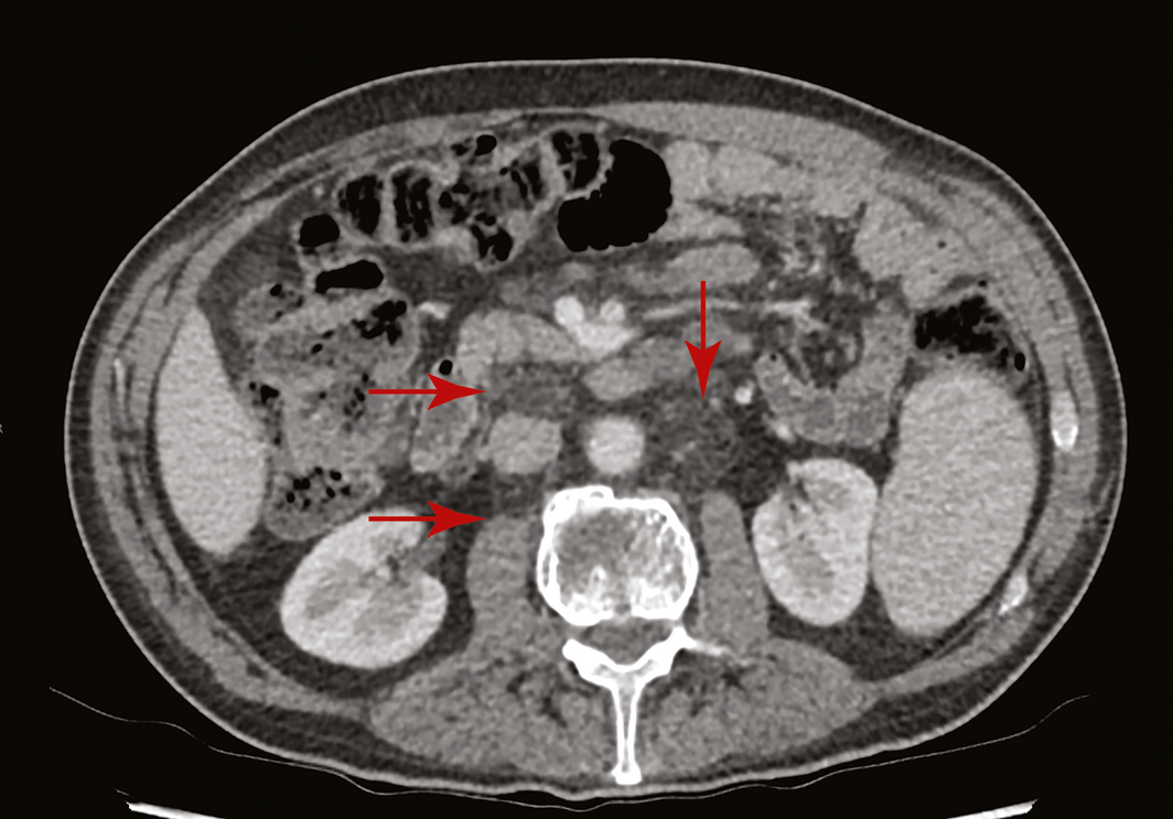
A CT scan of his abdomen had been performed one week before this presentation, for re‐assessment of his lymphadenopathy. While the mesenteric and retroperitoneal lymph node enlargement and splenomegaly remained stable over 3 years, it was noted that the lymph nodes were of abnormally low fat density attenuation (Box 1). Given this, the differential diagnosis was broadened from haematological malignancy to include atypical infection and rarer causes such as Whipple disease. Gastroenterology consultation was requested.
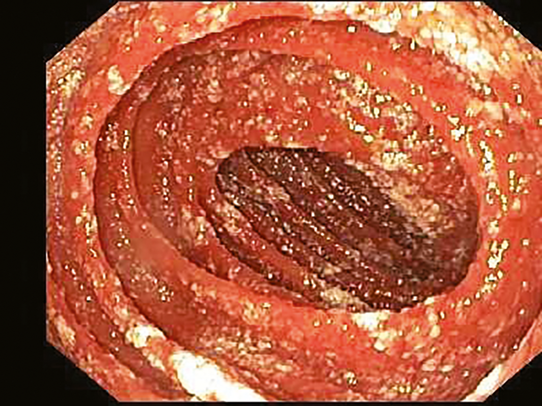
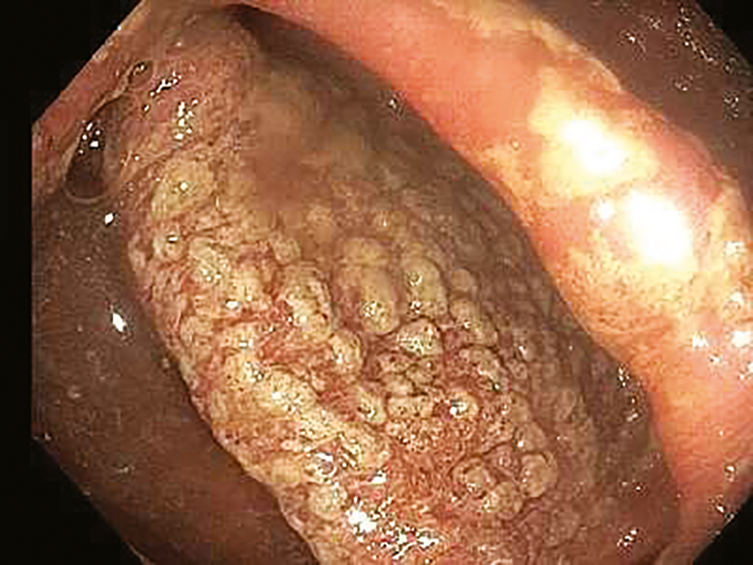
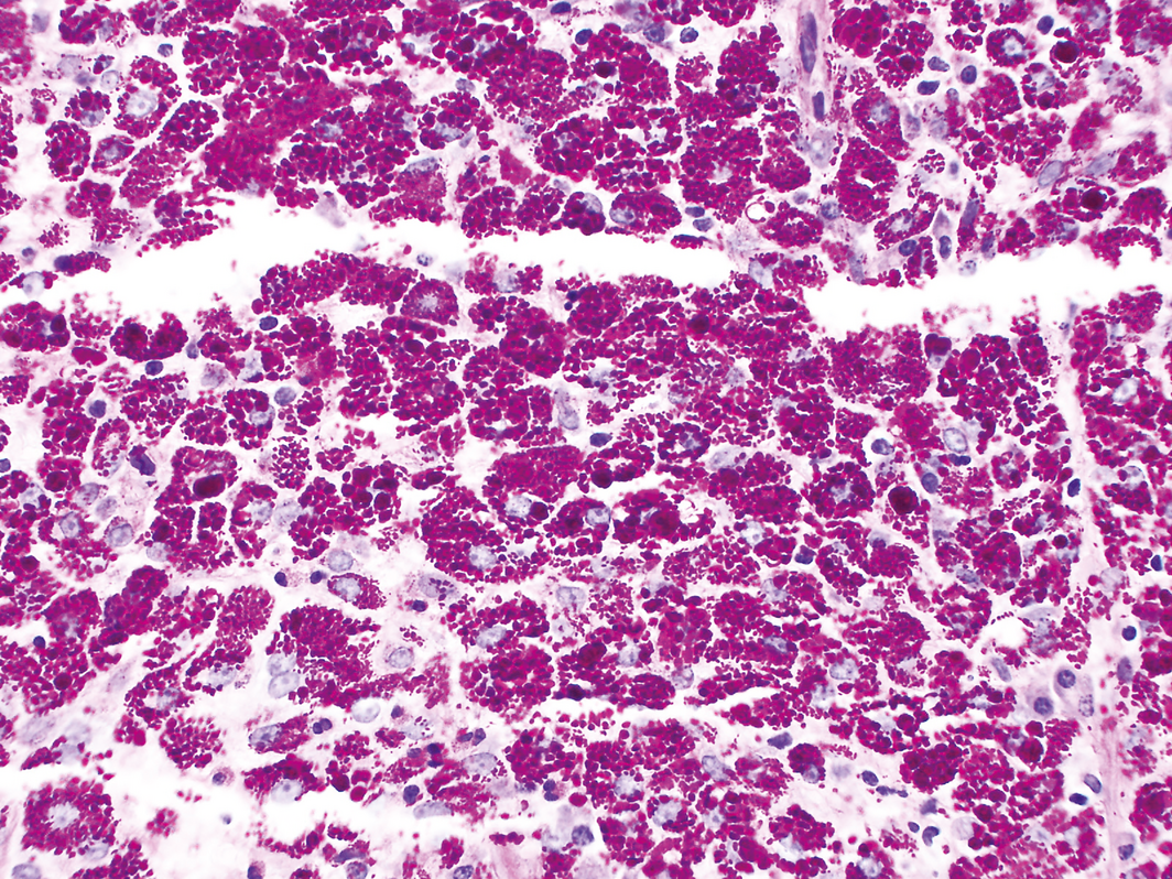
On enteroscopy and colonoscopy, ulceration with white plaque‐like lesions was identified throughout the gastrointestinal tract consistent with the endoscopic appearance of Whipple disease (Box 2 and Box 3) The duodenum and terminal ileum were affected, as was the colon from the sigmoid to the caecum with rectal sparing. The oesophagus and stomach were not affected and the jejunum was not examined. Histology and immunohistochemistry from biopsy samples revealed periodic acid–Schiff–diastase staining, supporting a diagnosis of Whipple disease (Box 4), and Tropheryma whipplei polymerase chain reaction results were positive.
Antibiotic therapy commenced with 2 g ceftriaxone for 2 weeks, extended to 4 weeks after a transoesophageal echocardiogram identified a small mitral valve vegetation. The patient's gastrointestinal symptoms resolved within 48 hours of treatment. After 4 weeks, his antibiotic therapy was changed to oral sulfamethoxazole–trimethoprim for 12 months (standard therapy for this condition). The patient had total resolution of his symptoms within 2 weeks of starting therapy and remained symptom free at the time of writing (12 months after starting therapy).
Discussion
Whipple disease is a rare multisystem condition caused by infection by the gram‐positive bacillus Tropheryma whipplei. While the organism is ubiquitous, the actual incidence of Whipple disease is exceedingly low at one per million people, with our patient fitting the highest risk demographic profile.1 Susceptibility to infection is thought to be genetic, resulting in cell mediated and macrophage immune defect, as yet incompletely understood.1,2 The clinical presentation of this disease is non‐specific and diagnosis is therefore difficult and often delayed. The earliest manifestation is arthralgia, followed by gastrointestinal symptoms some months or years later. Our patient's history of polyarthralgia and a recent admission with pulmonary oedema may be attributable to Whipple disease.
If left unrecognised and untreated, the disease is progressive and can be fatal;3 timely recognition and treatment is therefore crucial. Standard treatment is 2–4 weeks of ceftriaxone followed by one year of trimethoprim–sulfamethoxazole, and relapse rates are 17–35%.4 Anaemia and malnutrition are found in almost all cases, while less common complications include endocarditis, myocarditis, skin hyperpigmentation, dementia, confusion and cerebellar ataxia, with neurological involvement usually manifesting late in the disease process.
Whipple disease is classically described in the radiology literature by its features on barium small bowel follow‐through studies.5,6 However, with technical advances and increasing availability of CT and magnetic resonance imaging, small bowel barium contrast studies are becoming less frequently performed and unlikely to be utilised unless there is pre‐existing clinical suspicion for specific small bowel pathology.
Low attenuation lymphadenopathy is a recognised feature of Whipple disease on CT imaging,5,6 although the literature is scarcer, presumably due to relatively less frequent use of cross‐sectional imaging in the past. The low fat density of the lymph nodes is thought to be a consequence of lymphatic obstruction and intranodal deposition of fat.4 It is important to consider other important disease processes that could produce this appearance, including the more commonly encountered coeliac disease, as well as abdominal tuberculosis. Other intestinal opportunistic infections, lymphoma and metastases are also considerations.
Lessons from practice
- Whipple disease is a rare systemic disease caused by Tropheryma whipplei infection.
- Diagnosis is often challenging due to the non‐specific nature of symptoms.
- Low attenuation lymphadenopathy is a distinctive imaging finding for Whipple disease which can aid this diagnosis. It is now likely to be more frequently encountered with increasing use of computed tomography. The primary differential diagnoses for this imaging appearance include coeliac disease, mycobacterial infection and malignancy.
- Prognosis improves with early recognition, diagnosis and treatment.
Box 1 – Computed tomography scan showing abnormally enlarged, low attenuation retroperitoneal lymph nodes (arrows) approaching the density of the adjacent abdominal fat
Box 2 – Endoscopic image of the duodenum demonstrating the classic white plaque appearance of Whipple disease
Competing interests
No relevant disclosures.
References
- Schneider T, Moos V, Loddenkemper C, et al. Whipple's disease: new aspects of pathogenesis and treatment. Lancet Infect Dis 2008; 8: 179–190.
- Marth T, Roux M, von Herbay A, et al. Persistent reduction of complement receptor 3 alpha‐chain expressing mononuclear blood cells and transient inhibitory serum factors in Whipple's disease. Clin Immunol Immunopathol 1994; 72: 217.
- Chears WC, Hargrove MD, Verner JV, et al. Whipple's disease. Am J Med 1961; 30: 226–234.
- Durand DV, Lecomte C, Cathébras P, et al. Whipple disease. Clinical review of 52 cases. The SNFMI Research Group on Whipple Disease. Société Nationale Française de Médecine Interne. Medicine (Baltimore) 1997; 76: 170.
- Horton KM, Fishman EK. Uncommon inflammatory diseases of the small bowel: CT findings. Am J Roentgenol 1998; 170: 385–388.
- Johnson CD, Schmit GD. Mayo Clinic gastrointestinal imaging review. Rochester, MN: Mayo Clinic Scientific Press, 2005: 225–226.
Provenance: Not commissioned; externally peer reviewed.
Early-Onset Colorectal Cancer With Liver-Only Metastases: A Retrospective Cohort Study Integrating Prospectively Collected Real-World Clinical and Molecular Data From an Australian National Database (2009–2024) to Guide Treatment Planning
Savio G. Barreto, Christos S. Karapetis, Shahid Ullah, Matthew Burge, Susan Caird, Angus Campbell, Azim Jalali, Ross Jennens, Muhammad A. Khattak, Belinda Lee, Stephanie H. Lim, Shehara Mendis, Louise Nott, Timothy J. Price, Jeremy D. Shapiro, Jeanne Tie, Javier Torres, Colin Williams, Rachel Wong, Vanessa Wong, Peter Gibbs
Australian Pathways for Specialist Pain Management and Early Palliative Care for People With Pancreatic Cancer: Developed Using a Community Consensus Approach
Jennifer Philip, Melanie R. Lovell, Kylee Bellingham, Gail Garvey, Gregory B. Crawford, Nicole M. Rankin, Kara Burns, Isabel Young, Vivienne Milch, Dorothy Keefe, Katrina Anderson, James Lawson, Meinir Krishnasamy
Cause‐specific mortality among Queensland people with cirrhosis, by cirrhosis aetiology and decompensation status, 2007–22: a retrospective cohort study
Vikas Bhasker, Jessica R Fong, Paul J Clark, Gunter F Hartel, Richard Skoien, James O’Beirne, Elizabeth E Powell, Patricia C Valery
Clinical practice guidelines for hepatocellular carcinoma surveillance for people at high risk in Australia: summary of recommendations
Jacob George, Nicole L Allard, Stuart K Roberts, Leon A Adams, Jane Davies, Behzad Hajarizadeh, Jennifer H MacLachlan, Suzanne E Mahady, Rosalie Altus, Catherine Brown, David C Fry, Belinda Greenwood‐Smith, Natali Smud, Patricia C Valery, Nafisa Yussf, Kate Broun, Denise Campbell, Karen Canfell, Chelsea Carle Harrison, Victoria Freeman, Paul Grogan, Catherine Holliday, Suzanne Hughes, Anna Kelly, Cathelijne Kemenade, Claire Latumahina, Amanda McAtamney, Megan Varlow, Joachim Worthington, Susan Yuill, Eleonora Feletto
Assessment of metabolic dysfunction‐associated fatty liver disease in primary care: a consensus statement summary
Leon A Adams, William W Kemp, Kate R Muller, Elizabeth E Powell, Stuart K Roberts, Luis Calzadilla Bertot, Stephanie Best, Gary Deed, Jon D Emery, Samantha L Hocking, Graham R Jones, John S Lubel, Sinead Sheils, Stephen M Twigg, Gerald F Watts, Jacob George
Treatment outcomes for people with hepatitis C referred to tertiary care in Victoria, 2021–22: a retrospective observational study
Elly Layton, Nicole Matthews, Brendan Quinn, Nasra Higgins, Gabrielle Lindeman, Mielle Abbott, Jennifer MacLachlan, Elizabeth Birbilis, Margaret E Hellard, Joseph Doyle, Benjamin C Cowie, Mark Stoové