




Medical education
Lessons from practice
Volume 209 - Issue 8
Subclavian vein thrombosis with internal jugular vein extension in an Australian rules football player
Authors: John WP Wong, Fiona WY Lai and Ian Wilson
Med J Aust 2018; 209 (8): 335-336. || doi: 10.5694/mja18.00335
Published online: 15 October 2018
Published online: 15 October 2018
A 40-year-old female Australian rules football player presented to a rural emergency department with a 3-hour history of swelling and pain in her right upper limb
Clinical record
A 40-year-old female Australian rules football player presented to a rural emergency department with a 3-hour history of swelling and pain in her right upper limb. She denied chest pain, dyspnoea, cough, fevers or other symptoms. There was no history of cigarette smoking, oral contraceptive use, trauma, family history of coagulopathy or other significant past medical history. She was right-hand dominant and reported training most days and participating in competitive matches and team practices 3 days a week, with no recent increase in training intensity.
Vital signs were within reference range. Physical examination revealed a diffuse swelling and dusky discolouration of the entire right arm to the axilla. The right biceps measured 5 cm more than the left. Range of motion and neurovascular assessment of the upper limbs were normal. There was no evidence of lymphadenopathy.
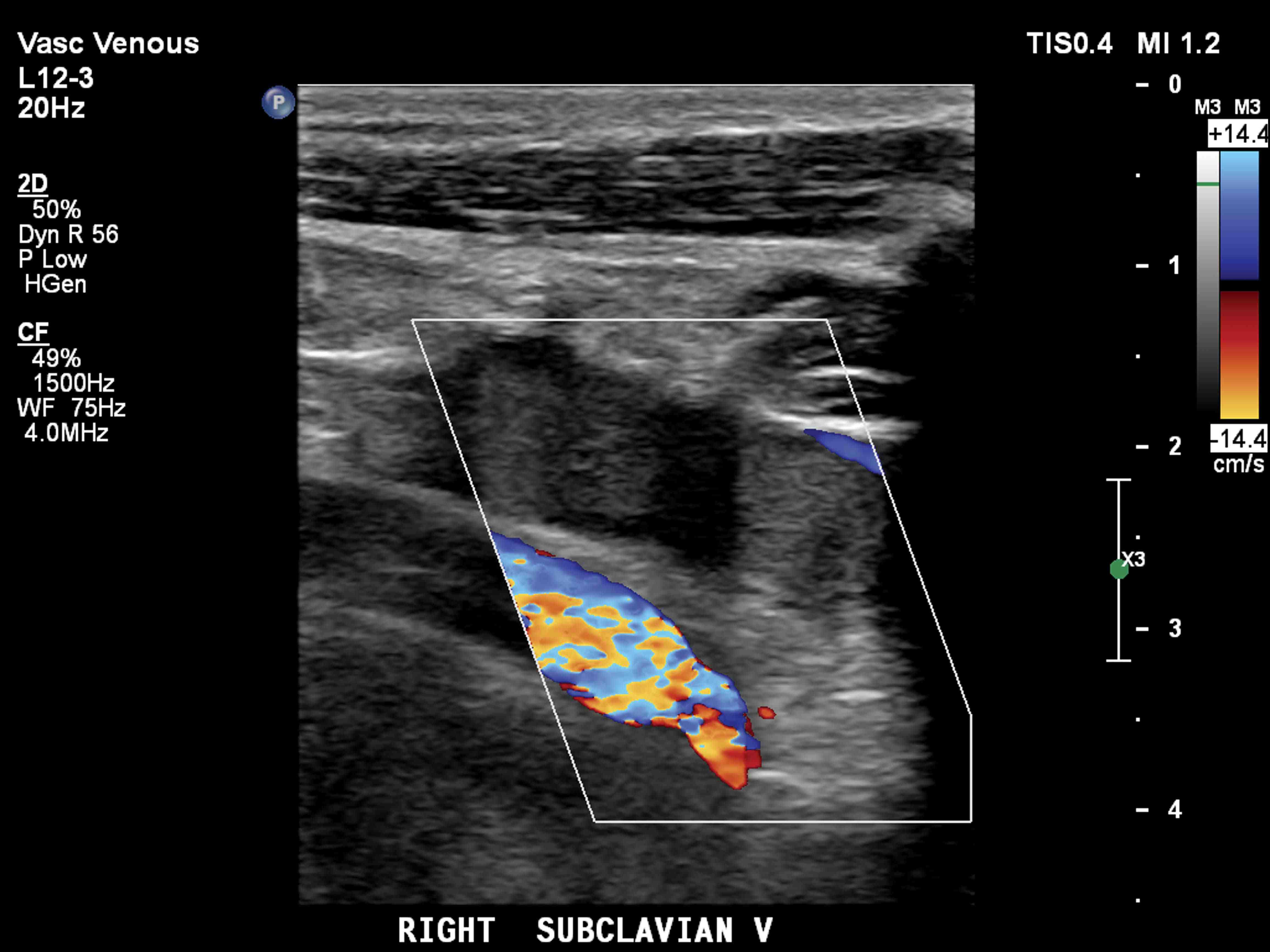
Given the concern of an upper extremity deep vein thrombosis (UEDVT), the patient was started on subcutaneous enoxaparin 1.5 mg/kg once daily. Ultrasonography was performed when first available the following day, confirming an extensive occlusive thrombus throughout the right subclavian vein that projected into the internal jugular vein (Box).
A complete blood count and basic metabolic panel were within normal limits. An extensive thrombophilic work-up included prothrombin time, partial thromboplastin time, anticardiolipin antibody, protein C activity, protein S activity, antithrombin III assay, factor V assay, prothrombin gene mutation G20210A, and lupus anticoagulant, which were all within the normal range. Computed tomography scan of the chest showed no evidence of venous thoracic outlet syndrome.
A diagnosis of Paget–Schroetter syndrome was made by exclusion. After 48 hours, subcutaneous enoxaparin was ceased and rivaroxaban 15 mg twice daily was commenced, and subsequently decreased to 20 mg once daily after 3 weeks of treatment. Upon review at one month, the patient reported improvement in swelling and was advised to return to normal activities, including her part-time work as a physiotherapist, in a graduated fashion. After 6 months of anticoagulation, ultrasonography confirmed resolution of the right upper extremity thrombus and rivaroxaban was ceased.
UEDVT accounts for 2–10% of all cases of deep vein thrombosis,1,2 and is a rare but important clinical entity with potential for considerable morbidity. The complications of UEDVT that may be associated with significant long term morbidity include pulmonary embolism, post-thrombotic syndrome and thrombotic recurrence.
UEDVT is classified as primary or secondary based on its pathogenesis. Secondary causes account for most cases, and are commonly due to placement of central venous catheters, malignancy or a hypercoagulable state, including ovarian hyperstimulation syndrome or exogenous oestrogen use. Primary UEDVT is a much rarer disorder (two per 100 000 persons per year), and refers to either Paget–Schroetter syndrome or idiopathic UEDVT.3
Paget–Schroetter syndrome, also termed effort-induced thrombosis, refers to axillary-subclavian vein thrombosis brought on by strenuous activities of the upper extremities, and it usually occurs in otherwise young and healthy individuals. It is believed that sustained and vigorous upper extremity movements, commonly following sporting activities, such as repetitive overhead arm movements and hyperabduction, which occur when playing Australian rules football, cause repetitive microtrauma of the vascular intima of the subclavian vein and its surrounding structures, leading to the activation of the coagulation cascade. Often, anatomical abnormalities involving the thoracic outlet lead to external venous compression and further contribute to the development of thrombosis, which has led to Paget–Schroetter syndrome being categorised as a venous variant of thoracic outlet syndrome.4 While screening for thrombophilic disorders is often done after a diagnosis of Paget–Schroetter syndrome, there is a lack of published literature to support it. One small study concluded that a hypercoagulable state played a large role in idiopathic, non-effort related UEDVT but not in Paget–Schroetter syndrome.5
Paget–Schroetter syndrome occurring in the absence of a predisposing cause has been described in only a few reported cases. Furthermore, the involvement of the internal jugular vein is atypical. While a favourable outcome was achieved in our patient with direct oral anticoagulant therapy, there is still a lack of consensus regarding the efficacy of direct oral anticoagulants and the optimum duration of anticoagulation with UEDVT.
Moreover, little is known about the incidence and predictors of complications following Paget–Schroetter syndrome. Current clinical care is predominantly guided by single-centre studies and case series and, importantly, by our knowledge of the treatment of lower extremity deep vein thrombosis. Of note, a systematic review of 432 patients with UEDVT found that 15% (range, 7–46%) developed post-thrombotic syndrome, although there was significant heterogeneity between studies in the diagnostic criteria for post-thrombotic syndrome and the proportion of patients with secondary causes included.6
Prospective studies are needed to guide management and to characterise the long term sequelae of Paget–Schroetter syndrome as young and active individuals are preferentially affected, making any residual disability significant. Absence of anatomical obstruction or other predisposing factors and extension of thrombotic material into the internal jugular vein make this case unique.Lessons from practice
Upper extremity deep vein thrombosis (UEDVT) is a rare but potentially fatal condition and should be considered in young physically active patients presenting with unilateral swelling of the arm.
Common causes of UEDVT are indwelling central venous catheters, hypercoagulable states and the thoracic outlet syndrome.
Paget–Schroetter syndrome is a cause of UEDVT and occurs following microtrauma to the subclavian vein after strenuous activities involving repetitive overhead arm movements and hyperabduction. It also more commonly affects young and active individuals.
Complications of UEDVT include pulmonary embolism, post-thrombotic syndrome and recurrence, all of which may cause significant morbidity and disability, especially if diagnosis is delayed.
Competing interests
No relevant disclosures.
References
- Esmon CT. Basic mechanisms and pathogenesis of venous thrombosis. Blood Rev 2009; 23: 225-229.
- Bernardi E, Pesavento R, Prandoni P. Upper extremity deep venous thrombosis. Semin Thromb Hemost 2006; 32: 729-736.
- Lindblad B, Tengborn L, Bergqvist D. Deep vein thrombosis of the axillary-subclavian veins: epidemiologic data, effects of different types of treatment and late sequelae. Eur J Vasc Surg 1998; 2: 161-165.
- Alla VM, Natarajan N, Kaushik M, et al. Paget–Schroetter syndrome: review of pathogenesis and treatment of effort thrombosis. West J Emerg Med 2010; 11: 358-362.
- Héron E, Lozinguez O, Alhenc-Gelas M, et al. Hypercoaguable states in primary upper-extremity deep vein thrombosis. Arch Intern Med 2000; 160: 382-386.
- Elman EE, Kahn SR. The post-thrombotic syndrome after upper extremity deep venous thrombosis in adults: a systematic review. Thromb Res 2006; 117: 609-614.
Splenic injury in severe cases of the zoonoses Q fever and rickettsial infection: diagnostic challenges
Ashleigh Drury, Philippa Harrison, Aiveen Bannan
Health and economic benefits of improving pre‐hospital identification of stroke in Australian women: a modelling study
Thomas Gadsden, Lei Si, Emily R Atkins, Cheryl Carcel, Xia Wang, Stephen Jan, Mark Woodward, Laura E Downey
Updating the diagnosis and management of elevated serum ferritin levels in the era of routine ferritin testing of blood donors by Australian Red Cross Lifeblood
Gary D Zhang, James Chen, Daniel M Johnstone, Martin B Delatycki, Katie Allen, John K Olynyk
Early cardiovascular collapse after envenoming by snakes in Australia, 2005–2020: an observational study (ASP‐31)
Geoffrey K Isbister, Katherine Z Isoardi, Angela L Chiew, Shane Jenkins, Nicholas A Buckley
Updated recommendations for warfarin reversal in the setting of four‐factor prothrombin complex concentrate
Danielle Robinson, James McFadyen, Eileen Merriman, Chee Wee Tan, Ross Baker, Huyen Tran
Updating the diagnosis and management of iron deficiency in the era of routine ferritin testing of blood donors by Australian Red Cross Lifeblood
Gary D Zhang, Daniel Johnstone, Michael F Leahy, John K Olynyk