




Volume 208 - Issue 11
Foreign tick smuggling rickettsia evades Australian border control
Authors: Sadid F Khan, Mythili Tadepalli, John Stenos, Stephen R Graves and Tony M Korman
Med J Aust 2018; 208 (11): 505. || doi: 10.5694/mja17.01201
Published online: 18 June 2018
Published online: 18 June 2018
To the Editor:
Tick-borne infectious diseases, including rickettsial infections, acquired in Australia or after international travel remain a diagnostic challenge.1
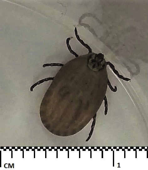
A 68-year-old man presented with umbilical pain 10 days after returning from a 2-month camping trip through the south-west of the United States (ie, Texas, New Mexico, Arizona, Colorado and Utah). On examination, a live tick was detected and removed from the patient’s umbilicus (Box).
The umbilical pain resolved after tick removal. There was no development of fevers, constitutional symptoms, or rash to suggest a tick-borne illness. Laboratory investigations were unremarkable. He was educated about the signs and symptoms of tick-borne illnesses and prescribed a single dose of doxycycline 200 mg for prophylaxis due to his high risk exposure.
The tick was identified as Dermacentor andersoni (Rocky Mountain wood tick), which is endemic to North America and not known to occur in Australia.2 D. andersoni adult ticks are principal vectors of Rickettsia rickettsii (the cause of Rocky Mountain spotted fever), and are associated with transmission of other pathogens to humans, including Colorado tick fever virus and Francisella tularensis (the cause of tularemia).2,3 Although isolated from D. andersoni ticks, transmission of Coxiella burnetii (the cause of Q fever) is uncommon. D. andersoni is not known to transmit Lyme disease.2
Analysis of the tick for rickettsial DNA was positive. No Borrelia DNA was detected. Rickettsia was isolated in cell culture and identified as Rickettsia peacockii based on sequencing of the 17kDa, OmpB, gltA and Sca4 genes. R. peacockii is a member of the spotted fever group of rickettsiae.3,4 The presence of R. peacockii in ticks is correlated with reduced prevalence of R. rickettsii.2,3 R. peacockii is closely related to R. rickettsii, and deletion or mutation of genes, possibly resulting in loss of virulence in R. peacockii, have been identified.3 R. peacockii is not known to be a pathogen of humans or other animals.3,4
Rickettsial serology 10 weeks after the tick bite showed detectable antibodies (titre, 1/256), predominantly to the spotted fever group of Rickettsia, compatible with exposure to R. peacockii identified in the tick. Unfortunately, definitive seroconversion or a rising antibody titre was not able to be demonstrated as no earlier sera were available for parallel testing. Pre-existing antibodies from a distant rickettsial exposure from his tick-prone lifestyle (history of extensive international camping trips) cannot be excluded. The patient remains asymptomatic 9 months later and is still an avid traveller.
Tick-borne rickettsial infections in Australia include Queensland tick typhus (Rickettsia australis), Flinders Island spotted fever and Australian spotted fever (Rickettsia honei) and Q fever transmitted by ticks including Ixodes spp., Amblyomma triguttatum and Bothriocroton hydrosauri.1
With increasing international travel, recognition of tick-borne rickettsial diseases is becoming more important. Dermacentor ticks have been detected on livestock exported from North America into Europe.5 This case shows the ability of human ectoparasites, and their potentially pathogenic bacteria, to bypass stringent Australian quarantine controls. Further studies of Australian and imported tick-borne infections are required to increase understanding of these emerging infectious diseases.
Competing interests
No relevant disclosures.
References
- Graves SR, Stenos J. Tick-borne infectious diseases in Australia. Med J Aust 2017; 206: 320-324.
- Dergousoff SJ, Gajadhar AJ, Chilton NB. Prevalence of Rickettsia species in Canadian populations of Dermacentor andersoni and D. variabilis. Appl Environ Microbiol 2009; 75: 1786-1789.
- Felsheim RF, Kurtti TJ, Munderloh UG. Genome sequence of the endosymbiont Rickettsia peacockii and comparison with virulent Rickettsia rickettsii: identification of virulence factors. PLoS One 2009; 4: e8361.
- Kurtti TJ, Simser JA, Baldridge GD, et al. Factors influencing in vitro infectivity and growth of Rickettsia peacockii (Rickettsiales: Rickettsiaceae), an endosymbiont of the Rocky Mountain wood tick, Dermacentor andersoni (Acari, Ixodidae). J Invertebr Pathol 2005; 90: 177-186.
- Lillehaug A, Mehl R, Gjerde B. Importation of Dermacentor albipictus into Europe. Vet Rec 2002; 151: 94-95.
Costs of Delivering the Hep B PAST Model of Care in Remote Australia: A Retrospective Pre–Post Study
Hoa Nguyen, Anh Le Tuan Nguyen, Paula Binks, Melita McKinnon, Emily Vintour-Cesar, Karen Wills, Nicola Stephens, Benjamin Cowie, Joshua S. Davis, Yuejen Zhao, Peter Nihill, Julie A. Campbell, Andrew J. Palmer, Jane Davies, Barbara de Graaff
Buruli Ulcer Acquired From Ringtail Possum Rescue
Daniel P. O'Brien, Naomi Clarke, Sara Bonnici, Mohammad A. Hussain
Q Fever Visceral Abscesses After Contact With Native Australian Marsupials
Samantha J. Laws, Enzo Binotto, James Stewart, Jennifer Ho, Trent Yarwood, Josh Hanson, Simon Smith
Hospital-Admitted Injection-Related Infections Among Incarcerated People Who Inject Drugs in Australia: A Retrospective Cohort Study
Andrew Palmer, Matthew Carter, Jeremy Yeo, Cecilia Shim, Jason Connor, Jeremy Hayllar, Gerald Holtmann, Naomi Moy, Elliott G. Playford, Naomi Runnegar, Paul J. Clark
The Live Attenuated Influenza Vaccine in Australia: An Additional Tool for Influenza Prevention
Cyra Patel, Alexis Pillsbury, Tran Nguyen, Xia Wang, Helen E. Quinn, Clayton K. Chiu, Allen C. Cheng, Katie L. Flanagan, Zhicheng Wang
Reflex Testing for Hepatitis D Infection: A Unique Opportunity to Reduce Hepatitis D-Related Chronic Liver Disease Deaths in Australia
Jessica Howell, Lauren Andersson, Miriam T. Levy, James O'Beirne, Leon Adams, Katharine Irvine, Avik Majumdar, Golo Ahlenstiel, Kathy Jackson, Krispin Hajkowicz, Joseph Doyle, Jane Davies, Sarah Cherian, Wayne Dimech, Alexander J. Thompson