




Medical education
Lessons from practice
Volume 207 - Issue 2
Not all that wheezes is asthma
Authors: Amy SM Wong, Bing Mei Teh, Glen Burgess and Michael CS Ho
Med J Aust 2017; 207 (2): 59-60. || doi: 10.5694/mja16.00561
Published online: 17 July 2017
Published online: 17 July 2017
Lessons from practice•Diagnosis of adult-onset asthma should be made with caution.•The presence of an inhaled foreign body in the upper airway may be misdiagnosed as asthma.•Direct visualisation with an endoscope is required to exclude the presence of a foreign body.
Clinical record
An 80-year-old woman presented with a history of worsening cough, dyspnoea and wheeze, and was hospitalised for an infective exacerbation of asthma. She reported a 17-year history of mild non-productive cough. She was diagnosed with adult-onset asthma 8 years earlier, when her exacerbations responded to bronchodilator therapy and corticosteroids and subsequent respiratory function tests showed a 21% bronchodilator response. Serial chest x-rays and computed tomography (CT) scans were normal except for stable pulmonary nodules, which were under close surveillance by her respiratory physician. Relevant past medical history included hypertension, pacemaker for complete heart block, hay fever, excision of cutaneous melanoma and laparotomy for small bowel obstruction. She had no known allergies, was a lifelong non-smoker and lived at home with her husband and pet budgie.
Three weeks before her presumed acute exacerbation of asthma, she reported choking on croutons in tomato soup. The admitting respiratory physician noted bilateral end expiratory wheezes on auscultation and no stridor. She improved with bronchodilators and corticosteroids and was discharged with outpatient pulmonary rehabilitation. However, she required further courses of corticosteroids from her general practitioner due to persistent dyspnoea, cough and wheeze. At her respiratory follow-up, examination revealed loud inspiratory and expiratory wheezes emanating from her throat to her chest. Alternative diagnoses of vocal cord dysfunction or upper airway lesions were considered, and she was referred to an otolaryngologist 8 months after her choking episode.
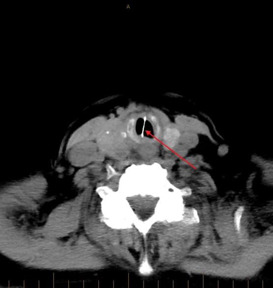
Ear, nose and throat examination was normal. However, further examination using flexible nasendoscopy showed an abnormal midline lesion in the larynx just visible in the subglottis. She had an urgent CT neck scan, which revealed a thin, hyperdense lesion, measuring 14 × 22 × 1 mm in the subglottic region (Box 1). This was not seen on previous chest x-rays or CT scans of the chest.
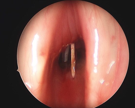
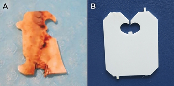
Emergency direct microlaryngoscopy was performed under general anaesthesia using percutaneous transtracheal ventilation. A hard whitish lesion was found firmly buried vertically in the trachea, mostly covered in granulation tissue in the midline just below the vocal cords (Box 2). The remaining trachea was normal. The foreign body was removed under direct visualisation after semi-transection of the lesion without complications, and resembled half a plastic bread clip with chipped edges (Box 3). The foreign body was covered in extensive granulation tissue, suggesting a prolonged impaction.
Following the removal of the foreign body and administration of oral steroids, the patient’s symptoms and exercise tolerance improved to her pre-morbid level of function.
We highlight here the aspects of this case which affect management, including differential diagnoses that should be considered. The patient had non-specific pulmonary nodules that may contribute to her cough. Metastases to the lungs or airways should be considered with her history of melanoma. In addition, she could have developed a delayed hypersensitivity reaction to her pet budgie. A diagnosis of adult-onset asthma in a 72-year-old lifelong non-smoker is unusual. With a choking history and persistent symptoms despite treatment, endoscopy was warranted to exclude an inhaled foreign body. The inhaled foreign body was firmly embedded in the subglottis and covered in extensive granulation tissue. The surrounding fibrosis suggests it could have been there for many months — the expiry date on the bread clip was not visible to confirm this. Her inspiratory flow volume curve also did not show the classic plateau or squaring in subglottic obstruction, which may have aided the diagnosis.
Ingestion or inhalation of plastic bread clips and its complications have been reported in the literature on several occasions.1,2 Foreign body inhalations, although prevalent in the paediatric population, are rare in adults.3 The sphincteric function of the larynx efficiently protects the lower respiratory tract; therefore, accidental inhalation of a foreign body is less likely than the chance of swallowing.4 Laryngotracheal foreign bodies are rare and potentially life-threatening. The commonest symptoms are choking, followed by a protracted cough and occasionally wheezing.3 Physical examination findings include fever, stridor, sternocostal retractions and reduced breath sounds. Radiographic imaging may be helpful if it targets the correct area, if the object is radiopaque or if there are signs of hyperexpansion on expiration. Plastic foreign bodies often do not produce a signal on x-ray-based imaging and, hence, this procedure cannot reliably exclude a foreign body.5
Flexible nasendoscopy is easily performed by otolaryngologists under local anaesthesia in the office and is generally safe and well tolerated. Diagnostic laryngoscopy or bronchoscopy may be indicated in patients where the diagnosis remains uncertain.4 Foreign body removal should be performed under direct visualisation with flexible or rigid endoscopy3 and airway equipment readily available. Open surgery for extraction should only be performed as a last resort.
Lessons from practice
-
Adult-onset asthma is a diagnosis that needs to be made with caution, particularly in older people. When there are persistent or worsening symptoms despite conventional therapy, further investigation with imaging and endoscopy is warranted.
-
The presence of an inhaled foreign body in the upper airway is an emergent, potentially life-threatening condition that requires careful, rapid evaluation and management. Diagnosis is often difficult and it may be misdiagnosed as asthma.
-
Negative-imaging studies do not exclude the presence of a foreign body in the airway, particularly if it is made of plastic. Direct visualisation with an endoscope is warranted.
-
Flexible nasendoscopy can be performed easily by otolaryngologists in the consultation room, and is generally safe and well tolerated. If no foreign bodies are seen, diagnostic laryngoscopy or bronchoscopy under general anaesthesia may be indicated.
Competing interests
No relevant disclosures.
References
- Bhardwaj N, Kanhere HA, Maddern GJ. Small bowel obstruction caused by a bread clip: a time and time again problem. ANZ J Surg 2016; 86: 94-95.
- Beer TW. Fatalities from bread tag ingestion. Med J Aust 2002; 176: 506.
- Swanson KL, Edell ES. Tracheobronchial foreign bodies. Chest Surg Clin N Am 2001; 11: 861-872.
- Kothari S, Vijay A. A rare glottic foreign body with unusual presentation. World Articles in Ear, Nose and Throat 2013; 6.
- Ricketti PA, Ricketti AJ, Cleri DJ, et al. A 41-year-old male with cough, wheeze, and dyspnea poorly responsive to asthma therapy. Allergy Asthma Proc 2010; 31: 355-358.
Provenance: Not commissioned; externally peer reviewed.
Barriers and Facilitators to Timely Detection and Optimal Management of Chronic Wet Cough in Aboriginal Children Across Australia From the Perspectives of Carers of Aboriginal Children: A Qualitative Study
Gloria T. Y. Lau, Pam Laird, Robyn Aitken, Melanie Barwick, Liam Bedford, Emily R. Bowden, Jamie Everingham, Angela Fuery, Reece Griffin, Neha Jain, Mina Kinghorn, Renae Knapp, Gabrielle B. McCallum, Peter Morris, Richard Norman, Peter C. Richmond, Aarti Saiganesh, Slade Sibosado, Cameron Taylor, Maree Toombs, Roz Walker, Joan Wilson, Anne B. Chang, André Schultz
Barriers and Facilitators to Timely Detection and Optimal Management of Chronic Wet Cough in Aboriginal Children Across Australia From the Perspective of Health Care Providers: A Qualitative Study
Gloria T. Y. Lau, Pamela Laird, Robyn Aitken, Melanie Barwick, Liam Bedford, Emily R. Bowden, Corey Dalton, Jamie Everingham, Angela Fuery, Reece Griffin, Neha Jain, Mina Kinghorn, Renae Knapp, Gabrielle B. McCallum, Peter Morris, Richard Norman, Peter C. Richmond, Aarti Saiganesh, Slade Sibosado, Maree Toombs, Roz Walker, Joan Wilson, Anne B. Chang, André Schultz
Respiratory Syncytial Virus Disease Burden and Evidence Gaps in Australia Before Immunisation Introduction: A Retrospective Population-Based Study Using 2016–2019 Hospitalisation and 2023 Laboratory Surveillance Data
Chisato Imai-Boulter, Jean Li-Kim-Moy, Clayton Chiu, Sanjay Jayasinghe, Kristine Macartney
Not Gone but Often Forgotten: Action Needed to Address Tuberculosis in Aboriginal and Torres Strait Islander Communities
Nikkita Joshua, Emma L. Smith, Eunice (Cheeky) Love, Christopher P. Lowbridge, Nicole Tukana, Amy Bowden, Megan A. Campbell, Dawn Casey, James Ward
Uneven Ground: Survival Differences Among Victorian Lung Cancer Patients by Location of Residence (2011–2023): A Retrospective Cohort Study
Evangeline Samuel, Eldho Paul, Mike Lloyd, Sanuki Tissera, Craig Underhill, Sagun Parakh, Phillip Parente, Inger Olesen, Javier Torres, Katharine See, Gavin M. Wright, David Langton, Thomas John, Matthew Conron, James Bartlett, Nicola Atkin, Nikolajs Zeps, Susan V. Harden, Wasek Faisal, John R. Zalcberg, Rob G. Stirling
Treatment and Survival Outcomes for Indigenous and Non-Indigenous Australians Within the Victorian Lung Cancer Registry: A Retrospective Cross-Sectional Cohort Study
Melanie Wong, Mike Lloyd, Jessie Zeng, Sanuki Tissera, Kalinda E. Griffiths, Justine Clark, Jonathan Gillies, Lisa Briggs, Jacqueline Lesage, Tom Wood, Craig Underhill, Sagun Parakh, Louis B. Irving, Wasek Faisal, Rob Blum, Gary E. Richardson, Phillip Parente, Michelle Caldecott, Inger Olesen, Javier Torres, Evangeline Samuel, Christopher Lyne, Katharine See, David Langton, Thomas John, Gavin Wright, Matthew Conron, James Bartlett, Golsa Adabi, Maggie Moore, Susan Harden, Zoe K. McQuilten, John R. Zalcberg, Rob Stirling