




Medical education
Lessons from practice
Volume 206 - Issue 11
A rash diagnosis
Authors: Alana Christensen, Anushia Ashokan and David L Gordon
Med J Aust 2017; 206 (11): 483-484. || doi: 10.5694/mja16.00937
Published online: 19 June 2017
Published online: 19 June 2017
Interpreting positive cytomegalovirus serology requires caution, and follow-up testing is important
Clinical record
A 46-year-old man presented with a rash over his trunk, neck and limbs. A viral exanthem was suspected and an initial diagnosis of acute cytomegalovirus (CMV) infection was made on the basis of serology results (positive IgM, no quantitation indicated, and negative IgG). Additional laboratory tests showed an elevated C-reactive protein level (26 mg/L; reference interval [RI], 0–10 mg/L) and erythrocyte sedimentation rate (26 mm/h; RI, < 20 mm/h), and normal routine haematology and biochemistry results, apart from a slight elevation of the globulin level (45 g/L; RI, 20–40 g/L). However, the rash persisted, the patient noticed hair loss and he developed worsening visual impairment, which prompted an urgent ophthalmology referral 4 months after initial presentation. On ophthalmological review, the patient was noted to have decreased visual acuity bilaterally (right eye, 6/15; left eye, 6/24) and right optic disc swelling. Syphilis serology was subsequently performed and the results (Treponema pallidum particle agglutination [TPPA] positive and rapid plasma reagin [RPR] reactive [1 : 32]) prompted referral to the infectious diseases unit.
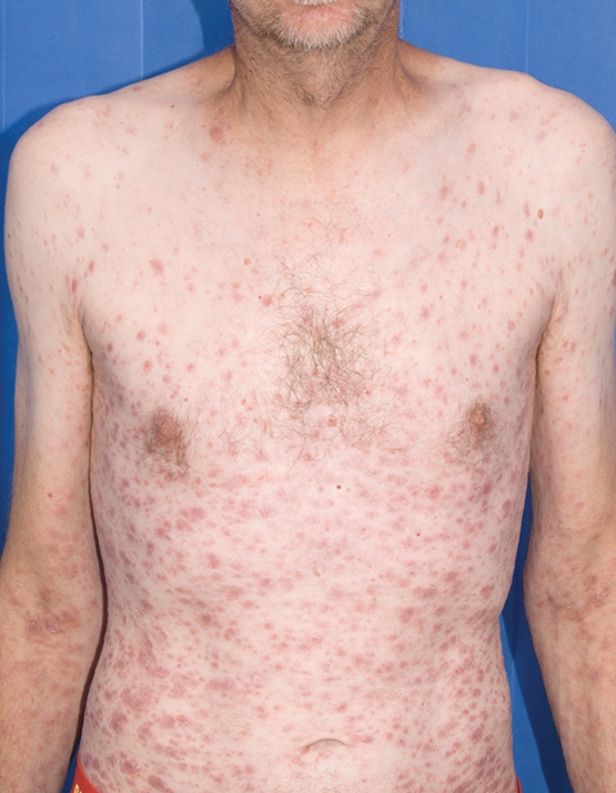
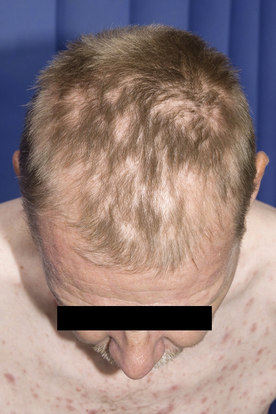
Clinical examination at presentation to the infectious diseases unit revealed widespread symmetrical erythematous papulo-squamous plaques over his trunk, neck and limbs (Box 1), with several small plaques on the plantar aspect of his left hand and left foot. He also had patchy alopecia (Box 2) and bilateral inguinal lymphadenopathy. At this time, the patient also reported unprotected intercourse with casual male partners. His HIV test result was negative and he had not been tested for syphilis previously. Retrospective testing of the earlier serum positive for CMV IgM revealed positive TPPA and reactive RPR (1 : 64). A punch biopsy of the rash showed a plasma cell-rich granulomatous process spanning the dermis, which supported the diagnosis of syphilis, likely consistent with tertiary syphilis. He was treated with a 15-day course of intravenous benzylpenicillin with concomitant prednisolone in the first 36 hours (given the optic involvement) to reduce the likelihood of a Jarisch–Herxheimer reaction. Over the treatment course, the patient remained well and his rash started to settle. On follow-up 3 months after treatment, his alopecia had completely resolved and his rash had significantly improved.
This case exemplifies the consequences of diagnosis based on the performance of a test, which may not have been indicated from the clinical presentation, compounded by a false-positive laboratory result. First, the presentation was not typical of CMV infection. In immunocompetent adults, the most common clinical presentation of CMV infection is a self-limiting mononucleosis-like syndrome1 characterised by fevers and malaise. Predominant skin manifestations are rare in CMV infection in immunocompetent patients; however, transient rubelliform rashes, generally lasting no longer than a few days,2 and maculopapular eruptions may occur.3 Exposure to ampillicin or related β-lactam antibiotics is associated with the development of a generalised maculopapular rash in patients with CMV mononucleosis, as is also the case for Epstein–Barr virus mononucleosis.2,3 In this patient, the persistent and atypical rash could have prompted other diagnoses to be explored earlier.
Second, the significance of the CMV IgM serology result is highly questionable. While the initial laboratory reported CMV serology (chemiluminescent immunoassay) as IgM positive and IgG negative without any quantitation, there was an appropriate comment suggesting repeat testing in 7–14 days to confirm IgG seroconversion, which did not occur in this case. After further retrospective clarification from the laboratory, the patient’s CMV IgM level was only 23 U/mL (RI, < 17 U/mL; equivocal, 18–22 U/mL). In our experience, positive CMV (and indeed other) IgM serology results that are near the defined cut-off values are most commonly false-positive results, and are rarely confirmed with IgG seroconversion on subsequent testing. We would also recommend that laboratories add additional cautionary comments and semi-quantitative data or sample to cut-off ratios for weakly positive IgM results; this is of particular importance in interpretation of CMV serology during pregnancy.
Interpretation of CMV-specific IgM results may also be problematic, as IgM may persist for up to 1 year after acute infection (in the presence of IgG), thus limiting its specificity for acute CMV infection.3 CMV IgM false-positive results have also been reported in patients with rheumatoid factor,3 other infections such as Epstein–Barr virus4 and antiphospholipid syndrome,5 which further highlights the need for paired specimens to confirm seroconversion in suspected cases. This case emphasises that positive test results should be cautiously interpreted in the context of the patient’s clinical presentation and that positive IgM results should be confirmed with subsequent IgG seroconversion.Lessons from practice
Predominant skin manifestations are rare in cytomegalovirus (CMV) infection in immunocompetent patients.
A positive CMV (and indeed other) IgM serology results that are near the defined cut-off values are most commonly false-positive results and are rarely confirmed with IgG seroconversion on subsequent testing.
To confirm a diagnosis, follow-up testing of positive IgM serology results to demonstrate IgG seroconversion is essential.
Competing interests
No relevant disclosures.
Acknowledgements
We thank the patient for permission to publish this case.
References
- Lancini D, Faddy HM, Flower R, Hogan C. Cytomegalovirus disease in immunocompetent adults. Med J Aust 2014; 201: 578-580.
- Lesher JL. Cytomegalovirus infections and the skin. J Am Acad Dermatol 1988; 18: 1333-1338.
- Cohen JI, Corey GR. Cytomegalovirus infection in the normal host. Medicine (Baltimore) 1985; 64: 100-114.
- Miendje Deyi Y, Goubau P, Bodéus M. False-positive IgM antibody tests for cytomegalovirus in patients with acute Epstein–Barr virus infection. Eur J Clin Microbiol Infect Dis 2000; 19: 557-560.
- De Carolis S, Santucci S, Botta A, et al. The relationship between TORCH complex false positivity and obstetric outcome in patients with antiphospholipid syndrome. Lupus 2012; 21: 773-775.
Provenance: Not commissioned; externally peer reviewed.
Costs of Delivering the Hep B PAST Model of Care in Remote Australia: A Retrospective Pre–Post Study
Hoa Nguyen, Anh Le Tuan Nguyen, Paula Binks, Melita McKinnon, Emily Vintour-Cesar, Karen Wills, Nicola Stephens, Benjamin Cowie, Joshua S. Davis, Yuejen Zhao, Peter Nihill, Julie A. Campbell, Andrew J. Palmer, Jane Davies, Barbara de Graaff
Buruli Ulcer Acquired From Ringtail Possum Rescue
Daniel P. O'Brien, Naomi Clarke, Sara Bonnici, Mohammad A. Hussain
Q Fever Visceral Abscesses After Contact With Native Australian Marsupials
Samantha J. Laws, Enzo Binotto, James Stewart, Jennifer Ho, Trent Yarwood, Josh Hanson, Simon Smith
Hospital-Admitted Injection-Related Infections Among Incarcerated People Who Inject Drugs in Australia: A Retrospective Cohort Study
Andrew Palmer, Matthew Carter, Jeremy Yeo, Cecilia Shim, Jason Connor, Jeremy Hayllar, Gerald Holtmann, Naomi Moy, Elliott G. Playford, Naomi Runnegar, Paul J. Clark
The Live Attenuated Influenza Vaccine in Australia: An Additional Tool for Influenza Prevention
Cyra Patel, Alexis Pillsbury, Tran Nguyen, Xia Wang, Helen E. Quinn, Clayton K. Chiu, Allen C. Cheng, Katie L. Flanagan, Zhicheng Wang
Reflex Testing for Hepatitis D Infection: A Unique Opportunity to Reduce Hepatitis D-Related Chronic Liver Disease Deaths in Australia
Jessica Howell, Lauren Andersson, Miriam T. Levy, James O'Beirne, Leon Adams, Katharine Irvine, Avik Majumdar, Golo Ahlenstiel, Kathy Jackson, Krispin Hajkowicz, Joseph Doyle, Jane Davies, Sarah Cherian, Wayne Dimech, Alexander J. Thompson