




Medical education
Snapshot
Volume 206 - Issue 10
Expectorate the unexpected
Authors: David J Russell and Francis A Ponnuthurai
Med J Aust 2017; 206 (10): 431. || doi: 10.5694/mja16.00877
Published online: 5 June 2017
Published online: 5 June 2017
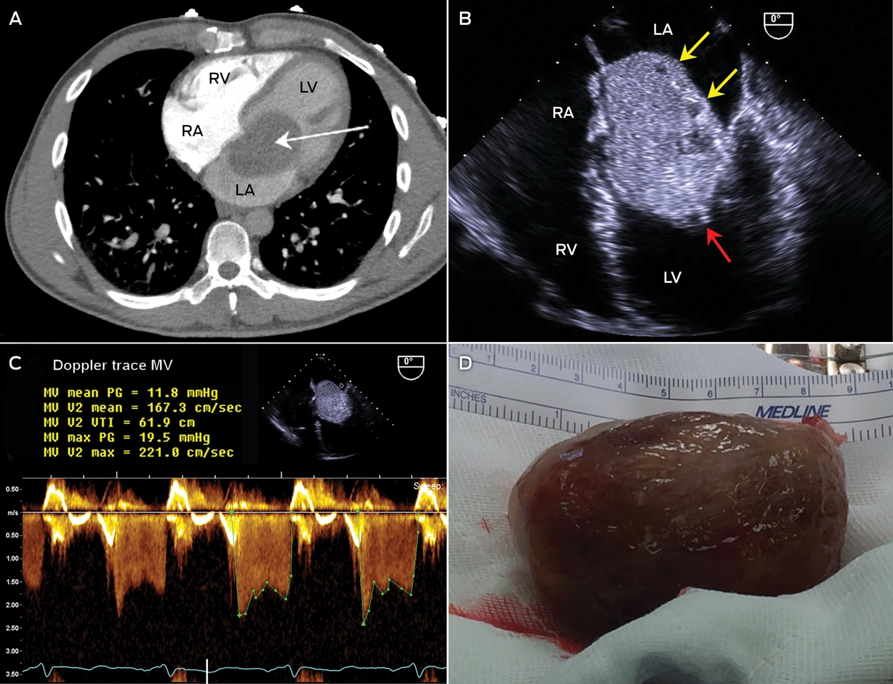
A 43-year-old man underwent a computed tomography pulmonary angiography for suspected pulmonary embolism after he presented with an episode of orthopnoea associated with blood-stained expectorate. The only finding was a left atrial mass (Figure, A, arrow). Further history revealed 6 months of progressive postural presyncope, malaise and weight loss of 3 kg.
Echocardiography showed a circumscribed 7.0 × 4.5 cm heterogeneous mass, with areas of cystic degeneration (Figure, B, yellow arrows) and frond-like extensions (∼ 3 mm; Figure, B, red arrow), consistent with a myxoma. The mass was attached to the interatrial septum via a small stalk and prolapsed through the mitral valve, causing significant mitral inlet obstruction; the mean gradient was approximately 12 mmHg (Figure, C). The mass was successfully removed (Figure, D).
Competing interests
No relevant disclosures.
Fatty Liver Disease in Australia: A Narrative Review on the Epidemiology, Natural History, Prognostication and Management in People With Metabolic Dysfunction
Karl Vaz, Daniel Clayton-Chubb, William W. Kemp, Stuart K. Roberts, Ammar Majeed
Sex Differences in Management and Outcomes of People With ST-Elevation Myocardial Infarction, New South Wales, 2011–2020: A Retrospective Cohort Study
Adam C. Livori, Jedidiah I. Morton, Dianna J. Magliano, Jonathan E. Shaw
Rethinking Rheumatic Fever Diagnosis: Progress, Pitfalls and the Path Towards a Definitive Test
Anna P. Ralph, Rachel Webb, Tom Parks, Nicole J. Moreland, Asha C. Bowen, Benjamin Jones, Emma Ndagire, Andrea Z. Beaton
Sex Differences in Management and Outcomes of People With ST-Elevation Myocardial Infarction, New South Wales, 2011–2020: A Retrospective Cohort Study
Samia Kazi, Simone Marschner, Clara K. Chow
Distribution of Cardiovascular Disease Risk Based on the Updated 2023 Guideline-Recommended Australian Cardiovascular Disease Risk Algorithm and Comparison With the 2012 Algorithm: An Observational Study
Nina Lazarevic, Meghana Bhat, Grace Joshy, Danielle C. Butler, Mark Woodward, Anushka Patel, Rod T. Jackson, Garry Jennings, Rosemary Wyber, Ellie Paige, Emily Banks
They Brought the Hospital to Me
Hugh N. Burrill