




Volume 202 - Issue 10
General practitioner-referred magnetic resonance imaging for musculoskeletal conditions: not a substitute for plain x-ray
Authors: Sam L Francis, Nicole Williams and Peter J Cundy
Med J Aust 2015; 202 (10): 526-527. || doi: 10.5694/mja14.01403
Published online: 1 June 2015
Published online: 1 June 2015
MRI is mostly unhelpful in diagnosing paediatric musculoskeletal complaints
To the Editor: In November 2012, the Australian Government extended requesting rights for Medicare-eligible magnetic resonance imaging (MRI) scans to general practitioners for “a small set of clinically appropriate indications”1 in patients under the age of 16 years. The purpose was
avoiding exposure of children to unnecessary radiation associated with other types of diagnostic imaging like computed tomography (CT) scans.1
With musculoskeletal MRI, a Medicare-eligible scan is performed “following radiographic examination”.1
Plain x-ray, rather than MRI, remains the gold standard for diagnosing most musculoskeletal conditions in children, and we report two cases where the initial use of MRI rather than x-ray led to a delay in diagnosis.
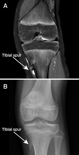
First, an 11-year-old girl was investigated for a 3-month history of left knee pain and locking. An ultrasound and MRI scan (Box 1, A) were performed, although an x-ray was not done. As no cause for the symptoms was identified on the ultrasound or MRI, the patient was referred to an orthopaedic surgeon, who requested an x-ray (Box 1, B). This showed a large proximal tibial spur tethering the semitendinosis tendon. This was poorly visualised on MRI and had not been reported. The patient's symptoms resolved after excision of the spur.2
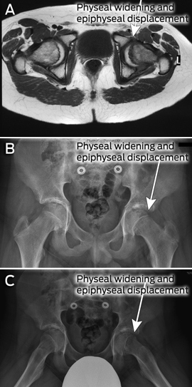
Second, a 13-year-old boy with a 3-week history of left hip pain and limp underwent MRI (Box 2, A) without a prior x-ray for suspected Perthes disease. The report stated “left hip joint effusion, without other identifiable abnormality”. When symptoms persisted, the patient was referred to a paediatric emergency department. The magnetic resonance images were reviewed and a slipped capital femoral epiphysis (SCFE) was evident (but had not been reported). Standard hip x-rays showed an obvious SCFE (Box 2, B and C). X-ray is always the initial investigation of choice for Perthes disease and SCFE (MRI should be reserved for equivocal cases), and SCFE is a far more common diagnosis than Perthes disease during adolescence.3
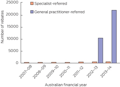
Over 20 000 GP-referred Medicare-rebated MRI scans were performed in patients under the age of 16 in the 2013–14 financial year (Box 3). The number of referrals per GP in Australia was 0.44 in 2012–13 and doubled to 0.90 in 2013–14.4,5
In contrast, specialist referrals for paediatric MRI were 0.31 scans per specialist in 2007–08 and 0.37 in 2013–14;4,5 the number of MRI scans referred per specialist has not altered appreciably.
While it is important to decrease radiation from CT scans in children under 16 years, it is also important to remember that MRI is mostly unhelpful in diagnosing paediatric musculoskeletal complaints — most of which can be confirmed on plain x-ray after taking an accurate history and examining the patient. Due to the significant cost to the health care system of MRI, we suggest that evidence of prior x-ray imaging should be a requirement before Medicare funds an MRI scan.
Competing interests
No relevant disclosures.
References
- Australian Government. Department of Health. Improving access to magnetic resonance imaging (MRI) services fact sheet. http://www.health.gov.au/internet/main/publishing.nsf/Content/di-factsheet-mri (accessed Sep 2014).
- Fraser RK, Nattrass GR, Chow CW, Cole WG. Pes anserinus syndrome due to solitary tibial spurs and osteochondromas. J Pediatr Orthop 1996; 16: 247-248. 2
- Jarrett DY, Matheney T, Kleinman PK. Imaging SCFE: diagnosis, treatment and complications. Pediatr Radiol 2013; 43 Suppl 1: S71-S82. lefthere
- Australian Government. Department of Human Services. Medicare Australia statistics. All Medicare items in “Group: I5 Magnetic resonance imaging, Subgroup: 33 for specified conditions - person under the age of 16 yrs” processed from July 2007 to June 2014. https://www.medicareaustralia.gov.au/cgi-bin/broker.exe?_PROGRAM=sas.mbs_item_standard_report.sas&_SERVICE=default&_DEBUG=0&VAR=services&STAT=count&PTYPE=finyear&START_DT=200707&END_DT=201406&RPT_FMT=by+time+period+and+state&DRILL=ag&GROUP=I0533 (accessed Sep 2014).
- Australian Institute of Health and Welfare. Workforce data. Medical workforce 2012 and 2013 detailed tables. http://www.aihw.gov.au/workforce-data (accessed Mar 2015).
Patterns of Osteoporosis Treatment Initiation Following Low-Trauma Fracture: A Population-Based Retrospective Cohort Study in New South Wales, Australia, Using Linked Administrative Health Data
Mike Lin, Huy Nguyen, Thach Tran, Robert D. Blank, Dana Bliuc, Jacqueline R. Center
Rapid Growth in Diagnostic CT Knee Imaging in Australia: Implications for Imaging Stewardship
Hanwen Chen, Rachelle Buchbinder, Sean Docking, Ian A Harris
Spinal cord stimulation patterns of care, re‐interventions, and costs for private health insurers, Australia, 2011–22: a retrospective observational study
Caitlin MP Jones, Christopher G Maher, Rachelle Buchbinder, Ian A Harris, Chung‐Wei Christine Lin, Christopher Hayes, Alexandra Gorelik
The incidence of and risk factors for hospitalisations and amputations for people with diabetes‐related foot ulcers in Queensland, 2011–19: an observational cohort study
Yuqi Zhang, Susanna M Cramb, Steven M McPhail, Rosana Pacella, Jaap J Netten, Ewan M Kinnear, Peter A Lazzarini
Bone health perspectives among Indigenous people: a qualitative study
Troy Walker (Yorta Yorta), Karan P Singh, Vanessa Gan, Brooke Conley (Ngiyampaa), Jessica Bravo, Nigel Smith (Weilwan), April Clarke (Eastern Maar, Kirrae Whurrung, Djap Wurrung), Jackson Baker, Louise J Maple‐Brown, Robin M Daly, Jennifer Browne, Jesse Zanker, Cat Shore‐Lorenti, David Scott, Peter R Ebeling, Ayse Zengin
I am a Civil War
Jennifer G Mowbray