




Volume 194 - Issue 7
Community-acquired Klebsiella pneumoniae liver abscesses — an “emerging disease” in Australia
Authors: Kudzai N Kanhutu, Jeffrey J Post, Kate R Clezy and Hong Y L Foo
Med J Aust 2011; 194 (7): 374. || doi: 10.5694/j.1326-5377.2011.tb03017.x
Published online: 4 April 2011
Published online: 4 April 2011
To the Editor: Further to the recent article by Anstey and colleagues on community-acquired Klebsiella pneumoniae liver abscesses,1 we report two similar cases at our hospital in late 2010.
Case 1: A 55-year-old Indonesian-born man was referred from general practice in October 2010 with a 5-day history of fever and progressive epigastric pain. He did not have diabetes, but did have dyslipidaemia. He had migrated from Indonesia in the 1980s; his most recent visit to Indonesia was in January 2010, for 3 weeks.
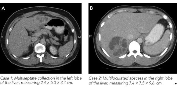
As he had mildly deranged liver function test results, he was investigated with abdominal ultrasound and computed tomography (CT). Both showed a large multiseptate collection in the left lobe of the liver (Box, A). The liver collection was drained under radiological guidance, yielding a pure growth of K. pneumoniae. Urine culture was also positive for an identical isolate of K. pneumoniae. This man had a rapid clinical response to percutaneous drainage and was discharged on oral ciprofloxacin therapy.
Case 2: A 25-year-old Indonesian-born man presented to our emergency department in early November 2010 after 2 days of headache, high fever and abdominal cramps, culminating in an acute confusional state. He had no significant medical or surgical history and had last visited Indonesia in March 2010, for 2 weeks. Initial therapy and investigations were aimed at excluding a diagnosis of meningitis. Results of a CT scan of the brain and of cerebrospinal fluid analysis were unremarkable.
The patient remained acutely unwell and developed diarrhoea and right upper quadrant abdominal pain. Blood cultures were positive for K. pneumoniae within 48 hours of admission. Abdominal CT showed a large multiloculated abscess in the right lobe of the liver (Box, B). The liver abscess aspirate grew a pure culture of K. pneumoniae. The patient responded to treatment with ceftriaxone and large-volume percutaneous drainage.
In both these cases, an antibiotic sensitive mucoid strain of K. pneumoniae was cultured.
These cases add weight to the possibility raised by Anstey and colleagues that community-acquired K. pneumoniae liver abscess is indeed an emerging phenomenon in Australia. Further, the extended length of time between our patients’ travel to Indonesia and the clinical presentation (9 and 8 months, respectively) is suggestive of local (Australian) acquisition of the disease. Clinicians should consider abdominal imaging in cases of bacteraemia due to K. pneumoniae.
References
- Anstey JR, Fazio TN, Gordon DL, et al. Community-acquired Klebsiella pneumoniae liver abscesses — an “emerging disease” in Australia. Med J Aust 2010; 193: 543-545. 0_IGBEJDHB
Costs of Delivering the Hep B PAST Model of Care in Remote Australia: A Retrospective Pre–Post Study
Hoa Nguyen, Anh Le Tuan Nguyen, Paula Binks, Melita McKinnon, Emily Vintour-Cesar, Karen Wills, Nicola Stephens, Benjamin Cowie, Joshua S. Davis, Yuejen Zhao, Peter Nihill, Julie A. Campbell, Andrew J. Palmer, Jane Davies, Barbara de Graaff
Buruli Ulcer Acquired From Ringtail Possum Rescue
Daniel P. O'Brien, Naomi Clarke, Sara Bonnici, Mohammad A. Hussain
Q Fever Visceral Abscesses After Contact With Native Australian Marsupials
Samantha J. Laws, Enzo Binotto, James Stewart, Jennifer Ho, Trent Yarwood, Josh Hanson, Simon Smith
Hospital-Admitted Injection-Related Infections Among Incarcerated People Who Inject Drugs in Australia: A Retrospective Cohort Study
Andrew Palmer, Matthew Carter, Jeremy Yeo, Cecilia Shim, Jason Connor, Jeremy Hayllar, Gerald Holtmann, Naomi Moy, Elliott G. Playford, Naomi Runnegar, Paul J. Clark
The Live Attenuated Influenza Vaccine in Australia: An Additional Tool for Influenza Prevention
Cyra Patel, Alexis Pillsbury, Tran Nguyen, Xia Wang, Helen E. Quinn, Clayton K. Chiu, Allen C. Cheng, Katie L. Flanagan, Zhicheng Wang
Reflex Testing for Hepatitis D Infection: A Unique Opportunity to Reduce Hepatitis D-Related Chronic Liver Disease Deaths in Australia
Jessica Howell, Lauren Andersson, Miriam T. Levy, James O'Beirne, Leon Adams, Katharine Irvine, Avik Majumdar, Golo Ahlenstiel, Kathy Jackson, Krispin Hajkowicz, Joseph Doyle, Jane Davies, Sarah Cherian, Wayne Dimech, Alexander J. Thompson