




Volume 194 - Issue 7
Takotsubo cardiomyopathy associated with alcohol withdrawal
Authors: Angus G Thompson and Joseph Hung
Med J Aust 2011; 194 (7): 373. || doi: 10.5694/j.1326-5377.2011.tb03015.x
Published online: 4 April 2011
Published online: 4 April 2011
To the Editor: A 61-year-old man presented to the emergency department (ED) of a tertiary hospital seeking treatment for alcohol withdrawal after 36 hours of abstinence. He reported central chest pain radiating to the jaw and left arm that had been present for 2 hours before his arrival at the hospital. He had no history of cardiac disease and no known risk factors for coronary artery disease.
An electrocardiogram (ECG) showed sinus tachycardia with T-wave inversion in leads V1, V2 and V3. Two hours after the patient’s arrival at the ED, he tested positive for troponin-T. Over the next hour, his ECG showed development of ST elevation of 1–2 mm in leads V3, V4 and V5. Because of severe alcohol withdrawal, his clinical status precluded urgent coronary angiography; and after treatment with diazepam was commenced, the ST elevation that was evident no longer met criteria for urgent angiography. The patient was given standard medical therapy for acute coronary syndrome, including aspirin, clopidogrel and intravenous heparin, while in the ED, along with ongoing diazepam for alcohol withdrawal.
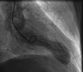
He was later admitted to the coronary care unit with a diagnosis of acute coronary syndrome. The next day, an ECG showed development of widespread T-wave inversion in leads V1 to V5. The dynamic ECG changes were not associated with ongoing chest pain. On Day 3 of the patient’s admission, coronary angiography showed normal coronary arteries, and ventriculography showed apical ballooning of the left ventricle, consistent with a diagnosis of takotsubo cardiomyopathy (Box). Treatment with an angiotensin-converting enzyme inhibitor and a β-blocker was commenced. Three months later, follow-up echocardiography showed a return to normal regional and global left ventricular function.
Takotsubo cardiomyopathy takes its name from a traditional Japanese octopus trap that has a similar shape to the abnormally contracting left ventricle seen with this condition.1 Typical findings in a patient with takotsubo cardiomyopathy are chest pain associated with emotional or physical stress, with ST segment changes on electrocardiography and apical ballooning on ventriculography, which is generally expected to resolve within weeks to months; troponin level may or may not be positive. The mechanism of this condition has not yet been determined, but it appears likely that it is due to hyperadrenergic-hypercatecholaminergic states (such as alcohol withdrawal) resulting in localised or diffuse coronary vasospasm.2 Takotsubo cardiomyopathy has only rarely been associated with alcohol withdrawal, and has once been reported in a patient with alcohol withdrawal associated with seizures.3,4
References
- Sato H, Tateishi H, Uchida T, et al. Tako-tsubo-like left ventricular dysfunction due to multivessel coronary spasm. In: Kodama K, Haze K, Hori M. Clinical aspect of myocardial injury: from ischemia to heart failure [Japanese]. Tokyo: Kagakuhyoronsha Publishing Co, 1990: 56-64. 0_CHDEEGAI
- Nef HM, Mollman H, Akashi YJ, Hamm CW. Mechanisms of stress (Takotsubo) cardiomyopathy. Nat Rev Cardiol 2010; 7: 187-193. 0_CBBEFCDI
- Suzuki K, Osada N, Akasi YJ, et al. An atypical case of “Takotsubo cardiomyopathy” during alcohol withdrawal: abnormality in the transient left ventricular wall motion and a remarkable elevation in the ST segment. Intern Med 2004; 43: 275-276. 0_CBBJBGGA
- Mitchell SA, Crone RA. Takotsubo cardiomyopathy: a case report. J Am Soc Echocardiog 2006; 19: 1190. 0_CBBHJDCB
Fatty Liver Disease in Australia: A Narrative Review on the Epidemiology, Natural History, Prognostication and Management in People With Metabolic Dysfunction
Karl Vaz, Daniel Clayton-Chubb, William W. Kemp, Stuart K. Roberts, Ammar Majeed
Sex Differences in Management and Outcomes of People With ST-Elevation Myocardial Infarction, New South Wales, 2011–2020: A Retrospective Cohort Study
Adam C. Livori, Jedidiah I. Morton, Dianna J. Magliano, Jonathan E. Shaw
Rethinking Rheumatic Fever Diagnosis: Progress, Pitfalls and the Path Towards a Definitive Test
Anna P. Ralph, Rachel Webb, Tom Parks, Nicole J. Moreland, Asha C. Bowen, Benjamin Jones, Emma Ndagire, Andrea Z. Beaton
Sex Differences in Management and Outcomes of People With ST-Elevation Myocardial Infarction, New South Wales, 2011–2020: A Retrospective Cohort Study
Samia Kazi, Simone Marschner, Clara K. Chow
Distribution of Cardiovascular Disease Risk Based on the Updated 2023 Guideline-Recommended Australian Cardiovascular Disease Risk Algorithm and Comparison With the 2012 Algorithm: An Observational Study
Nina Lazarevic, Meghana Bhat, Grace Joshy, Danielle C. Butler, Mark Woodward, Anushka Patel, Rod T. Jackson, Garry Jennings, Rosemary Wyber, Ellie Paige, Emily Banks
They Brought the Hospital to Me
Hugh N. Burrill