




Volume 193 - Issue 3
Subconjunctival dog heartworm
Authors: Elaine W Chong, Harsha Sheorey, Cheng Hean Lo, David M Spratt and Enrique Graue-Hernández
Med J Aust 2010; 193 (3): 184. || doi: 10.5694/j.1326-5377.2010.tb03845.x
Published online: 2 August 2010
Published online: 2 August 2010
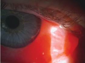
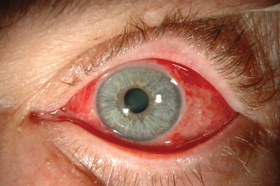
To the Editor: In February 2009, a 68-year-old man presented to the Royal Victorian Eye and Ear Hospital within hours of developing an itchy, red left eye. The patient, who was otherwise healthy, lived in suburban Melbourne, usually with his pet dogs, but the last of his dogs had recently died. The patient was unsure if all his dogs had been dewormed regularly because he spends about 6 months a year in Europe. General inspection of the eye suggested subconjunctival haemorrhage. However, slit-lamp examination showed a mobile, tightly coiled structure within the subconjunctival blood. It grew increasingly agitated with higher slit-lamp light intensity (Box, A). Assessment of the patient’s visual acuity and the anterior and posterior chambers of the eyes was unremarkable. Blood tests revealed a positive filarial serology and eosinophilia.
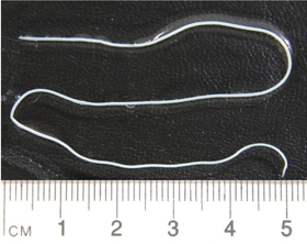
The patient was transferred to the operating theatre and, under topical anaesthesia, a 5 mm conjunctival incision was made and the mobile structure removed (Box, B and C). The patient was discharged with a prescription for prednisolone acetate 1% and chloramphenicol 0.5% eye drops (one drop four times a day). He made a full recovery.
The extracted specimen was reviewed by one of us (D M S). The 150 mm worm was identified as a young adult female filarioid nematode, Dirofilaria immitis (commonly named dog heartworm) after comparisons with laboratory specimens of D. immitis and Pelecitus roemeri. Infection with either P. roemeri (kangaroo and wallaby knee worm) or Loa loa (loiasis) was excluded. Our specimen did not have lateral alae and the distance from anus to tail was shorter than would be expected for the kangaroo worm. In addition, the patient had never been to Africa where loiasis is endemic to several countries.
Subconjunctival dog heartworm is rare, but its incidence is increasing in parts of the world.1,2 Dogs are the natural hosts and transmission to humans occurs through mosquito bites of the skin (into which the third-stage infective larva may escape). For an unknown reason, the worm sometimes takes an abnormal migratory route and ends up in the eye of the host. Ophthalmic cases have been reported in dogs.3,4 Careful measures to exterminate mosquitoes and deworm dogs and cats are important in limiting its transmission. Surgical extraction is the definitive treatment and further treatment with systemic anthelmintics is unnecessary.5 Humans are non-natural hosts for this parasite and, therefore, its life cycle cannot be completed within the human body. When a larva does evade the human immune system, as in the case of our patient, the chances of another larva being present elsewhere in an immunocompetent person seems remote. Furthermore, unless the larva becomes clinically apparent, it would be impossible to find.
Acknowledgements
We thank our patient for consenting to have details of his case published.
References
- Rouhette H, Marty P, Zur C, et al. Ocular filariasis: not strictly tropical. Ophthalmologica 1999; 213: 206-208.
- Stringfellow GJ, Francis IC, Coroneo MT, Walker J. Orbital dirofilariasis. Clin Experiment Ophthalmol 2002; 30: 378-380. 0_i1092991
- Lavers DW, Spratt DM, Thomas C. Dirofilaria immitis from the eye of a dog. Aust Vet J 1969; 284-286. 0_i1092993
- Spratt DM, Mallet G, Derrington PC, Thomas C. Male Dirofilaria immitis from the eye of a dog. Aust Vet J 1971; 47: 124. 0_i1092995
- Ruiz-Moreno JM, Bornay-Llinares FJ, Prieto Maza G, et al. Subconjunctival infection with Dirofilaria repens: serological confirmation of cure following surgery. Arch Ophthalmol 1998; 116: 1370-1372. 0_i1092999
Costs of Delivering the Hep B PAST Model of Care in Remote Australia: A Retrospective Pre–Post Study
Hoa Nguyen, Anh Le Tuan Nguyen, Paula Binks, Melita McKinnon, Emily Vintour-Cesar, Karen Wills, Nicola Stephens, Benjamin Cowie, Joshua S. Davis, Yuejen Zhao, Peter Nihill, Julie A. Campbell, Andrew J. Palmer, Jane Davies, Barbara de Graaff
Buruli Ulcer Acquired From Ringtail Possum Rescue
Daniel P. O'Brien, Naomi Clarke, Sara Bonnici, Mohammad A. Hussain
Q Fever Visceral Abscesses After Contact With Native Australian Marsupials
Samantha J. Laws, Enzo Binotto, James Stewart, Jennifer Ho, Trent Yarwood, Josh Hanson, Simon Smith
Hospital-Admitted Injection-Related Infections Among Incarcerated People Who Inject Drugs in Australia: A Retrospective Cohort Study
Andrew Palmer, Matthew Carter, Jeremy Yeo, Cecilia Shim, Jason Connor, Jeremy Hayllar, Gerald Holtmann, Naomi Moy, Elliott G. Playford, Naomi Runnegar, Paul J. Clark
The Live Attenuated Influenza Vaccine in Australia: An Additional Tool for Influenza Prevention
Cyra Patel, Alexis Pillsbury, Tran Nguyen, Xia Wang, Helen E. Quinn, Clayton K. Chiu, Allen C. Cheng, Katie L. Flanagan, Zhicheng Wang
Reflex Testing for Hepatitis D Infection: A Unique Opportunity to Reduce Hepatitis D-Related Chronic Liver Disease Deaths in Australia
Jessica Howell, Lauren Andersson, Miriam T. Levy, James O'Beirne, Leon Adams, Katharine Irvine, Avik Majumdar, Golo Ahlenstiel, Kathy Jackson, Krispin Hajkowicz, Joseph Doyle, Jane Davies, Sarah Cherian, Wayne Dimech, Alexander J. Thompson