




Volume 187 - Issue 11
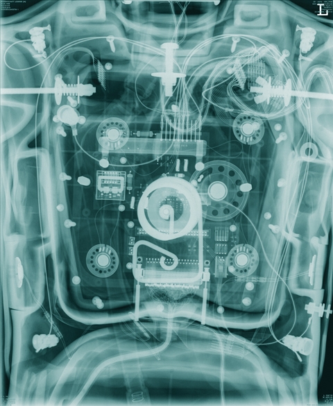
Recently in the emergency department: chest x-ray of a repeatedly resuscitated object
Authors: Wolfgang Lederer, Martin Breiteneder, Michael Rieger and Christoph J Schlimp
Med J Aust 2007; 187 (11): 695. || doi: 10.5694/j.1326-5377.2007.tb01484.x
Published online: 3 December 2007
Published online: 3 December 2007
“Kelly Laerdal”, age unknown, but repeatedly resuscitated, was scheduled for a chest x-ray to investigate the metallic contents of the thorax, with the aim of understanding why resuscitation manikins might differ in their susceptibility to electromagnetic interference in electrocardiogram recording.1
Besides some minor electronic components, the image shows a prominent cardiac massage pressure point, gives a hint of shoulder joint junctions, and shows exactly where the auscultation regions (covered by loudspeakers) are located — a “must know” for every advanced cardiac life support student.
References
- Schlimp CJ, Breiteneder M, Lederer W. Safety aspects for public access defibrillation using automated external defibrillators near high-voltage power lines. Acta Anaesthesiol Scand 2004; 48: 595-600. 0_BEIIHFGA
Reducing Nitrous Oxide Emissions Across the Melbourne Biomedical Precinct
Ross Robertson, Andrew Downey, Daryl Williams, Bjorn Makein, Ben Dunne, Tugce Ozturk, Ying Gu, Rebecca McIntyre
Multimorbidity Clusters Among People Aged 65 Years and Over in Australia: A Nationwide Cross-Sectional Data Linkage Study
Weisi Chen, Christine Y. Lu, Sarah N. Hilmer, Alice A. Gibson, Edwin C. K. Tan
When ‘Liver Enzymes’ Are Not Hepatic: Late-Onset Pompe Disease
Shauna Madigan, Georgina England, Wayne Rankin
West Nile virus Kunjin subtype in rural NSW
Emily Gibson, Megan Whitley, Peter Murray, Linda Hueston, Jane Bennett, Raguharan Kathiresu, David N Durrheim
The impact of the Breast Screen NSW transition from film to digital mammography, 2002–2016: a linked population health data analysis
Rachel Farber, Nehmat Houssami, Katy J L Bell