




Medical education
Lessons from practice
Volume 222 - Issue 3
Human case of diphyllobothriasis in Australia
Authors: Sadid F Khan, Anson V Koehler, Alistair Tinson, Maidhili Chinnappan, Harsha Sheorey and Robin B Gasser
Med J Aust 2025 || doi: 10.5694/mja2.52579
Published online: 3 February 2025
Published online: 3 February 2025
In 2024, a 30-year-old female resident of Melbourne, Australia, with no past medical history presented to the Royal Melbourne Hospital
Clinical record
In 2024, a 30‐year‐old female resident of Melbourne, Australia, with no past medical history presented to the Royal Melbourne Hospital. During an acute episode of diarrhoea, the patient passed part of a worm that she collected, placed in a jar, and presented to the emergency department.
This occurred on the background of a history of travel to more than 30 countries in Europe, Asia and the Americas between 2013 and 2024 (Supporting Information, figure 1). She reported eating fermented Baltic Sea herring (surströmming) in Sweden and cured salmon (gravlax) in Norway in 2020, in addition to consuming fresh fish and other seafood (ceviche) in South America (2023), Japan (2013) and, potentially, in Australia.
At the time of presentation, her symptoms of diarrhoea had resolved. The patient reported a history of intermittent right lower quadrant abdominal pain over a period of about seven years for which a cause had not been established. Laboratory investigations, including eosinophil count, were unremarkable.
The macroscopic examination of the worm specimen (40cm in length) revealed the segmented strobila of a tapeworm that was consistent with a pseudophyllidean cestode (Box 1, A).1 The segments (proglottids) were mature and gravid (ie, contained eggs); no scolex was found. Microscopic examination of operculated eggs expressed from individual segments were ovoid and about 70μm×40μm in size (Box 1, B).1 Based on morphological features and morphometrics, the worm was proposed to be a species of Dibothriocephalus.
To refine the identification, genetic analysis of genomic DNA isolated from this specimen2 was undertaken by polymerase chain reaction‐based sequencing of part of the mitochondrial gene cytochrome c oxidase subunit 1 (cox1) (Supporting Information, appendix 1). A phylogenetic analysis of the sequence obtained here (accession No. PP474259) and 31 other representative cox1 sequences revealed that the sequence (PP474259) clustered with, and was identical to, cox1 sequences derived from individual specimens of Dibothriocephalus latus from Chile and Switzerland. These sequences all clustered, with strong nodal support (97–100%), with other cox1 sequences from other D. latus specimens from Europe and South America (Box 2).
Following the diagnosis of diphyllobothriasis, the patient received a single dose (10mg/kg) of praziquantel, and further tapeworm segments were reported by the patient to have been expelled within two days. Although up to 40% of patients with D. latus infection can develop B12 deficiency,1 no abnormality was seen in this case. Following treatment, the patient experienced clinical improvement. One month after praziquantel treatment, a faecal examination showed no evidence of Dibothriocephalus or other worm eggs.
Discussion
D. latus (fish or broad tapeworm), previously Diphyllobothrium latum, and related species are the largest tapeworms infecting humans and can grow up to 25m in length.1 Although most people infected are asymptomatic, about 20% experience diarrhoea and abdominal pain.1 Rare complications include intestinal obstruction from massive infection and biliary obstruction caused by migrating proglottids.1
D. latus has an aquatic life cycle (Box 1, C) involving small crustacean copepods as a first intermediate host, subsequently ingested by a freshwater fish as the second intermediate host. The second larval (plerocercoid) stage develops in the tissue of the infected fish; thus, piscivorous animals, including humans, become infected by eating raw or undercooked fish. The parasite does not survive cooking, freezing or curing when adequately performed.1,3,4 Infection with this species is usually acquired in endemic countries of the Northern Hemisphere (Asia, Europe and North America).1 Imported cases, which shed large numbers of eggs in stool, pose a risk of introduction into non‐endemic regions, especially those with poor sanitary infrastructure.1 This has been demonstrated for D. latus in South America, likely through introduction from Europe.5,6 D. latus is not known to be endemic in Australian waters, but cases of infection could still occur in Australia through the consumption of fish imported from countries where D. latus is endemic. Autochthonous infection from Australian fish with other pseudophyllidean cestodes remains very rarely reported,7 though potentially underappreciated.4,8
Accurate identification supports better understanding of geographical distribution, host preferences, and impacts on human health and the fish industry. Phylogenetic determination of whether this patient acquired their infection from Europe or South America was not possible. However, the epidemiological risk, symptomatology, and molecular findings favour acquisition from raw fish consumption infected with tapeworms closely related to those found in Nordic Europe.
Timely diagnosis and treatment of human cases is central to preventing the potential for D. latus to become established in a non‐endemic country or region. In many high income countries (including Australia), the awareness and diagnostic expertise regarding zoonotic fish parasites are lacking among medical and laboratory practitioners.8,9 Infection risk of zoonotic fish‐borne pathogens, such as D. latus, can be influenced by converging factors, such as changes in climate and water temperatures, potentially leading to alterations in the geographical range of parasites, global demand on diminishing fish stocks, use of aquaculture associated with risk of parasite introduction or spread, and global mobility of infected people and fish worldwide. Dietary habits linked to the consumption of raw and undercooked fish and a lack of standardised food inspection, storage and processes are additional contributing factors.1,3,4
Lessons from practice
- Multiple converging factors place humans at risk of infection with fish parasites. History of raw fish consumption is an important epidemiological risk when assessing patients.
- Clinician education and awareness of fish parasites are required to facilitate timely patient diagnosis and treatment, and to mitigate ecological risk.
- Diagnostic laboratories need to maintain competency in the identification of fish parasites. If required, expert consultation should be sought to ensure timely and accurate identification.
- Diagnosis using molecular methods is suggested to avoid misidentification, and to generate better epidemiological understanding to inform risks to human health and economy.
Box 1 – Key aspects relating to the present clinical case
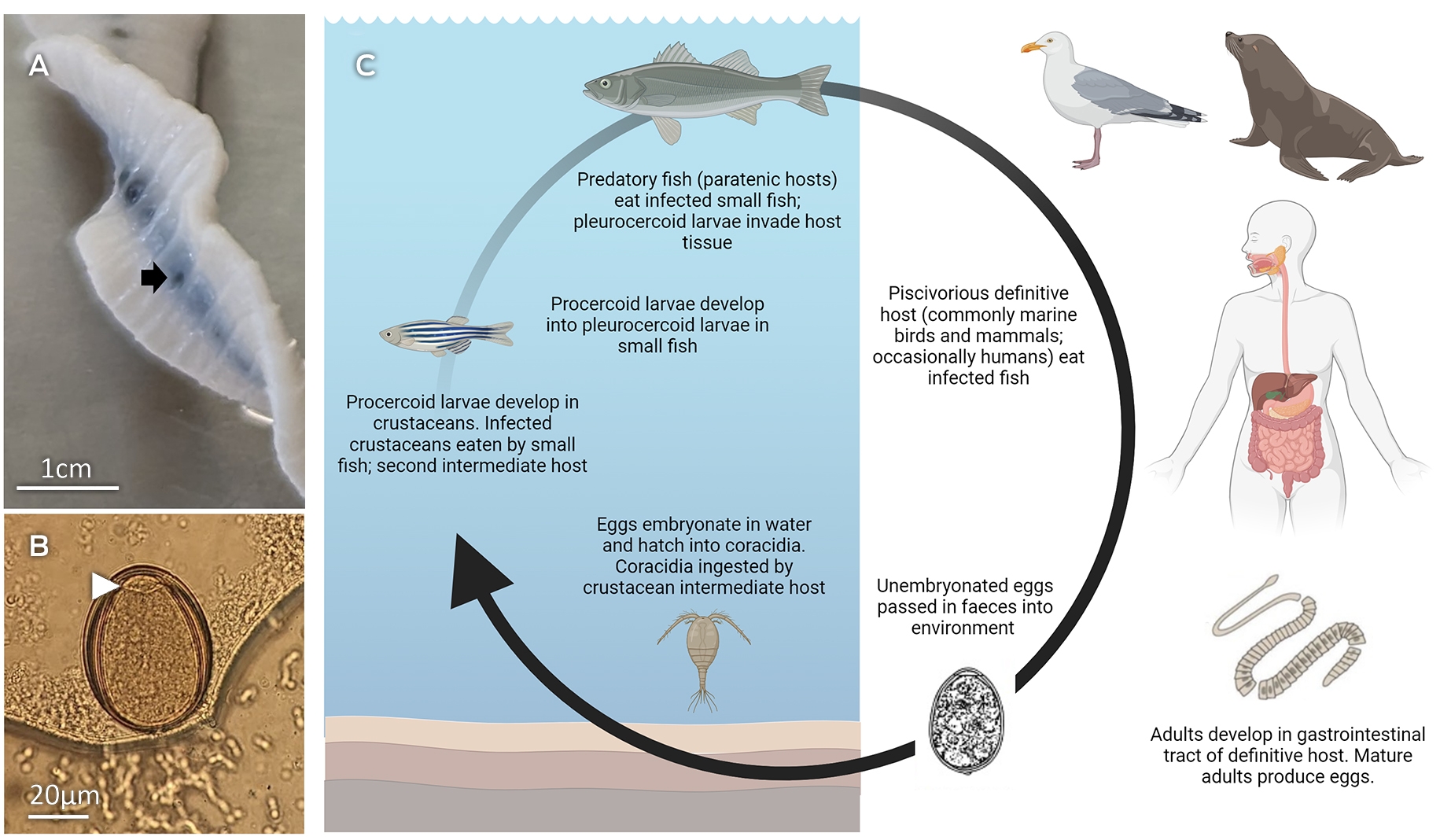
(A) Worm specimen (40cm×1.5cm) expelled from this patient during a bout of diarrhoea; each segment (proglottid ~3mm×15mm) of this specimen had a centrally located uterus, a dark “zipper‐like” pattern under the strobila and a ventrally located uterine pore/opening (arrow), consistent with a pseudophyllidean cestode, most commonly Dibothriocephalus spp. (B) Eggs expelled from a segment (via the uterine pore) were operculated (arrowhead) and~70×40 μm in size. (C) Life cycle of Dibothriocephalus latus showing transmission (via the egg) from the piscivorous host (eg, human, dog, cat, seal, birds) to the first intermediate host — a crustacean (including copepod) — in which the first larval stage (procercoid) develops. The infected crustacean is ingested by the second intermediate host — a freshwater fish — in which the second larvae stage (plerocercoid) develops in tissues and can remain alive for the lifespan of the fish. The human and other piscivorous definitive hosts become infected by eating raw or undercooked fish; here, the adult worm can grow to a length of 25m and produces eggs within four to six weeks.1
Box 2 – Phylogenetic analysis of mitochondrial cytochrome c oxidase subunit 1 gene (cox1; 443 bp) sequence data derived from the worm specimen from the present human case (bold type) and also representative Dibothriocephalus latus specimens from different countries*
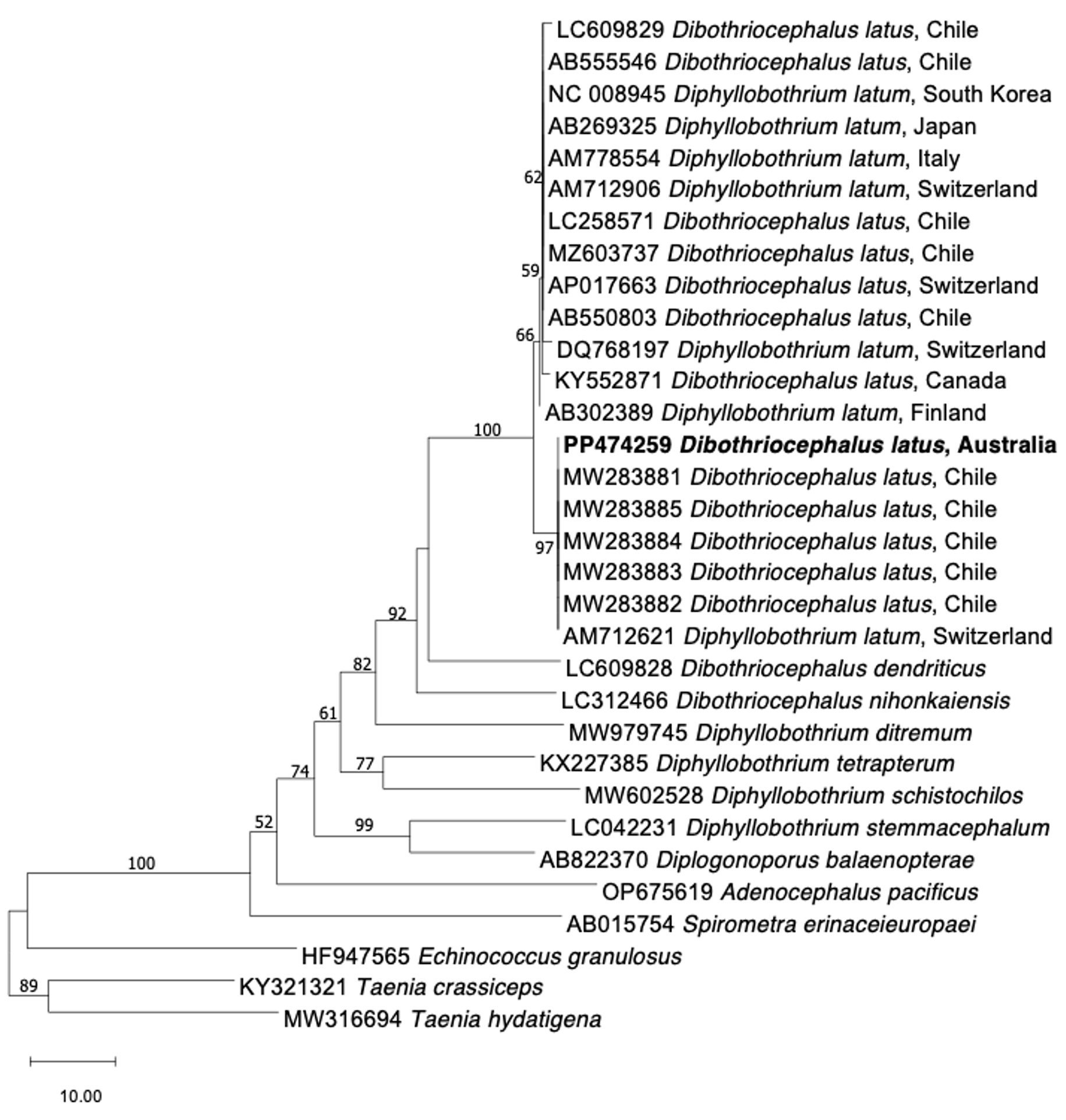
* GenBank accession numbers indicated on the tree constructed using the neighbour‐joining distance method.2 Nodal support is given in bootstrap percentages. Members of the genus Taenia were used as outgroups.
Competing interests
No relevant disclosures.
Acknowledgements
The molecular work performed at the laboratory of the University of Melbourne Veterinary School was funded through a grant from the Australian Research Council (LP220200614 to RBG). No specific funding for this study was provided.
References
- Scholz T, Garcia HH, Kuchta R, Wicht B. Update on the human broad tapeworm (genus Diphyllobothrium), including clinical relevance. Clin Microbiol Rev 2009; 22: 146‐160.
- Koehler AV, Robson JMB, Spratt DM, et al. Ocular filariasis in human caused by Breinlia (Johnstonema) annulipapillata nematode, Australia. Emerg Infect Dis 2021; 27: 297‐300.
- Williams W, Hernandez‐Jover M, Shamsi S. A critical appraisal of global testing protocols for zoonotic parasites in imported seafood applied to seafood safety in Australia. Foods 2020; 9: 448.
- Shamsi S. Seafood‐borne parasites in Australia: human health risks, fact or fiction? Microbiology Australia 2020; 41: 33‐37.
- Radačovská A, Bazsalovicsová EČ, Šoltys K, et al. Unique genetic structure of the human tapeworm Dibothriocephalus latus from the Alpine lakes region — a successful adaptation? Parasitology 2022; 149: 1106‐1118.
- Torres P, Yera H. Diphyllobothriidae. In: Rose JB, Jiménez‐Cisneros B; editors. Global Water Pathogen Project. Part 3: Specific excreted pathogens: environmental and epidemiology aspects. Michigan (USA): Michigan State University, 2018.
- Moore CV, Thompson R, Jabbar A, et al. Rare human infection with Pacific broad tapeworm Adenocephalus pacificus, Australia. Emerg Infect Dis 2016; 22: 1510‐1512.
- Shamsi S, Sheorey H. Seafood‐borne parasitic diseases in Australia: are they rare or underdiagnosed? Intern Med J 2018; 48: 591‐596.
- Seal A, Harding C, Shamsi S. A preliminary report on the awareness and knowledge of seafood‐borne parasitic diseases among medical doctors in Australia. Parasitol Int 2020; 74: e101993.
Provenance: Not commissioned; externally peer reviewed.
Costs of Delivering the Hep B PAST Model of Care in Remote Australia: A Retrospective Pre–Post Study
Hoa Nguyen, Anh Le Tuan Nguyen, Paula Binks, Melita McKinnon, Emily Vintour-Cesar, Karen Wills, Nicola Stephens, Benjamin Cowie, Joshua S. Davis, Yuejen Zhao, Peter Nihill, Julie A. Campbell, Andrew J. Palmer, Jane Davies, Barbara de Graaff
Buruli Ulcer Acquired From Ringtail Possum Rescue
Daniel P. O'Brien, Naomi Clarke, Sara Bonnici, Mohammad A. Hussain
Q Fever Visceral Abscesses After Contact With Native Australian Marsupials
Samantha J. Laws, Enzo Binotto, James Stewart, Jennifer Ho, Trent Yarwood, Josh Hanson, Simon Smith
Hospital-Admitted Injection-Related Infections Among Incarcerated People Who Inject Drugs in Australia: A Retrospective Cohort Study
Andrew Palmer, Matthew Carter, Jeremy Yeo, Cecilia Shim, Jason Connor, Jeremy Hayllar, Gerald Holtmann, Naomi Moy, Elliott G. Playford, Naomi Runnegar, Paul J. Clark
The Live Attenuated Influenza Vaccine in Australia: An Additional Tool for Influenza Prevention
Cyra Patel, Alexis Pillsbury, Tran Nguyen, Xia Wang, Helen E. Quinn, Clayton K. Chiu, Allen C. Cheng, Katie L. Flanagan, Zhicheng Wang
Reflex Testing for Hepatitis D Infection: A Unique Opportunity to Reduce Hepatitis D-Related Chronic Liver Disease Deaths in Australia
Jessica Howell, Lauren Andersson, Miriam T. Levy, James O'Beirne, Leon Adams, Katharine Irvine, Avik Majumdar, Golo Ahlenstiel, Kathy Jackson, Krispin Hajkowicz, Joseph Doyle, Jane Davies, Sarah Cherian, Wayne Dimech, Alexander J. Thompson